Carbohydrate Metabolism Flashcards

1
Q

Tissue Specialization

A

In multi-cellular organisms:

  • Different tissues assume specialized roles and play different roles in metabolism.
  • Some may lack one or more of the basic catabolic pathway (ex. brain)
  • Some may carry out unique functions and exhibit special pathways and processes (ex. liver)
  • Functional differences between tissues frequently reflects a differing regulation of metabolism and/or the occurance of different isozymes.
2
Q

Systemic Coordination

of

Metabolism

A

Specialized metabolism of different tissues requires regulation and integration by hormones.

Systemically circulated hormones can be used to coordinate the metabolic activities of a wide range of tissues simultaneously.

Most short term responses of the target tissues to hormone binding are the result of regluation of protein phosphorylation.

Three important hormones involved in short-term metabolic regulation are:

  1. Epinephrine: released from the adrenal medula in response to stress.
  2. Glucagon: released from the alpha cells of the pancreas in response to low blood glucose and insulin levels.
  3. Insulin: released from the beta celsl of the pancreas in response to high blood glucose levels.
3
Q

Glucagon Mechanism

A

Acts via its G-protein coupled receptors which utilizes cAMP as a second messenger.

4
Q

Epinephrine Mechanism

A
  1. Can act via a G-protein coupled receptor utilizing cAMP as the secondary messenger.
    • Many regulatory pathways act via cAMP and PKA.
  2. Can act via α-adrenergic receptors (also G-protein coupled) utilizing both diacylglycerol (DAG) and inositol triphosphate (IP3) as secondary messengers.
    • Note that after DAG is cleaved from PIP2 it will remain in the plasma membrane.
    • Relative number of enzymes affected via the Ca2+/calmodulin protein kinase pathway is less compared to PKA.
5
Q

Insulin Mechanism

A
  • Functions through a tyrosine-kinase receptor.
  • Can also utilize phosphatidylinositol-3,4,5-P3 (PIP3) as a secondary messenger.
  • Protein kinase B (serine kinase) is responsible for many of the metabolic effects of insulin including:
    • Activation of glycogen synthase by inhibition of glycogen synthase kinase 3.
    • Activation of glycolysis in muscle.
    • Inhibition of lipolysis in adipose tissue.
6
Q

Glucose Uptake

A
  • Glucose transported across cell membranes by:
    1. Facilitated diffusion via GLUT transports down their concentration gradients.
    2. In renal and intestinal epithelium, transported against its concentration gradient by Na+-glucose co-transporters (SGLTs)
7
Q

GLUT transporters

A
  • Glucose-dependent tissues such as RBC’s and brain have low Km insulin-independent GLUT1 or GLUT3 transports respectively.
  • In peripheral tissues such as muscle which are glucose-independent, GLUT4 transports have a low KM but is insulin-dependent ⇒ allows cross regulation.
  • The liver which does not rely on glucose for energy, uses the GLUT2 transporters have a high KM for glucose but is insulin-independent.
    • Limits glucose uptake to conditions when blood glucose levels are high.
    • Allows the transporter to act as a sensor of high blood glucose levels.

* Normal fasting blood glucose levels are 3.9 - 5.5 mmol/L.

8
Q

GLUT 4 Regulation

A
  • Located in muscle and adipose tissue.
  • Relatively low Km for glucose so would transport around normal fasting levels.
  • Insulin-dependent glucose transporter:
    • When insulin is absent, the transporters are removed from the plasma membrane and sequestered into vesicles.
      • Functional but not in the membrane.
    • Insulin signaling stimulates the movement of the transporter from internal stores to the plasma membrane.
  • In skeletal muscle, exercise stimulates GLUT4 translocation to the plasma membrane through AMP-activated protein kinase (AMPK) via unknown mechanism.
    • Long-term exercise also increases the amount of GLUT4 in the muscle cell.
9
Q

Mechanism of Insulin

GLUT4 Activation

A
  1. Binding of insulin to the α-subunit of its receptor activates a tyrosine kinase domain resulting in auto-cross-phosphorylation of tyrosine residues in the β-subunits.
  2. Negative charge of the phosphates causes IRS (insulin receptor substrate) proteins to bind to the β-subunit.
  3. IRS proteins phosphorylated at two Tyr residues by the kinase activity of activated insulin receptor.
  4. Phosphorylated-IRS dissociate from the receptor then bind to and activate proteins with SH2 domains i.e. PI-3-kinase (Phosphatidylinositol-3-kinase).
  5. PI-3-kinase phosphorylates PIP2 to PIP3.
  6. PIP3 activates PDK-1 (phosphoinositide-dependent kinase).
  7. PDK-1 activates downstream effectors Akt and PKB which results in the movement of GLUT4 to the cell surface in adipose and muscle, increasing glucose uptake.

Akt/PKB also:

  • phosphorylates and inactivates GSK3 (glycocen synthase kinase 3) resulting in increased glycogenesis.
  • Activate amino acid uptake and protein synthesis
  • Increase lipid synthesis
  • Inhibit gluconeogenesis
  • decrease cAMP levels by activating phosphodiesterase
  • various gene expression modulations both +/-
    • Increases protein synthesis by activating a kinase (mTOR) that ultimately results in the activation of eIF4 and EF2 from protein translation.
10
Q

Glucose Phosphorylation

A
  • Phosphorylation of glucose prevents back diffuse out of the cell via the transporter and commits it for use in that cell.
  • Hexokinases
    • Found in tissues such as muscle and brain.
    • Has a low KM for glucose
      • Can phosphorylate other monosaccharides but affinity for glucose considerably higher
    • Show product inhibition by glucose-6-phosphate.
  • Glucokinase
    • Found in liver and pancreatic β-cells
    • Has a high KM for glucose
    • Shows no direct product inhibition
11
Q

Glucokinase Kinetics

A
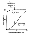
  • Despite being monomeric, glucokinase displays sigmoidal kinetics towards glucose.
  • The inflection point of the glucokinase enzyme curve is such that small changes in blood glucose levels causes significant changes in enzymatic activity.
  • When blood glucose levels are high the hepatic glucokinase becomes significantly more active.
  • Hexokinase, however, is fully saturated a normal concentrations of blood glucose.
12
Q

Glucokinase Regulation

A

Glucokinase activity is not directly regulated by its product but is indirectly regulated.

  • In hepatocytes, glucokinase binds to GKRP (glucokinase regulatory protein) which acts as a competitive inhibitor.
  • Glucokinase-GKRP complex is translocated into the nucleus where glucokinase is held in an inactive state.
  • Fructose-6-phosphate strongly stimulates this association.
    • F-6-P is in equilibrium with G-6-P
  • Fructose-1-phosphate or high glucose concentrations reverse the inhibition by triggering dissociation of the complex.

Transcription of the glucokinase gene is up-regulated in response to insulin ⇒ hormonal control.

13
Q

Regulation of glucose utilization

A

Cells must both take up and then phosphorylate glucose in order to utilize it for metabolic proesses.

Tissue-specific regulation of these two processes alows control of glucose utilization in a manner specific to the needs and function of the tissue.

  • Tissues which are glucose-dependent are controlled by product negative feedback only.
  • Tissues which are glucose-independent are controlled by blood glucose concentration and product inhibition.
  • Glucose-producing tissues are controlled by blood glucose concentrations only.
14
Q

Role of Glucose-6-Phosphate

A

G-6-P lies at a branch point for several pathways of carbohydrate metabolism.

Its production and utilization by various metabolic pathways are key regulatory points.

15
Q

Redox Balance

A

NADH produced during glycolysis must be recycled back into NAD+ for glycolysis to continue.

In aerobic conditions, reduction equivalents of NADH are shuttled into the mitochondria to undergo oxidative phosphorylation.

In anerobic conditions, pyruvate is converted to lactate via homolactic fermentation with the regeneration of NAD+.

16
Q

Malate-Aspartate Shuttle

A
  1. Electrons are transferred from cytosolic NADH to oxaloacetate forming malate and NAD+.
  2. Malate enters the mitochondrial inner membrane via malate/α-ketoglutarate transporter.
  3. Inside the matrix, malate is reoxidized by malate dehydrogenase and NAD+ to form OAA and NADH.
  4. OAA is converted to aspartate via a transamination reaction.
  5. Aspartate is transported back to the cytosol via a glutamate/aspartate transporter.
  6. In the cytosol the aspartate undergoes transamination to reform OAA.

*Shuttle is readily reversible: important in gluconeogenesis.

17
Q

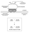
Glycerol-3-Phosphate Shuttle

A

Couples the cytosolic oxidation of NADH with the mitochondrial reduction of FAD.

  1. Cytoplasmic NADH utilized by glycerol-3-phosphate dehydrogenase to convert dihydroxyacetone phosphate to glycerol-3-phosphate.
  2. Glycerol-3-phosphate is then converted back to DHAP by mitochondrial version of the dehydrogenase which resides on the inner mitochondrial membrane. FAD is reduced to FADH2.
  3. Electrons from FADH2 are trasferred to the electron carrier Q which enters the respiratory chain as QH2.
18
Q

Homolactate Fermentation

A

Pyruvate converted to lacate under anaerobic conditions.

Concomitant oxidation of NADH to NAD+ restores redox balance in the cytoplasm and enables glycolysis to continue.

Lactate enters the blood and is ultimately reconverted to glucose in the liver via gluconeogenesis.

Excess H+ and lactate inhibits glycolysis.

19
Q

PFK1

(6-Phosphofructo-1-kinase)

Regulation and Effectors

A

Most important regulatory step in glycolysis because:

  • It is the commitment step for the glycolytic pathway.
  • It is allosterically modulated by many metabolic intermediates and products.
20
Q

Mechanism for the Allosteric Regulation

of

6-phosphofructo-1-kinase

(PFK1)

A

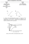
Allosteric effectors act by influencing the equilibrium between the active and inhibited forms of the enzyme by binding preferentially to one form or the other and stabilizing that form.

  • Fructose-6-Phosphate
    • Shows postive cooperativity in binding of PFK1 ⇒ reflected by the sigmoidal shape of [F-6-P] to Vo curve.
  • ATP
    • Vo initially rises with increasing [ATP] then falls again.
    • ATP acts at two different sites:
      • Is a substrate for the reaction
      • Is a negative heterotropic allosteric effector ⇒ ATP inhibits F-6-P binding
    • Since ATP affects substrate binding and not the Vmax it is K-type regulation.
21
Q

Role of

Fructose-2,6-Bisphosphate

A

Fructose-2,6-bisphosphate (F-2,6-P2) is a positive heterotropic effector of PFK1.​

F-2,6-P2 is is synthesized and degraded by a multifunctional enzyme with both:

  • kinase activity ⇒ PFK2
  • phosphatase activity
22
Q

PFK2

(6-phosphofructo-2-kinase)

Isozymes

&

Regulation by Tissue

A

Multifunctional enzyme reponsible for synthesis and degradation of fructose-2,6-bisphosphate:

PFK2 & F26Pase

Phosphorylation of either domain inhibits its catalytic activity.

Liver:

PFK2 enzyme a substrate for cAMP-dependent PKA.

Phosphorylation site for liver PFK2 isozyme lies within the kinase domain.

Phosphorylation → inhibits kinase activity & stimulates phosphatase activity.

Heart:

F26Pase enzyme is a substrate for PKB (Akt).

Phosphorylation site for heart PFK2 isozyme lies within the phosphatase domain.

Phosphorylation → inhibits phosphatase activity & stimulates kinase activity.

Skeletal Muscle:

PFK2/F26Pase isozyme has no phosphorylation sites.

Is not covalently regulated.

23
Q

Hepatic PFK1 Regulation

A
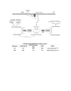
  • Hepatic PFK1 is primarily regulated by the [F-2,6-P2]
  • The primary function of F-2,6-P2 in the liver is to make PFK1 sensitive to regulation by glucagon and other hormones.
  • The liver does not consume glucose as fuel during times of need–rather it makes glucose for use by other tissues.
24
Q

Tissue Differences in the control of PFK1

A

Liver:

low [glucose]blood → high [glucagon] → [cAMP] increases → high [PKA] → PFK-2 inhibited → low [F-2,6-P2] → PFK-1 inhibited → glycolysis decreases

Heart:

Biochem test → flight or flight response → high [epinephrine] → high [PKA] or [PKB/Akt] → PFK-2 activated → high [F-2,6-P2] → PFK-1 activated → glycolysis increases

Skeletal Muscle:

ATP utilization → high [AMP] → PFK-1 activated → glycolysis increases

In a resting cell:

With an energy charge of 0.8-0.9 (ΔGATP = -14 - -15 kcal/mole)

PFK-1 would be strongly inhibited by ATP

25
Q

Pyruvate Kinase Deficiency

A
  • PK defects inhibit glycolysis therefore affects tissues which rely heavily on it for energy
    • RBC → causes chronic hemolytic anemia
  • Mutations can affect:
    • Enzyme stability
    • Substrate binding/turnover
    • Response to allosteric effectors
26
Q

Pyruvate Kinase

Isozymes

A

There are 4 tissue specific isozymes of pyruvate kinase:

L is found in the liver and a small amount in the kidney.

R is found in RBC and hematopoietic tissues.

M1 found in muscle, heart, and brain.

M2 predominates in all remaining tissues.

The L & R isoforms originate from a common gene and result from alternate promoters which result in different N-terminals.

  • L isozyme contains a serine at residue 11 which R lacks and is therefore subject to phosphorylative regulation.

The M1 and M2 isoforms also originate from a common gene and result from alternative splicing.

27
Q

Pyruvate Kinase

Hepatic Isozyme

A

Regluation of PK is important in the liver because it is also the site of gluconeogenesis.

  • Homo-tetrametric allosteric enzyme
  • Each subunit has four domains which work cooperatively in the tetramer to enduce changes affecting activity
    • Positive effectors:
      • Fructose-1,6-Bisphosphate
      • PEP
    • Negative effector:
      • ATP
  • The liver isoform of PK contains a serine residue in the N-domain which is not present in other forms.
    • This residue is a substrate for phosphorylation by cAMP-dependent PKA
      • Phosphorylation weakens substrate binding ⇒ increasing KM
      • Phosphorylation also increases PK’s sensitivity to allosteric regulations
      • A-form (active) = unphosphorylated
      • B-form (inactive) = phosphorylated

The allosteric and covalent effects are additive.

28
Q

Pyruvate Kinase

Transcriptional Regulation

A

Mediated through

carbohydrate response element binding protein (ChREBP)

  • Fasting state:
    • Glucagon → cAMP → PKA → phosphorylated ChREBP
    • Phosphorylated ChREBP is blocked from entering the nucleus and is inactive.
  • High glucose:
    • Glucose → xylulose-5-phosphate via the Pentose Phosphate Pathway
    • X-5-P activates Protein Phosphatase 2A (PP2A)
    • PP2A dephosphorylates ChREBP allowing it to enter the nucleus
    • In the nucleus ChREBP activates PK transcription as well as transcription of genes for fatty acid biosynthesis enzymes
29
Q

Pentose Phosphate Pathway

aka

Hexose Monophosphate Pathway

Basics

A
  • Alternate pathway from glucose-6-phosphate.
  • In the liver up to 30% of glucose can be oxidized this way.
  • Primary function is generation of NADPH
    • Important in neutralization of ROS
    • Used to support fatty acid biosynthesis
  • Serves as a source for ribose-5-phosphate synthesis
    • Used in synthesis of nucleic acids
  • Pathway can be divided into two sections:
    • Oxidative
    • Non-oxidative
30
Q

Pentose Phosphate Pathway

Oxidative Steps

A
  1. Glucose-6-phosphate is converted to 6-phosphoglucono-δ-lactone by glucose-6-phosphate dehydrogenase.
    • NADPH is produced
  2. Reaction made irreversible by the splitting of the lactone ring of the product by 6-phosphoglucolactonase to produce 6-phosphogluconate.
  3. 6-phosphogluconate decarboxylated in the second oxidative step by 6-phosphogluconate dehydrogenase to for ribulose-5-phosphate.
    • NADPH produced.
    • Made irreversible by release of CO2
  4. Ribulose-5-phosphate can be interconverted to ribose-5-phosphate by phosphopentose isomerase which can then be converted to PRPP and used in nucleic acid synthesis.
31
Q

Pentose Phosphate Pathway

Non-oxidative steps

A
  1. Ribulose-5-phosphate (5c) can be converted to ribose-5-phosphate (5c) by phosphopentose isomerase OR to xylulose-5-phosphate (5c) by phosphopentose epimerase.
  2. Transketolase (+TPP) transfers a 2-C subunit from xylulose-5-phosphate to ribose-5-phosphate to produce glyceraldehyde-3-phosphate and sedoheptulose-7-phosphate. (5 + 5 → 7 + 3)
  3. Transaldolase transfers a 3-C subunit from sedoheptulose-7-P to glyceraldehyde 3-P producing fructose-6-P and erythrose-4-P. (7 + 4 → 6 + 4)
  4. Transketolase (+TPP) can then transfer a 1-C subunit from erythrose-4-phosphate to xylulose-5-phosphate to form fructose-6-phosphate and glyceraldehyde-3-phosphate.
32
Q

Transketolase

Pathology

A

Enzymes requires thiamine pyrophosphate (TPP) conzyme.

Thiamine deficiency can be diagnosed by:

  • abnormally low transketolase activity in erythrocytes
  • large stimulation of transketolase activity in vitro following addition of thiamine ⇒ TPP effect

Wernicke-Karsakoff Syndrome

  • Seen in certain malnourished chronic alcoholic patients
  • Caused by thiamine deficiency
33
Q

Glutathione

A
  • Glutathione and the glutathione peroxidase system is the principal antioxidant defense system in mammalian cells
  • Glutathione is a tripeptide which contains a central Cys residue.
    • Reduced form (GSH)
    • Oxidized form (GSSG)
34
Q

Glucose-6-Phosphate dehydrogenase

(G6PD)

Deficiency

A
  • Most common of all enzyme deficiency-related diseases
  • X-linked
  • Function but more so half-life of mutant variants drastically reduced
  • G6PD deficiency cuts off cell’s supply of NADPH
  • Affects RBC’s most who cannot produce new proteins
  • Oxidative stress leads to hemolysis and anemia
    • Infection most common
    • Drugs, chemicals, certain foods
      • Fava beans
      • Anti-malarials
      • Certain antibiotics
      • Naphthalene
  • Heterozygotes have some resistance against malaria
    • Therefore gene prevalent in the Mediterranean and Africa
35
Q

Metabolism of other Monosaccharides

A
  • Glucose preferred but cells will also utilize other sugars
  • Fructose and galactoase are significant in the diet
  • Mannose important components of glycoproteins
  • Metabolism of other sugars are fed into glycolytic pathways
  • Sugars must be phosphorylated by the cell before they can be used.
    • Hexokinase/glucokinase can phosphorylate other monnosaccharides but their KM are significantly higher than glucoses
    • Galactokinase in most cells
    • Fructokinase in liver
36
Q

Fructose Metabolism

A
  1. Fructokinase (found primarily in the liver) phosphorylates fructose at the 1 position producting fructose-1-phosphate
    • No mechanism exists to convert F-1-P to G-1-P
  2. Aldolase B (liver isozyme of aldolase) can split both F-1-P and F-1,6-P2 to form dihydroxyacetone phosphate (DHAP) and glyceraldehyde.
  3. Glyceraldehyde formed can be:
    • Phosphorylated by triose kinase to form glyceraldehyde-3-phosphate → enter glycolysis
    • Reduced to glycerol by alcohol dehydrogenase then phosphorylated by glycerol kinase to form glycerol-3-phosphate which is then converted to DHAP by glycerol phosphate dehydrogenase.
    • DHAP converted to GAP by isomerase.
37
Q

Essential Fructosuria

A
  • Due to deficiency of fructokinase
  • Relatively benign
  • Leads to accumulation of fructose in the urine
38
Q

Hereditary Fructose Intolerance

(HFI)

A
  • Autosomal recessive
  • Absence of aldolase B (liver isoform)
    • Results in accumulation of fructose-1-P in the liver
    • Depletes levels of ATP and Pi
      • Low Pi levels inhibit glycogenolysis
      • Low ATP levels inhibit gluconeogenesis
      • Low Pi activates AMP deaminase in muscle
        • Results in increased purine catabolism and hyperuricemis → gout
      • Low Pi prevents phosphorylation of ADP so adenylate kinase will convert 2 ADP → ATP + AMP
        • AMP degraded to urate
  • Symptoms include:
    • Vomiting
    • Hypoglycemia
    • Jaundice
    • Metabolic acidosis
    • Coma
39
Q

Galactose Metabolism

A

Most tissues are cabable of metabolizing galactose.

  1. Galactokinase phosphorylates galactose at the 1 position producing galactose-1-P.
  2. Galactose-1-phosphate uridylyltransferase (GALT) switches galactose for glucose from UDP-glucose producing UDP-galactose.
  3. UDP-galactose can interconverted to UDP-glucose by UDP-glucose-4-epimerase.
40
Q

Uridylyltransferase (GALT) deficiency

A
  • Most common cause of galactosemia
  • Symptoms include:
    • Failure to thrive
    • Liver damage
    • Bleeding
    • Sepsis
    • Cataracts - later on
  • If galactose restricted diet provided within the first 10 days the most severe complications can be avoided:
    • Neonatal death
    • Liver failure
    • Intellectual disability
  • Children with galactosemia remain at risk for developmental delays and problems with speech and mother function.
41
Q

Galactokinase Deficiency

A
  • Rare disorder
  • Morbidity limited to cataract formation
42
Q

Gluconeogenesis

Basics

A
  • The synthesis of glucose from smaller precursors such as:
    • pyruvate
    • lactate
    • glycerol
    • most amino acids
      • Alanine ⇒ pyruvate
      • Glutamine ⇒ α-ketoglutarate
      • Except: leucine and lysine
  • Serves to stabilize glucose levels in the blood during fasting after glycogen stores have been depleted
  • Occurs in the liver and to some extent the kidneys
  • Requires specialized gluconeogenic enzymes to bypass the irreversible steps of glycolysis
43
Q

Gluconeogenesis

Bypass 1

A
  1. Pyruvate + HCO3- → oxaloacetate
    by pyruvate carboxylase + ATP

Reaction is anaplerotic because it yields a citric acid cycle intermediate OAA.
2. Oxaloacetate → PEP + CO2
by phosphoenolpyruvate carboxykinase (PEPCK) + GTP

Required to bypass the highly exergonic reaction catalyzed by pyruvate kinase during glycolysis.

44
Q

Pyruvate Carboxylase

Mechanism

A
  • Biotin-containing multifunctional protein
  • Biotin coenzyme linked to a lysine residue

During course of reaction:

  1. Carbon dioxide is first bound to the biotin in one active site forming a high energy carboxy-biotin species.
  2. Long flexible side chain swings the carboxy-biotin to the second active site.
  3. Carboxy-group transferred to pyruvate.
45
Q

Pyruvate Carboxylase

Regulation

A

Allosterically activated by acetyl CoA.

Acetyl CoA is a product of fatty acid metabolism.

Links gluconeogenesis and fat catabolism.

Acetyl CoA also inhibits pyruvate dehydrogenase by activating PDH kinase.

Decreased glycolysis, increased gluconeogensis, and increased fatty acid catabolism by the liver when blood glucose low.

Compartmentalized in the mitochrondria.

46
Q

Gluconeogenesis

Bypass 2

A

Fructose-1,6-bisphosphatefructose-6-phosphate

by fructose-1,6-bisphosphatase

By passes the reaction of PFK1 in glycolysis.

fructose-1,6-bisphosphatase

Allosterically inhibited by F-2,6-P2 and AMP

Same allosteric activators of PFK1 = Reciprocal regulation

47
Q

Gluconeogensis

Bypass 3

A

Glucose-6-phosphateglucose

by glucose-6-phosphatase

By passes reaction of hexokinase/glucokinase.

Allows glucose to leave the cell and enter the blood.

48
Q

Glucose-6-phosphatase

A

Glucose-6-Phosphatase is a membrane bound enzyme located in the ER.

Active site faces into the lumen of ER.

Glucose-6-P must enter then glucose and Pi must leave.

Deficiency causes glycogen storage disease Ia.

Found only in liver and kidney.

There is no direct control of the enzyme but has a high KM for glucose-6-P so only functions when concentrations high.

49
Q

Reciprocal Regulation

of

Glycolysis and Gluconeogensis

A

Glycolysis converts 1 glucose into 2 molecules of pyruvate.

Produces 2 NADH and 2 ATP

Gluconeogenesis converts 2 pyruvates into 1 glucose

Consumes 2 NADH and 6 ATP or GTP

Two processes are tightly regulated so that only once may proceed at a time.

Allosteric Regulation

Important sites for control are the irreversible reactions

  • pyruvate kinase - F-1,6-P2 (+) , ATP (-)
    pyruvate carboxylase - Acetyl CoA (+), ADP (-)
  • phosphofructokinase - AMP (+) , F-2,6-P2 (-)
    fructose bisphosphatase - AMP (-) , F-2,6-P2 (-)

Hormonal Regulation

  • Glucagon → increased cAMP → phosphorylation of hepatic pyruvate kinase and PFK 2 → decrease glycolysis
  • Insulin → decreased cAMP → increase hepatic glycolysis

Transcriptional Regulation

  • Glucagon → stimulates expression of PEPCK and maybe glucose-6-phosphatase through phosphorylation of cAMP response element (CREB) by PKA
  • Insulin → stimulates expression of PFK1, pyruvate kinase, PFK2, enzymes of glycolysis
50
Q

Compartmentalization of Gluconeogenesis

A

Gluconeogenesis requires both mitochrondrial and cytosolic enzymes.

Pyruvate carboxylase is a mitochondrial enzyme while the other gluconeogenic enzymes are largely cytoplasmic.

Pyruvate must be transported into the mitochondria where it is converted to OAA.

OAAmito ⇒ Malatemito ⇒ Malatecyto ⇒ OAAcyto

Gluconeogenesis consumes NADH in the cytosol in the conversion of 1,3-bisphosphoglycerate to glyceraldehyde-3-P but the NADH/NAD+ ratio is normally very low so glycolysis usually preferred.

51
Q

Gluconeogenesis from lactate

A
  • Occurs in the liver
  • Lactate converted to pyruvate in the cytosol by LDH
    • Yields NADH ⇒ export of reducing equivalents fro mthe mitochondria is not nescessary
  • Pyruvate enters mitochondria where it is converted to OAA by pyruvate carboxylase
  • OAA can either:
    • Be converted to aspartate by transamination in mitochondria ⇒ leave mitochondria via aspartate shuttle ⇒ converted back to OAA in the cytosol
    • Be converted to PEP by mitochrondrial PEP carboxykinase ⇒ PEP leaves mitochondria
52
Q

The Cori Cycle

A

Recycles glucose carbons from lactate in order to maintain blood glucose levels.

Lacate produced via anaerobic glycolysis travels to the liver.

There it is used for gluconeogenesis and resulting glucose is released back in to the blood.

(Liver hopes the next time the glucose will be used oxidatively for more energy.)