MRCS Part B Anatomy Flashcards

1
Q

Popliteal Fossa: Which structures are demonstrated by labels 1, 2, and 3?

A

The structures are:

  1. Tibial nerve
  2. Common peroneal nerve
  3. Sural nerve

Boundaries of the popliteal fossa

Laterally: Biceps femoris above, lateral head of gastrocnemius and plantaris below

Medially: Semimembranosus and semitendinosus above, medial head of gastrocnemius below

Floor: Popliteal surface of the femur, posterior ligament of knee joint and popliteus muscle

Roof: Superficial and deep fascia

Contents:

Popliteal artery and vein

Small saphenous vein

Common peroneal nerve

Tibial nerve

Posterior cutaneous nerve of the thigh

Genicular branch of the obturator nerve

Lymph nodes

2
Q

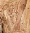
Popliteal Fossa: Which structures are demonstrated by labels 1, 2, 3, 4 and 5?

A

The structures are:

  1. Semitendinosus
  2. Semimembranosus
  3. Gracilis
  4. Gastrocnemius
  5. Biceps femoris

Boundaries of the popliteal fossa

Laterally: Biceps femoris above, lateral head of gastrocnemius and plantaris below

Medially: Semimembranosus and semitendinosus above, medial head of gastrocnemius below

Floor: Popliteal surface of the femur, posterior ligament of knee joint and popliteus muscle

Roof: Superficial and deep fascia

Contents:

Popliteal artery and vein

Small saphenous vein

Common peroneal nerve

Tibial nerve

Posterior cutaneous nerve of the thigh

Genicular branch of the obturator nerve

Lymph nodes

3
Q

Which structure is located deepest in the popliteal fossa?

A

The popliteal artery is located immediately adjacent to the posterior surface of the femur and the most deeply located structure. The vein lies superficial to it.

Popliteal fossa

Boundaries of the popliteal fossa

Laterally: Biceps femoris above, lateral head of gastrocnemius and plantaris below

Medially: Semimembranosus and semitendinosus above, medial head of gastrocnemius below

Floor: Popliteal surface of the femur, posterior ligament of knee joint and popliteus muscle

Roof: Superficial and deep fascia

Contents:

Popliteal artery and vein

Small saphenous vein

Common peroneal nerve

Tibial nerve

Posterior cutaneous nerve of the thigh

Genicular branch of the obturator nerve

Lymph nodes

4
Q

Femur: Please point out the key bony landmarks visible on this bone.

A

Being handed a femur in the anatomy section is a gift. A slick and well practised answer is essential. Begin your answer by naming the bone and orientating it. You should then move on to describe the key landmarks. Since you will have the bone in the exam you can use this as a prompt.
Salient points to identify include the head, neck and body, the greater and lesser trochanters, the adductor tubercle. You should also identify the condyles.

Femur

Extends from a rounded head, which articulates with the acetabulum down to the knee joint where the two large condyles at it’s inferior aspect articulate with the tibia.

The superior aspect comprises a head and neck which pass inferolaterally to the body and the two trochanters. These lie at the junction between the neck and the body.

The neck meets the body of the femur at an angle of 125o.

Developmentally, the neck is part of the body but is demarcated from it by a wide rough intertrochanteric crest, this continues inferomedially as a spiral line that runs below the lesser trochanter. Medially, the intertrochanteric line gives attachment to the inferior end of the iliofemoral ligament. The neck is covered by synovial membrane up to the intertrochanteric line. The posterior aspect of the neck is demarcated from the shaft by the intertrochanteric crest and only it’s medial aspect is covered by synovium and the joint capsule.

The greater trochanter has discernible surfaces that form the site of attachment of the gluteal muscles.Laterally, the greater trochanter overhangs the body and this forms part of the origin of vastus lateralis

Viewed anteriorly, the body of the femur appears rounded. Viewed laterally, it has an anterior concavity which gives fullness to the anterior thigh. Posteriorly, there is a ridge of bone, the linea aspera. The surface of the anterior aspect of the body forms the origin of the vastus intermedius. More medially, it forms the origin of vastus medialis.

The upper and middle aspects of the linea aspera form part of the origin of the attachments of the thigh adductors. Inferiorly, it spans out to form the bony floor of the popliteal fossa. At the inferior aspect of the popliteal surface the surface curves posteriorly to form the femoral condyles.

The structures that are attached to the inferior aspect of the linea aspera split with it as it approaches the popliteal fossa. Thus the vastus medialis and adductor magnus continue with the medial split and the biceps femoris and vastus intermedius along the lateral split.
Blood supply
The femur has a rich blood supply and numerous vascular foramina exist throughout it’s length. The blood supply to the femoral head is clinically important and is provided by the medial circumflex femoral and lateral circumflex femoral arteries (Branches of profunda femoris). Also from the inferior gluteal artery. These form an anastomosis and travel to up the femoral neck to supply the head.

5
Q

What is the usual angle between the femoral neck and the femoral shaft?

A

This can vary according to sex and age. However, it is typically around 125o.

Femur

Extends from a rounded head, which articulates with the acetabulum down to the knee joint where the two large condyles at it’s inferior aspect articulate with the tibia.

The superior aspect comprises a head and neck which pass inferolaterally to the body and the two trochanters. These lie at the junction between the neck and the body.

The neck meets the body of the femur at an angle of 125o.

Developmentally, the neck is part of the body but is demarcated from it by a wide rough intertrochanteric crest, this continues inferomedially as a spiral line that runs below the lesser trochanter. Medially, the intertrochanteric line gives attachment to the inferior end of the iliofemoral ligament. The neck is covered by synovial membrane up to the intertrochanteric line. The posterior aspect of the neck is demarcated from the shaft by the intertrochanteric crest and only it’s medial aspect is covered by synovium and the joint capsule.

The greater trochanter has discernible surfaces that form the site of attachment of the gluteal muscles.Laterally, the greater trochanter overhangs the body and this forms part of the origin of vastus lateralis

Viewed anteriorly, the body of the femur appears rounded. Viewed laterally, it has an anterior concavity which gives fullness to the anterior thigh. Posteriorly, there is a ridge of bone, the linea aspera. The surface of the anterior aspect of the body forms the origin of the vastus intermedius. More medially, it forms the origin of vastus medialis.

The upper and middle aspects of the linea aspera form part of the origin of the attachments of the thigh adductors. Inferiorly, it spans out to form the bony floor of the popliteal fossa. At the inferior aspect of the popliteal surface the surface curves posteriorly to form the femoral condyles.

The structures that are attached to the inferior aspect of the linea aspera split with it as it approaches the popliteal fossa. Thus the vastus medialis and adductor magnus continue with the medial split and the biceps femoris and vastus intermedius along the lateral split.
Blood supply
The femur has a rich blood supply and numerous vascular foramina exist throughout it’s length. The blood supply to the femoral head is clinically important and is provided by the medial circumflex femoral and lateral circumflex femoral arteries (Branches of profunda femoris). Also from the inferior gluteal artery. These form an anastomosis and travel to up the femoral neck to supply the head.

6
Q

Which muscle originates from the area marked number 1 and which two muscles insert into sites 2 and 3?

A

Vastus intermedius originates from area 1. Gluteus minimis inserts into area 2 and psoas major inserts into area 3.

Femur

Extends from a rounded head, which articulates with the acetabulum down to the knee joint where the two large condyles at it’s inferior aspect articulate with the tibia.

The superior aspect comprises a head and neck which pass inferolaterally to the body and the two trochanters. These lie at the junction between the neck and the body.

The neck meets the body of the femur at an angle of 125o.

Developmentally, the neck is part of the body but is demarcated from it by a wide rough intertrochanteric crest, this continues inferomedially as a spiral line that runs below the lesser trochanter. Medially, the intertrochanteric line gives attachment to the inferior end of the iliofemoral ligament. The neck is covered by synovial membrane up to the intertrochanteric line. The posterior aspect of the neck is demarcated from the shaft by the intertrochanteric crest and only it’s medial aspect is covered by synovium and the joint capsule.

The greater trochanter has discernible surfaces that form the site of attachment of the gluteal muscles.Laterally, the greater trochanter overhangs the body and this forms part of the origin of vastus lateralis

Viewed anteriorly, the body of the femur appears rounded. Viewed laterally, it has an anterior concavity which gives fullness to the anterior thigh. Posteriorly, there is a ridge of bone, the linea aspera. The surface of the anterior aspect of the body forms the origin of the vastus intermedius. More medially, it forms the origin of vastus medialis.

The upper and middle aspects of the linea aspera form part of the origin of the attachments of the thigh adductors. Inferiorly, it spans out to form the bony floor of the popliteal fossa. At the inferior aspect of the popliteal surface the surface curves posteriorly to form the femoral condyles.

The structures that are attached to the inferior aspect of the linea aspera split with it as it approaches the popliteal fossa. Thus the vastus medialis and adductor magnus continue with the medial split and the biceps femoris and vastus intermedius along the lateral split.
Blood supply
The femur has a rich blood supply and numerous vascular foramina exist throughout it’s length. The blood supply to the femoral head is clinically important and is provided by the medial circumflex femoral and lateral circumflex femoral arteries (Branches of profunda femoris). Also from the inferior gluteal artery. These form an anastomosis and travel to up the femoral neck to supply the head.

7
Q

What type of bone is marked by the letter “x”. What is it’s functional relevance?

A

The bone is the patella, it is a sesamoid bone contained within the tendon of quadriceps femoris. It plays an important role in reducing the work required to extend the knee joint.

Femur

Extends from a rounded head, which articulates with the acetabulum down to the knee joint where the two large condyles at it’s inferior aspect articulate with the tibia.

The superior aspect comprises a head and neck which pass inferolaterally to the body and the two trochanters. These lie at the junction between the neck and the body.

The neck meets the body of the femur at an angle of 125o.

Developmentally, the neck is part of the body but is demarcated from it by a wide rough intertrochanteric crest, this continues inferomedially as a spiral line that runs below the lesser trochanter. Medially, the intertrochanteric line gives attachment to the inferior end of the iliofemoral ligament. The neck is covered by synovial membrane up to the intertrochanteric line. The posterior aspect of the neck is demarcated from the shaft by the intertrochanteric crest and only it’s medial aspect is covered by synovium and the joint capsule.

The greater trochanter has discernible surfaces that form the site of attachment of the gluteal muscles.Laterally, the greater trochanter overhangs the body and this forms part of the origin of vastus lateralis

Viewed anteriorly, the body of the femur appears rounded. Viewed laterally, it has an anterior concavity which gives fullness to the anterior thigh. Posteriorly, there is a ridge of bone, the linea aspera. The surface of the anterior aspect of the body forms the origin of the vastus intermedius. More medially, it forms the origin of vastus medialis.

The upper and middle aspects of the linea aspera form part of the origin of the attachments of the thigh adductors. Inferiorly, it spans out to form the bony floor of the popliteal fossa. At the inferior aspect of the popliteal surface the surface curves posteriorly to form the femoral condyles.

The structures that are attached to the inferior aspect of the linea aspera split with it as it approaches the popliteal fossa. Thus the vastus medialis and adductor magnus continue with the medial split and the biceps femoris and vastus intermedius along the lateral split.
Blood supply
The femur has a rich blood supply and numerous vascular foramina exist throughout it’s length. The blood supply to the femoral head is clinically important and is provided by the medial circumflex femoral and lateral circumflex femoral arteries (Branches of profunda femoris). Also from the inferior gluteal artery. These form an anastomosis and travel to up the femoral neck to supply the head.

8
Q

Identify the structures A-H in the image below.

A

A Vastus medialis

B Sartorius

C Gracilis

D Gastrocnemius

E Semitendinosus

F Semimembranosus

G Rectus femoris

H Adductor magnus tendon

Femur

Extends from a rounded head, which articulates with the acetabulum down to the knee joint where the two large condyles at it’s inferior aspect articulate with the tibia.

The superior aspect comprises a head and neck which pass inferolaterally to the body and the two trochanters. These lie at the junction between the neck and the body.

The neck meets the body of the femur at an angle of 125o.

Developmentally, the neck is part of the body but is demarcated from it by a wide rough intertrochanteric crest, this continues inferomedially as a spiral line that runs below the lesser trochanter. Medially, the intertrochanteric line gives attachment to the inferior end of the iliofemoral ligament. The neck is covered by synovial membrane up to the intertrochanteric line. The posterior aspect of the neck is demarcated from the shaft by the intertrochanteric crest and only it’s medial aspect is covered by synovium and the joint capsule.

The greater trochanter has discernible surfaces that form the site of attachment of the gluteal muscles.Laterally, the greater trochanter overhangs the body and this forms part of the origin of vastus lateralis

Viewed anteriorly, the body of the femur appears rounded. Viewed laterally, it has an anterior concavity which gives fullness to the anterior thigh. Posteriorly, there is a ridge of bone, the linea aspera. The surface of the anterior aspect of the body forms the origin of the vastus intermedius. More medially, it forms the origin of vastus medialis.

The upper and middle aspects of the linea aspera form part of the origin of the attachments of the thigh adductors. Inferiorly, it spans out to form the bony floor of the popliteal fossa. At the inferior aspect of the popliteal surface the surface curves posteriorly to form the femoral condyles.

The structures that are attached to the inferior aspect of the linea aspera split with it as it approaches the popliteal fossa. Thus the vastus medialis and adductor magnus continue with the medial split and the biceps femoris and vastus intermedius along the lateral split.
Blood supply
The femur has a rich blood supply and numerous vascular foramina exist throughout it’s length. The blood supply to the femoral head is clinically important and is provided by the medial circumflex femoral and lateral circumflex femoral arteries (Branches of profunda femoris). Also from the inferior gluteal artery. These form an anastomosis and travel to up the femoral neck to supply the head.

9
Q

What is the name of the structure indicated by the arrow and which muscle inserts into it?

A

This is the adductor tubercle and is the point of insertion of the adductor magnus tendon.

10
Q

Identify the structures A-E.

A

A Tibialis anterior

B Peroneus longus

C Long head of biceps femoris

D Iliotibial tract

E Vastus lateralis

11
Q

Which structures insert or originate from areas 1 and 2 in the image above?

A
  1. Anterior cruciate ligament
    Anterior tibia to lateral intercondylar notch of femur
    Prevents tibia sliding anteriorly
  2. Posterior cruciate ligament
    Posterior tibia to medial intercondylar notch of femur
    Prevents tibia sliding posteriorly
12
Q

What is this structure?
Outline its key anatomical features.

A

This is the first cervical vertebra, also known as the atlas. Among the vertebral bodies it has a unique structure which reflects its important structural role. It consists of a ring of bone with slender anterior and posterior arches that are united by lateral bony masses that extend transversely as the two transverse processes that contain the foramen transversarium.

Atlas

The first cervical vertebra (Atlas) is a ring of bone consisting of two slender arches, one anterior and the other posterior. These are united on each side by a lateral mass on which is situated the articular facets and the transverse processes. Most of the body of the atlas joins the axis to form the dens, and the anterior arch and lateral masses represent that part of the body normally formed by the vertebral arch ossification. The facets articulate with the skull above and the axis below. The normal articular facets are missing. The foramen transversarium transmits the vertebral artery.

13
Q

What is the structure that attaches to the site marked with the “x” and what is its role?

What structure passes through the foramen transversarium?

A

This is the site of the attachment of the transverse ligament. It’s main role is to hold the dens in place.

The foramen transversarium transmits the vertebral artery, which enters it after passing along a groove on the superior aspect of the posterior arch.

Atlas

The first cervical vertebra (Atlas) is a ring of bone consisting of two slender arches, one anterior and the other posterior. These are united on each side by a lateral mass on which is situated the articular facets and the transverse processes. Most of the body of the atlas joins the axis to form the dens, and the anterior arch and lateral masses represent that part of the body normally formed by the vertebral arch ossification. The facets articulate with the skull above and the axis below. The normal articular facets are missing. The foramen transversarium transmits the vertebral artery.

14
Q

Identify the structures 1,2 and 3.

What are the main branches of structure 1?

Which nerves are closely related to the aortic arch near structure 3?

A

In this section of the aortic arch, the left subclavian arises close to the brachiocephalic trunk. The structures are therefore;

  1. Brachiocephalic trunk
  2. Left common carotid artery
  3. Left subclavian

The brachiocephalic artery branches to provide the right common carotid artery and right subclavian artery. It typically branches at the level of the right sternoclavicular joint.

The two nerves are the phrenic nerve, which continues inferiorly across the anterior surface of the pericardium and the vagus nerve, which then branches around the inferior aspect of the aorta to give rise to the left recurrent laryngeal nerve.

Aortic arch and brachiocephalic vein

The arch of the aorta lies in the superior mediastinum, opposite the lower half of the manubrium. It runs superiorly and then arches in a posterior direction. It continues to curve posteriorly and then towards the left side of the body on a level of T4 where it then becomes the descending aorta.

Anterior to the arch of the aorta lie the thymic remnants, lungs and pleura. On it’s left hand side the following structures lie between the aortic arch and the pleura; phrenic nerve, inferior cardiac branch of the left vagus, superior cervical cardiac branch of the of the left sympathetic nerve and the trunk of the left vagus. The left superior intercostal vein crosses it, superficial to the left vagus nerve and deep to the left phrenic nerve. Posteriorly, on the right hand side of the arch lie the deep cardiac plexus, trachea, left recurrent laryngeal nerve, left border of the oesophagus, thoracic duct and finally the vertebral column.

There are three main branches, brachiocephalic trunk, left common carotid and left subclavian arteries. The brachiocephalic vein is related anteriorly.

The two brachiocephalic veins are formed by the union of the internal jugular and subclavian veins. The right runs a near vertical course and is joined by the left brachiocephalic vein to form the superior vena cava. The veins have no valves.

15
Q

Identify the structures 1, 2 and 3.

How many valves are typically found in structures 1 and 2?

What are the tributaries of the left brachiocephalic vein?

A

Structures 1 and 2 are the right and left brachiocehphalic veins. They unite to form the superior vena cava (3).

The brachiocephalic veins (and indeed also the SVC) do not usually contain any valves. This is why the wave patterns of the JVP can be readily appreciated.

The subclavian vein, internal jugular vein, thoracic duct (which joins it at the angle of the junction of the internal jugular and subclavian veins), the vertebral, internal thoracic, inferior thyroid and superior intercostal veins of its own side.

Aortic arch and brachiocephalic vein

The arch of the aorta lies in the superior mediastinum, opposite the lower half of the manubrium. It runs superiorly and then arches in a posterior direction. It continues to curve posteriorly and then towards the left side of the body on a level of T4 where it then becomes the descending aorta.

Anterior to the arch of the aorta lie the thymic remnants, lungs and pleura. On it’s left hand side the following structures lie between the aortic arch and the pleura; phrenic nerve, inferior cardiac branch of the left vagus, superior cervical cardiac branch of the of the left sympathetic nerve and the trunk of the left vagus. The left superior intercostal vein crosses it, superficial to the left vagus nerve and deep to the left phrenic nerve. Posteriorly, on the right hand side of the arch lie the deep cardiac plexus, trachea, left recurrent laryngeal nerve, left border of the oesophagus, thoracic duct and finally the vertebral column.

There are three main branches, brachiocephalic trunk, left common carotid and left subclavian arteries. The brachiocephalic vein is related anteriorly.

The two brachiocephalic veins are formed by the union of the internal jugular and subclavian veins. The right runs a near vertical course and is joined by the left brachiocephalic vein to form the superior vena cava. The veins have no valves.

16
Q

What structure is this?
Identify the structures labeled 1-5.

A

This is the left lung
The structures are:
1. Lower left pulmonary vein
2. Oblique fissure
3. Groove for left subclavian artery
4. Left pulmonary artery
5. Upper left pulmonary vein

Left lung
Above the hilum is the furrow produced by the aortic arch, and then superiorly the groove accommodating the left subclavian artery; Behind the hilum and pulmonary ligament is a vertical groove produced by the descending aorta, and in front of this, near the base of the lung, is the lower part of the oesophagus.

The root of the left lung passes under the aortic arch and in front of the descending aorta.

Right lung
Above the hilum is the azygos vein; Superior to this is the groove for the superior vena cava and right innominate vein; behind this, and nearer the apex, is a furrow for the innominate artery. Behind the hilum and the attachment of the pulmonary ligament is a vertical groove for the oesophagus; In front and to the right of the lower part of the oesophageal groove is a deep concavity for the extrapericardiac portion of the inferior vena cava.

The root of the right lung lies behind the superior vena cava and the right atrium, and below the azygos vein.

The right main bronchus is shorter, wider and more vertical than the left main bronchus and therefore the route taken by most foreign bodies.

17
Q

What structures are likely to be located at sites 1,2 and 3?

A
  1. Descending thoracic aorta
  2. Heart
  3. Arch of aorta

Lung anatomy

The right lung is composed of 3 lobes divided by the oblique and transverse fissures. The left lung has two lobes divided by the oblique fissure.The apex of both lungs is approximately 4cm superior to the sterno-costal joint of the first rib. Immediately below this is a sulcus created by the subclavian artery.

Peripheral contact points of the lung

Base: diaphragm

Costal surface: corresponds to the cavity of the chest

Mediastinal surface: Contacts the mediastinal pleura. Has the cardiac impression. Above and behind this concavity is a triangular depression named the hilum, where the structures which form the root of the lung enter and leave the viscus. These structures are invested by pleura, which, below the hilum and behind the pericardial impression, forms the pulmonary ligament

Right lung
Above the hilum is the azygos vein; Superior to this is the groove for the superior vena cava and right innominate vein; behind this, and nearer the apex, is a furrow for the innominate artery. Behind the hilum and the attachment of the pulmonary ligament is a vertical groove for the oesophagus; In front and to the right of the lower part of the oesophageal groove is a deep concavity for the extrapericardiac portion of the inferior vena cava.

The root of the right lung lies behind the superior vena cava and the right atrium, and below the azygos vein.

The right main bronchus is shorter, wider and more vertical than the left main bronchus and therefore the route taken by most foreign bodies.

Left lung
Above the hilum is the furrow produced by the aortic arch, and then superiorly the groove accommodating the left subclavian artery; Behind the hilum and pulmonary ligament is a vertical groove produced by the descending aorta, and in front of this, near the base of the lung, is the lower part of the oesophagus.

The root of the left lung passes under the aortic arch and in front of the descending aorta.

Inferior borders of both lungs

6th rib in mid clavicular line

8th rib in mid axillary line

10th rib posteriorly

The pleura runs two ribs lower than the corresponding lung level.

18
Q

Identify the structures labeled 1-4.

A
  1. Foramen magnum
  2. Occipital condyle
  3. Jugular fossa
  4. Foramen lacerum

Foramen/Location/Contents:

  • Foramen ovale - Sphenoid bone - Otic ganglion, V3 (Mandibular nerve:3rd branch of trigeminal), Accessory meningeal artery, Lesser petrosal nerve, Emissary veins
  • Foramen spinosum - Sphenoid bone - Middle meningeal artery, Meningeal branch of the Mandibular nerve
  • Foramen rotundum - Sphenoid bone - Maxillary nerve (V2)
  • Foramen lacerum - carotid canal - Sphenoid boneBase of the medial pterygoid plate. Internal carotid artery* Nerve and artery of the pterygoid canal
  • Jugular foramen - Temporal bone - Anterior: inferior petrosal sinus
    Intermediate: glossopharyngeal, vagus, and accessory nerves.
    Posterior: sigmoid sinus (becoming the internal jugular vein) and some meningeal branches from the occipital and ascending pharyngeal arteries.
  • Foramen magnum - Occipital bone - Anterior and posterior spinal arteries, Vertebral arteries, Medulla oblongata
  • Stylomastoid foramen - Temporal bone, Stylomastoid artery, Facial nerve
  • Superior orbital fissure - Sphenoid bone, Oculomotor nerve (III), Recurrent meningeal artery, Trochlear nerve (IV), Lacrimal, frontal and nasociliary branches of ophthalmic nerve (V1), Abducent nerve (VI), Superior ophthalmic vein

*= In life the foramen lacerum is occluded by a cartilagenous plug. The ICA initially passes into the carotid canal which ascends superomedially to enter the cranial cavity through the foramen lacerum.

19
Q

What is the most important factor in maintaining the stability of the knee joint?

Outline the different roles played by these structures and outline the anatomical attachments

A

The main stability of the knee joint is maintained by 2 sets of strong ligaments; the cruciate ligaments and the collateral ligaments.

The cruciate ligaments provide antero-posterior stability, the collateral ligaments serve to limit both medial and lateral stressors.

Medial collateral ligament: Medial femoral epicondyle –> Tibial surface, the deeper aspect attaches to the medial meniscus - valgus stability

Lateral collateral ligament: Lateral femoral epicondyle –> Fibula - varus stability

Anterior cruciate ligament: Anterior intercondylar area of tibia –> Lateral femoral condyle in intercondylar notch - prevents tibia sliding anteriorly

Posterior cruciate ligament: Posterior intercondylar area of tibia –> Medial femoral condyle in intercondylar notch - prevents tibia sliding posteriorly

Patellar ligament: Central band of the tendon of quadriceps femoris, extends from patella to tibial tuberosity

Knee joint
The knee joint is a synovial joint, the largest and most complicated. It consists of two condylar joints between the femur and tibia and a sellar joint between the patella and the femur. The tibiofemoral articular surfaces are incongruent, however, this is improved by the presence of the menisci. The degree of congruence is related to the anatomical position of the knee joint and is greatest in full extension.

Knee joint compartments

  • Tibiofemoral: Comprised of the patella/femur joint, lateral and medial compartments (between femur condyles and tibia). Synovial membrane and cruciate ligaments partially separate the medial and lateral compartments
  • Patellofemoral: Ligamentum patellae. Actions: provides joint stability in full extension

Fibrous capsule
The capsule of the knee joint is a complex, composite structure with contributions from adjacent tendons.

Anterior fibresThe capsule does not pass proximal to the patella. It blends with the tendinous expansions of vastus medialis and lateralis

Posterior fibresThese fibres are vertical and run from the posterior surface of the femoral condyles to the posterior aspect of the tibial condyle

Medial fibresAttach to the femoral and tibial condyles beyond their articular margins, blending with the tibial collateral ligament

Lateral fibresAttach to the femur superior to popliteus, pass over its tendon to head of fibula and tibial condyle

Bursae

  • Anterior: Subcutaneous prepatellar bursa; between patella and skin, Deep infrapatellar bursa; between tibia and patellar ligament, Subcutaneous infrapatellar bursa; between distal tibial tuberosity and skin
  • Laterally: Bursa between lateral head of gastrocnemius and joint capsule, Bursa between fibular collateral ligament and tendon of biceps femoris, Bursa between fibular collateral ligament and tendon of popliteus
  • Medially: Bursa between medial head of gastrocnemius and the fibrous capsule, Bursa between tibial collateral ligament and tendons of sartorius, gracilis and semitendinosus, Bursa between the tendon of semimembranosus and medial tibial condyle and medial head of gastrocnemius
  • Posterior: Highly variable and inconsistent

Menisci
Medial and lateral menisci compensate for the incongruence of the femoral and tibial condyles.
Composed of fibrous tissue.
Medial meniscus is attached to the tibial collateral ligament.
Lateral meniscus is attached to the loose fibres at the lateral edge of the joint and is separate from the fibular collateral ligament. The lateral meniscus is crossed by the popliteus tendon.

Nerve supply
The knee joint is supplied by the femoral, tibial and common peroneal divisions of the sciatic and by a branch from the obturator nerve. Hip pathology pain may be referred to the knee.

Blood supply
Genicular branches of the femoral artery, popliteal and anterior tibial arteries all supply the knee joint.

20
Q

What nerves are at risk when performing an excision of the submandibular gland?

Where would you site an incision for a sub mandibular gland excision and why?

A

Mandibular branch of the facial nerve
Hypoglossal nerve
Lingual nerve

I would site the incision two to three finger breadths below the mandible to avoid injury to the marginal mandibular branch of the facial nerve

Submandibular gland

Relations of the submandibular gland

Superficial: Platysma, deep fascia and mandible, Submandibular lymph nodes, Facial vein (facial artery near mandible), Marginal mandibular nerve, Cervical branch of the facial nerve

Deep: Facial artery (inferior to the mandible), Mylohyoid muscle
Sub mandibular duct, Hyoglossus muscle, Lingual nerve, Submandibular ganglion, Hypoglossal nerve

Submandibular duct (Wharton’s duct): Opens lateral to the lingual frenulum on the anterior floor of mouth. , 5 cm length. Lingual nerve wraps around Wharton’s duct. As the duct passes forwards it crosses medial to the nerve to lie above it and then crosses back, lateral to it, to reach a position below the nerve.

Innervation

Sympathetic innervation- Derived from superior cervical ganglion

Parasympathetic innervation- Submandibular ganglion via lingual nerve

Arterial supply: Branch of the facial artery. The facial artery passes through the gland to groove its deep surface. It then emerges onto the face by passing between the gland and the mandible.

Venous drainage: Anterior facial vein (lies deep to the Marginal Mandibular nerve)

Lymphatic drainage: Deep cervical and jugular chains of nodes

21
Q

What are the effects of injury to the hypoglossal nerve and lingual nerves?

What are the effects of injury to the marginal mandibular branch of the facial nerve?

A

Injury to the lingual nerve will result in ipsilateral loss of somatic sensation to the anterior two thirds of the tongue. Injury to the hypoglossal nerve will result in atrophy the muscles of the tongue on the ipsilateral side. This can be clinically demonstrated by asking the patient to protrude their tongue, when this is done it will deviate to the side of the lesion.

This branch supplies risorius and the muscles of the lower lip. Therefore, injury to it will potentially result in an ipsilateral weakness of the lip.

All at risk during submandibular gland excision

Submandibular gland:
Relations of the submandibular gland

Superficial: Platysma, deep fascia and mandible, Submandibular lymph nodes, Facial vein (facial artery near mandible), Marginal mandibular nerve, Cervical branch of the facial nerve

Deep: Facial artery (inferior to the mandible), Mylohyoid muscle
Sub mandibular duct, Hyoglossus muscle, Lingual nerve, Submandibular ganglion, Hypoglossal nerve

Submandibular duct (Wharton’s duct): Opens lateral to the lingual frenulum on the anterior floor of mouth. , 5 cm length. Lingual nerve wraps around Wharton’s duct. As the duct passes forwards it crosses medial to the nerve to lie above it and then crosses back, lateral to it, to reach a position below the nerve.

Innervation

Sympathetic innervation- Derived from superior cervical ganglion

Parasympathetic innervation- Submandibular ganglion via lingual nerve

Arterial supply: Branch of the facial artery. The facial artery passes through the gland to groove its deep surface. It then emerges onto the face by passing between the gland and the mandible.

Venous drainage: Anterior facial vein (lies deep to the Marginal Mandibular nerve)

Lymphatic drainage: Deep cervical and jugular chains of nodes

22
Q

What cartilaginous structures constitute the larynx?

A

The larynx is a fibrocartilagenous structure that is comprised of 6 different cartilages.

Epiglottis- This broad structure is attached to the posterior aspect of the thyroid cartilage inferiorly. Superiorly, it projects posterior to the hyoid and tongue.

Thyroid- This shield shaped segment consist of two laminae that unite in the midline with the thyroid notch superiorly. Inferiorly, the horns articulate with the cricoid.

Cricoid- This forms a complete ring and is the only complete ring within the larynx. It articulates with both the thyroid and arytenoid cartilages.

Arytenoid- There are two arytenoids, the have a pyramidal shape and sit on the cricoid. Together, they are responsible for tension within the vocal cords.

Corniculate and cuneiform- There are two of each of these cartilaginous structures. Together, the sit in the aryepiglottic fold.

Larynx
The larynx lies in the anterior part of the neck at the levels of C3 to C6 vertebral bodies. The laryngeal skeleton consists of a number of cartilagenous segments. Three of these are paired; arytenoid, corniculate and cuneiform. Three are single; thyroid, cricoid and epiglottic. The cricoid cartilage forms a complete ring (the only one to do so).
The laryngeal cavity extends from the laryngeal inlet to the level of the inferior border of the cricoid cartilage.

Divisions of the laryngeal cavity

Laryngeal vestibule: Superior to the vestibular folds

Laryngeal ventricle: Lies between vestibular folds and superior to the vocal cords

Infraglottic cavity: Extends from vocal cords to inferior border of the cricoid cartilage

The vocal folds (true vocal cords) control sound production. The apex of each fold projects medially into the laryngeal cavity. Each vocal fold includes:

  • Vocal ligament
  • Vocalis muscle (most medial part of thyroarytenoid muscle)

The glottis is composed of the vocal folds, processes and rima glottidis. The rima glottidis is the narrowest potential site within the larynx, as the vocal cords may be completely opposed, forming a complete barrier.

Muscle/Origin/Insertion/Innervation/Action

Posterior cricoarytenoid: Posterior aspect of lamina of cricoid–> Muscular process of arytenoid. Recurrent Laryngeal - Abducts vocal fold

Lateral cricoarytenoid: Arch of cricoid–> Muscular process of arytenoid. Recurrent laryngeal - Adducts vocal fold

Thyroarytenoid: Posterior aspect of thyroid cartilage –> Muscular process of arytenoid. Recurrent laryngeal - Relaxes vocal fold

Transverse and oblique arytenoids: Arytenoid cartilage –> Contralateral arytenoid. Recurrent laryngeal - Closure of intercartilagenous part of the rima glottidis

Vocalis: Depression between lamina of thyroid cartilage –> Vocal ligament and vocal process of arytenoid cartilage. Recurrent laryngeal - Relaxes posterior vocal ligament, tenses anterior part

Cricothyroid: Anterolateral part of cricoid –> Inferior margin and horn of thyroid cartilage. External laryngeal - Tenses vocal fold

Blood supply
Arterial supply is via the laryngeal arteries, branches of the superior and inferior thyroid arteries. The superior laryngeal artery is closely related to the internal laryngeal nerve. The inferior laryngeal artery is related to the inferior laryngeal nerve. Venous drainage is via superior and inferior laryngeal veins, the former draining into the superior thyroid vein and the latter draining into the middle thyroid vein, or thyroid venous plexus.

Lymphatic drainage
The vocal cords have no lymphatic drainage and this site acts as a lymphatic watershed.

Supraglottic part: Upper deep cervical nodes

Subglottic part: Prelaryngeal and pretracheal nodes and inferior deep cervical nodes

The aryepiglottic fold and vestibular folds have a dense plexus of lymphatics associated with them and malignancies at these sites have a greater propensity for nodal metastasis.

23
Q

Please name the intrinsic muscles of the larynx.

A

The function of the intrinsic muscles is to alter the tension of the vocal cords during speech. They open the cords during inspiration and close them during swallowing. They comprise:

Posterior cricoarytenoid- Abducts the cords and externally rotates the arytenoids

Lateral cricoarytenoid- Adducts the cords and internally rotates the arytenoid

Interarytenoid- Adducts the cords and closes the glottis. It is unpaired.

Thyroarytenoid- Shortens the vocal cords by drawing arytenoids forward

Vocalis- Arises from the thyroarytenoid and alters vocal cord tension

Cricothyroid- Lifts the cricoid anteriorly towards the thyroid cartilage. It lengthens the vocal cords

Larynx
The larynx lies in the anterior part of the neck at the levels of C3 to C6 vertebral bodies. The laryngeal skeleton consists of a number of cartilagenous segments. Three of these are paired; arytenoid, corniculate and cuneiform. Three are single; thyroid, cricoid and epiglottic. The cricoid cartilage forms a complete ring (the only one to do so).
The laryngeal cavity extends from the laryngeal inlet to the level of the inferior border of the cricoid cartilage.

Divisions of the laryngeal cavity

Laryngeal vestibule: Superior to the vestibular folds

Laryngeal ventricle: Lies between vestibular folds and superior to the vocal cords

Infraglottic cavity: Extends from vocal cords to inferior border of the cricoid cartilage

The vocal folds (true vocal cords) control sound production. The apex of each fold projects medially into the laryngeal cavity. Each vocal fold includes:

  • Vocal ligament
  • Vocalis muscle (most medial part of thyroarytenoid muscle)

The glottis is composed of the vocal folds, processes and rima glottidis. The rima glottidis is the narrowest potential site within the larynx, as the vocal cords may be completely opposed, forming a complete barrier.

Muscle/Origin/Insertion/Innervation/Action

Posterior cricoarytenoid: Posterior aspect of lamina of cricoid–> Muscular process of arytenoid. Recurrent Laryngeal - Abducts vocal fold

Lateral cricoarytenoid: Arch of cricoid–> Muscular process of arytenoid. Recurrent laryngeal - Adducts vocal fold

Thyroarytenoid: Posterior aspect of thyroid cartilage –> Muscular process of arytenoid. Recurrent laryngeal - Relaxes vocal fold

Transverse and oblique arytenoids: Arytenoid cartilage –> Contralateral arytenoid. Recurrent laryngeal - Closure of intercartilagenous part of the rima glottidis

Vocalis: Depression between lamina of thyroid cartilage –> Vocal ligament and vocal process of arytenoid cartilage. Recurrent laryngeal - Relaxes posterior vocal ligament, tenses anterior part

Cricothyroid: Anterolateral part of cricoid –> Inferior margin and horn of thyroid cartilage. External laryngeal - Tenses vocal fold

Blood supply
Arterial supply is via the laryngeal arteries, branches of the superior and inferior thyroid arteries. The superior laryngeal artery is closely related to the internal laryngeal nerve. The inferior laryngeal artery is related to the inferior laryngeal nerve. Venous drainage is via superior and inferior laryngeal veins, the former draining into the superior thyroid vein and the latter draining into the middle thyroid vein, or thyroid venous plexus.

Lymphatic drainage
The vocal cords have no lymphatic drainage and this site acts as a lymphatic watershed.

Supraglottic part: Upper deep cervical nodes

Subglottic part: Prelaryngeal and pretracheal nodes and inferior deep cervical nodes

The aryepiglottic fold and vestibular folds have a dense plexus of lymphatics associated with them and malignancies at these sites have a greater propensity for nodal metastasis.

24
Q

Please name the extrinsic muscles of the larynx.

A

These attach the larynx to adjacent structures and serve to elevate or depress it. They comprise:
Sternothyroid, thyrohyoid and the inferior constrictor.

Larynx
The larynx lies in the anterior part of the neck at the levels of C3 to C6 vertebral bodies. The laryngeal skeleton consists of a number of cartilagenous segments. Three of these are paired; arytenoid, corniculate and cuneiform. Three are single; thyroid, cricoid and epiglottic. The cricoid cartilage forms a complete ring (the only one to do so).
The laryngeal cavity extends from the laryngeal inlet to the level of the inferior border of the cricoid cartilage.

Divisions of the laryngeal cavity

Laryngeal vestibule: Superior to the vestibular folds

Laryngeal ventricle: Lies between vestibular folds and superior to the vocal cords

Infraglottic cavity: Extends from vocal cords to inferior border of the cricoid cartilage

The vocal folds (true vocal cords) control sound production. The apex of each fold projects medially into the laryngeal cavity. Each vocal fold includes:

  • Vocal ligament
  • Vocalis muscle (most medial part of thyroarytenoid muscle)

The glottis is composed of the vocal folds, processes and rima glottidis. The rima glottidis is the narrowest potential site within the larynx, as the vocal cords may be completely opposed, forming a complete barrier.

Muscle/Origin/Insertion/Innervation/Action

Posterior cricoarytenoid: Posterior aspect of lamina of cricoid–> Muscular process of arytenoid. Recurrent Laryngeal - Abducts vocal fold

Lateral cricoarytenoid: Arch of cricoid–> Muscular process of arytenoid. Recurrent laryngeal - Adducts vocal fold

Thyroarytenoid: Posterior aspect of thyroid cartilage –> Muscular process of arytenoid. Recurrent laryngeal - Relaxes vocal fold

Transverse and oblique arytenoids: Arytenoid cartilage –> Contralateral arytenoid. Recurrent laryngeal - Closure of intercartilagenous part of the rima glottidis

Vocalis: Depression between lamina of thyroid cartilage –> Vocal ligament and vocal process of arytenoid cartilage. Recurrent laryngeal - Relaxes posterior vocal ligament, tenses anterior part

Cricothyroid: Anterolateral part of cricoid –> Inferior margin and horn of thyroid cartilage. External laryngeal - Tenses vocal fold

Blood supply
Arterial supply is via the laryngeal arteries, branches of the superior and inferior thyroid arteries. The superior laryngeal artery is closely related to the internal laryngeal nerve. The inferior laryngeal artery is related to the inferior laryngeal nerve. Venous drainage is via superior and inferior laryngeal veins, the former draining into the superior thyroid vein and the latter draining into the middle thyroid vein, or thyroid venous plexus.

Lymphatic drainage
The vocal cords have no lymphatic drainage and this site acts as a lymphatic watershed.

Supraglottic part: Upper deep cervical nodes

Subglottic part: Prelaryngeal and pretracheal nodes and inferior deep cervical nodes

The aryepiglottic fold and vestibular folds have a dense plexus of lymphatics associated with them and malignancies at these sites have a greater propensity for nodal metastasis.

25
Q

What is the main blood supply to the larynx?

What are the effects of bilateral recurrent laryngeal nerve injury?

A

The larynx is supplied by the superior and inferior laryngeal arteries. The superior laryngeal artery closely follows the superior laryngeal nerve and the inferior artery, the recurrent laryngeal nerve.

Following recurrent laryngeal nerve injury, the vocal cords sit in a position that is slightly narrower than the usual neutral position. The cords cannot be adducted for phonation or abducted to increase respiration and therefore produce inspiratory stridor.

Larynx
The larynx lies in the anterior part of the neck at the levels of C3 to C6 vertebral bodies. The laryngeal skeleton consists of a number of cartilagenous segments. Three of these are paired; arytenoid, corniculate and cuneiform. Three are single; thyroid, cricoid and epiglottic. The cricoid cartilage forms a complete ring (the only one to do so).
The laryngeal cavity extends from the laryngeal inlet to the level of the inferior border of the cricoid cartilage.

Divisions of the laryngeal cavity

Laryngeal vestibule: Superior to the vestibular folds

Laryngeal ventricle: Lies between vestibular folds and superior to the vocal cords

Infraglottic cavity: Extends from vocal cords to inferior border of the cricoid cartilage

The vocal folds (true vocal cords) control sound production. The apex of each fold projects medially into the laryngeal cavity. Each vocal fold includes:

  • Vocal ligament
  • Vocalis muscle (most medial part of thyroarytenoid muscle)

The glottis is composed of the vocal folds, processes and rima glottidis. The rima glottidis is the narrowest potential site within the larynx, as the vocal cords may be completely opposed, forming a complete barrier.

Muscle/Origin/Insertion/Innervation/Action

Posterior cricoarytenoid: Posterior aspect of lamina of cricoid–> Muscular process of arytenoid. Recurrent Laryngeal - Abducts vocal fold

Lateral cricoarytenoid: Arch of cricoid–> Muscular process of arytenoid. Recurrent laryngeal - Adducts vocal fold

Thyroarytenoid: Posterior aspect of thyroid cartilage –> Muscular process of arytenoid. Recurrent laryngeal - Relaxes vocal fold

Transverse and oblique arytenoids: Arytenoid cartilage –> Contralateral arytenoid. Recurrent laryngeal - Closure of intercartilagenous part of the rima glottidis

Vocalis: Depression between lamina of thyroid cartilage –> Vocal ligament and vocal process of arytenoid cartilage. Recurrent laryngeal - Relaxes posterior vocal ligament, tenses anterior part

Cricothyroid: Anterolateral part of cricoid –> Inferior margin and horn of thyroid cartilage. External laryngeal - Tenses vocal fold

Blood supply
Arterial supply is via the laryngeal arteries, branches of the superior and inferior thyroid arteries. The superior laryngeal artery is closely related to the internal laryngeal nerve. The inferior laryngeal artery is related to the inferior laryngeal nerve. Venous drainage is via superior and inferior laryngeal veins, the former draining into the superior thyroid vein and the latter draining into the middle thyroid vein, or thyroid venous plexus.

Lymphatic drainage
The vocal cords have no lymphatic drainage and this site acts as a lymphatic watershed.

Supraglottic part: Upper deep cervical nodes

Subglottic part: Prelaryngeal and pretracheal nodes and inferior deep cervical nodes

The aryepiglottic fold and vestibular folds have a dense plexus of lymphatics associated with them and malignancies at these sites have a greater propensity for nodal metastasis.

26
Q

PAROTID GLAND: Where do the secretions of the parotid gland drain?

Which structures pass through the parotid gland?

What is the lymphatic drainage of the parotid gland?

Which nerves supply the parotid gland?

On the image below, outline where you would place the incision for a superficial parotidectomy.

A
  • The secretions of the parotid gland pass into the oral cavity via Stensons duct whose oral opening is opposite the second upper molar tooth.
  • Structures passing through parotid gland: Facial nerve, External carotid artery, Retromandibular vein, Auriculotemporal nerve
  • It contains lymph nodes within the substance of the gland itself. It then drains to the deep cervical nodes.
  • Nerve Supply: Parasympathetic-Secretomotor, Sympathetic-Superior cervical ganglion, Sensory- Greater auricular nerve

The incision runs posterior to the mandible and up inferior to the tragus of the ear. Loss of cutaneous sensation to the ear lobe is therefore a risk of the procedure.

Anatomy of the parotid gland

Location: Overlying the mandibular ramus; anterior and inferior to the ear.

Salivary duct: Crosses the masseter, pierces the buccinator and drains adjacent to the 2nd upper molar tooth (Stensen’s duct).

Structures passing through the gland:

  • Facial nerve (Mnemonic: The Zebra Buggered My Cat; Temporal Zygomatic, Buccal, Mandibular, Cervical), External carotid artery, Retromandibular vein, Auriculotemporal nerve

Relations

Anterior: masseter, medial pterygoid, superficial temporal and maxillary artery, facial nerve, stylomandibular ligament

Posterior: posterior belly digastric muscle, sternocleidomastoid, stylohyoid, internal carotid artery, mastoid process, styloid process

Arterial supply: Branches of external carotid artery

Venous drainage: Retromandibular vein

Lymphatic drainage: Deep cervical nodes

Parasympathetic stimulation produces a water rich, serous saliva. Sympathetic stimulation leads to the production of a low volume, enzyme-rich saliva.

27
Q

What structures form the wall of the spermatic cord?

What are the contents of the spermatic cord?

From where does the testicular artery originate?

How does the lymphatic drainage of the testis and scrotum differ?

What is the gubernaculum?

A

The spermatic cord is formed by the vas deferens and covered by 3 layers:

  1. Internal spermatic fascia-Transversalis fascia
  2. Cremasteric fascia-From the fascial coverings of internal oblique
  3. External spermatic fascia-External oblique aponeurosis

Contents Spermatic Cord:

  • Vas deferens: transmits sperm and accessory gland secretion
  • Testicular artery; branch of abdominal aorta, supplies testis and epididymis
  • Artery of vas deferens: arises from inferior vesical artery
  • Cremasteric artery: arises from inferior epigastric artery
  • Pampiniform plexus: venous plexus, drains into right or left testicualr vein
  • Sympathetic nerve fibres: lies on arteries, the parasymp fibres lie on the vas
  • Genital branch of the genitofemoral nerve: supplies cremaster
  • Lymphatic vessels: drain to lumbar and para-aortic nodes

The pampiniform plexus is a rich network of veins that arise from the testis and epididymis and travel superiorly through the inguinal canal within the spematic cord.

Testicular artery originates from the aorta around a level of L2 and then travels through the retroperitoneum to reach the inguinal canal.

The testes drain primarily to the para-aortic lymph nodes and the scrotum drains to the inguinal lymph nodes. This is very relevant surgically, since surgical resection of testicular tumours should never be undertaken via a scrotal incision for fear of seeding the tumour into the inguinal nodal basin.

The gubernaculum is a ridge of fibrous tissue that forms early in embryonic development. It links the apex of the testis to the scrotum and as the foetus grows, so draws the testis into the scrotum. Occasionally, the gubernaculum may form abnormal connections and this then results in an ectopic testis.

Scrotum

Composed of skin and closely attached dartos fascia.

Arterial supply from the anterior and posterior scrotal arteries

Lymphatic drainage to the inguinal lymph nodes

Parietal layer of the tunica vaginalis is the innermost layer

Testes

The testes are surrounded by the tunica vaginalis (closed peritoneal sac). The parietal layer of the tunica vaginalis adjacent to the internal spermatic fascia.

The testicular arteries arise from the aorta immediately inferiorly to the renal arteries.

The pampiniform plexus drains into the testicular veins, the left drains into the left renal vein and the right into the inferior vena cava.

Lymphatic drainage is to the para-aortic nodes.

28
Q

Identify the structures labelled 1-7

A
  1. Adductor longus
  2. Pectineus
  3. Sartorious Muscle
  4. Rectus femoria
  5. Tensor fascia lata
  6. femoral vein
  7. Femoral artery
29
Q

What are the structures labelled 1 and 2?

What is the root value of the structure labelled 3?

A

1=Profunda femoris artery
2=Long saphenous vein

3= It is the femoral nerve and supplied by root segments L2, 3 and 4 of the lumbar plexus.

Femoral triangle anatomy

Superiorly: Inguinal ligament

Laterally: Sartorius

Medially: Adductor longus

Floor: Iliopsoas, adductor longus and pectineus

Roof: Fascia lata and Superficial fascia, Superficial inguinal lymph nodes (palpable below the inguinal ligament), Long saphenous vein

Contents: Femoral vein (medial to lateral), Femoral artery-pulse palpated at the mid inguinal point, Femoral nerve, Deep and superficial inguinal lymph nodes, Lateral cutaneous nerve, Great saphenous vein, Femoral branch of the genitofemoral nerve

30
Q

Identify the structures labelled 1-6

Which structures pass through the area labelled 4 in the diagram below?

Which structures are transmitted by the areas labelled 5 and 6 in the diagram below?

A
  1. Frontal bone
  2. Greater Wing of sphenoid
  3. Maxilla
  4. Superior Orbital Fissue: transmits recurrent meningeal artery, lacrimal nerve, trochlear nerve, abducens nerve, superior ophthalmic nerve, superior division of oculomotor nerve
  5. Optic Canal: optic nerve and ophthalmic artery
  6. Inferior Orbital fissure: maxillary nerve, inferior ophthalmic vein, zygomatic nerve
31
Q

What is the origin of the opthalmic artery?

What is the first branch of the opthalmic artery?

A

It is the first branch of the internal carotid artery after it emerges from the cavernous sinus. It branches to produce vessels supplying the orbit and the eye itself.

Orbital branches: Lacrimal artery, Supraorbital artery, Posterior ethmoidal artery, Anterior ethmoidal artery, Medial palpebral artery, Frontal artery, Dorsal nasal artery

Ocular branches: Long posterior ciliary arteries, Short posterior ciliary arteries, Anterior ciliary artery, Central retinal artery, Superior muscular artery, Inferior muscular artery

The central retinal artery is the first branch of the ophthalmic artery and is also one of the smallest. It supplies the internal structures of the eye, most notably the retina.

32
Q

Identify the structures labeled 1-8.

Describe the blood supply to 1.

Which structure attaches to 5.?

What is distinctive about 6.?

A

1Scaphoid: In some individuals, the scaphoid derives it’s entire blood supply from a nutrient foramina in it’s distal segment. This is from the carpal vessels via the palmar arch. In others, there are foramina in both segments. As a result, fractures that occur in this bone, may compromise the blood supply to its proximal aspect. This can result in avascular necrosis.

2Trapezium

3Trapezoid

4Capitate

5Hamate: has a hook on its palmar aspect which serves as the attachment for the flexor retinaculum.

6Pisiform (displaced laterally for display purposes) - it is a sesamoid bone located within the tendon of flexor carpi ulnaris.

7Triquetral

8Lunate

33
Q

Identify the structures labeled 1-5.

A

1Peroneus brevis tendon

2Peroneus tertius muscle

3Extensor digitorum longus tendon

4Extensor hallucis longus tendon

5Extensor hallucis brevis tendon

34
Q

Identify the labeled structures.

A

1Flexor retinaculum

2Tibialis posterior tendon

3Flexor digitorum longus tendon

4Flexor hallucis tendon

Structures at the medial malleolus: Tibialis posterior tendon, Flexor digitorum longus tendon, Posterior tibial artery, Tibial nerve, Tendon of flexor hallucis longus

35
Q

Identify the structures labeled 1-4

A

1Medial cuneiform

2Navicular

3Talus

4Calcaneum

36
Q

What is the name of the arrowed structure and what is its nerve supply and function?

A

It is the tendon of tibialis anterior. It is supplied by the deep peroneal nerve (L4 and 5 roots). It is a dorsiflexor of the ankle joint and an invertor of the foot.

Foot- anatomy

Arches of the foot
The foot is conventionally considered to have two arches.

The longitudinal arch is higher on the medial than on the lateral side. The posterior part of the calcaneum forms a posterior pillar to support the arch. The lateral part of this structure passes via the cuboid bone and the lateral two metatarsal bones. The medial part of this structure is more important. The head of the talus marks the summit of this arch, located between the sustentaculum tali and the navicular bone. The anterior pillar of the medial arch is composed of the navicular bone, the three cuneiforms and the medial three metatarsal bones.

The transverse arch is situated on the anterior part of the tarsus and the posterior part of the metatarsus. The cuneiforms and metatarsal bases narrow inferiorly, which contributes to the shape of the arch.

Intertarsal joints

Sub talar jointFormed by the cylindrical facet on the lower surface of the body of the talus and the posterior facet on the upper surface of the calcaneus. The facet on the talus is concave anteroposteriorly, the other is convex. The synovial cavity of this joint does not communicate with any other joint.

Talocalcaneonavicular jointThe anterior part of the socket is formed by the concave articular surface of the navicular bone, posteriorly by the upper surface of the sustentaculum tali. The talus sits within this socket

Calcaneocuboid jointHighest point in the lateral part of the longitudinal arch. The lower aspect of this joint is reinforced by the long plantar and plantar calcaneocuboid ligaments.

Transverse tarsal jointThe talocalcaneonavicular joint and the calcaneocuboid joint extend across the tarsus in an irregular transverse plane, between the talus and calcaneus behind and the navicular and cuboid bones in front. This plane is termed the transverse tarsal joint.

Cuneonavicular jointFormed between the convex anterior surface of the navicular bone and the concave surface of the the posterior ends of the three cuneiforms.

Intercuneiform jointsBetween the three cuneiform bones.

Cuneocuboid jointBetween the circular facets on the lateral cuneiform bone and the cuboid. This joint contributes to the tarsal part of the transverse arch.

Muscles of the foot

Muscle/Origin/Insertion/Nerve supply/Action

Abductor hallucis: Medial side of the calcaneus, flexor retinaculum, plantar aponeurosis –> Medial side of the base of the proximal phalanx. Medial plantar nerve - Abducts the great toe

Flexor digitorum brevis: Medial process of the calcaneus, plantar eponeurosis –> Via 4 tendons into the middle phalanges of the lateral 4 toes. Medial plantar nerve - Flexes all the joints of the lateral 4 toes except for the interphalangeal joint.

Abductor digit minimi: From the tubercle of the calcaneus and from the plantar aponeurosis –> Together with flexor digit minimi brevis into the lateral side of the base of the proximal phalanx of the little toe. Lateral plantar nerve - Abducts the little toe at the metatarsophalangeal joint.

Flexor hallucis brevis: From the medial side of the plantar surface of the cuboid bone, from the adjacent part of the lateral cuneiform bone and from the tendon of tibialis posterior –> Into the proximal phalanx of the great toe, the tendon contains a sesamoid bone. Medial plantar nerve - Flexes the metatarsophalangeal joint of the great toe.

Adductor hallucis: Arises from two heads. The oblique head arises from the sheath of the peroneus longus tendon, and from the plantar surfaces of the bases of the 2nd, 3rd and 4th metatarsal bones. The transverse head arises from the plantar surface of the lateral 4 metatarsophalangeal joints and from the deep transverse metatarsal ligament –> Lateral side of the base of the proximal phalanx of the great toe. Lateral plantar nerve - Adducts the great toe towards the second toe. Helps maintain the transverse arch of the foot.

Extensor digitorum brevis: On the dorsal surface of the foot from the upper surface of the calcaneus and its associated fascia –> Via four thin tendons which run forward and medially to be inserted into the medial four toes. The lateral three tendons join with hoods of extensor digitorum longus. Deep peroneal - Extend the metatarsophalangeal joint of the medial four toes. It is unable to extend the interphalangeal joint without the assistance of the lumbrical muscles.

Nerves in the foot

Lateral plantar nerve
Passes anterolaterally towards the base of the 5th metatarsal between flexor digitorum brevis and flexor accessorius. On the medial aspect of the lateral plantar artery. At the base of the 5th metatarsal it splits into superficial and deep branches.

Medial plantar nerve
Passes forwards with the medial plantar artery under the cover of the flexor retinaculum to the interval between abductor hallucis and flexor digitorum brevis on the sole of the foot.

Plantar arteries
Arise under the cover of the flexor retinaculum, midway between the tip of the medial malleolus and the most prominent part of the medial side of the heel.

Medial plantar artery. Passes forwards medial to medial plantar nerve in the space between abductor hallucis and flexor digitorum brevis.Ends by uniting with a branch of the 1st plantar metatarsal artery.

Lateral plantar artery. Runs obliquely across the sole of the foot. It lies lateral to the lateral plantar nerve. At the base of the 5th metatarsal bone it arches medially across the foot on the metatarsals

Dorsalis pedis artery
This vessel is a direct continuation of the anterior tibial artery. It commences on the front of the ankle joint and runs to the proximal end of the first metatarsal space. Here is gives off the arcuate artery and continues forwards as the first dorsal metatarsal artery. It is accompanied by two veins throughout its length. It is crossed by the extensor hallucis brevis

37
Q

Identify the labeled structures in the image below

A

1Gonadal vein

2Gonadal artery

3Left renal vein

4Left renal artery

5Left ureter

Each kidney is about 11cm long, 5cm wide and 3cm thick. They are located in a deep gutter alongside the projecting vertebral bodies, on the anterior surface of psoas major. In most cases the left kidney lies approximately 1.5cm higher than the right. The upper pole of both kidneys approximates with the 11th rib (beware pneumothorax during nephrectomy). On the left hand side the hilum is located at the L1 vertebral level and the right kidney at level L1-2. The lower border of the kidneys is usually alongside L3.

Fascial covering
Each kidney and suprarenal gland is enclosed within a common layer of investing fascia, derived from the transversalis fascia. It is divided into anterior and posterior layers (Gerotas fascia).

Renal structure
Kidneys are surrounded by an outer cortex and an inner medulla which usually contains between 6 and 10 pyramidal structures. The papilla marks the innermost apex of these. They terminate at the renal pelvis, into the ureter.
Lying in a hollow within the kidney is the renal sinus. This contains:
1. Branches of the renal artery
2. Tributaries of the renal vein
3. Major and minor calyces’s
4. Fat

Structures at the renal hilum
The renal vein lies most anteriorly, then renal artery (it is an end artery) and the ureter lies most posterior.

Relations:

  • Posterior: Quadratus lumborum, diaphragm, psoas major, transversus abdominis
  • Anterior: Right - Hepatic flexure of colon. Left - Stomach, Pancreatic tail
  • Superior: Right - Liver, adrenal gland. Left - Spleen, adrenal gland
38
Q

Anterior Anatomy of the Neck: Identify the structures labeled in the prosection below.

What are the major tributaries of structure B? What structures overlie the structure labeled B?

What is the main inflow to structure C?

Where would you insert a needle to gain access to the internal jugular vein for the purposes of central venous access?

A

A Facial artery

B External jugular vein: its major tributaries are posterior division of the retromandibular veins and the posterior auricular veins. It commences either within the parotid gland or near the angle of the mandible. The external jugular vein lies beneath the platysma muscle, the superficial fascia of the neck and the skin.

C Internal jugular vein: its main inflow is the venous drainage of the cranial cavity which is via the sigmoid sinus.

I would insert the needle between the sternal and clavicular heads of the sternocleidomastoid muscle. Although this low approach is associated with a greater risk of pneumothorax, it avoids the set of valves that lie superior to the bulb of the internal jugular vein on which guidewires can become entrapped.

Anterior triangle

Boundaries: Anterior border of the Sternocleidomastoid, Lower border of mandible, Anterior midline

Sub triangles (divided by Digastric above and Omohyoid)

  • Muscular triangle: Neck strap muscles
  • Carotid triangle: Carotid sheath
  • Submandibular Triangle (digastric)

Contents of the anterior triangle

Digastric triangle: Submandibular gland, Submandibular nodes, Facial vessels, Hypoglossal nerve

Muscular triangle: Strap muscles, External jugular vein

Carotid triangle: Carotid sheath (Common carotid, vagus and internal jugular vein), Ansa cervicalis

Nerve supply to digastric muscle:

Anterior: Mylohyoid nerve

Posterior: Facial nerve

Veins
Veins of the neck are either superficial or deep to the deep fascia of the neck.Superficial tributaries of the external jugular vein drain a much smaller tissue volume than the tributaries of the internal jugular vein.

The external jugular veins drains the superficial aspects of the scalp and neck. It commences as the union of the posterior divisions of the retromandibular and posterior auricular veins near the angle of the mandible either below or within the parotid. It then descends superficially from there to the mid aspect of the clavicle. It is covered by the platysma, superficial fascia and skin.

The anterior jugular vein commences near the hyoid bone by the confluence of the superficial mandibular veins. It descends between the midline and the anterior border of the sternocleidomastoid. It eventually descends deep to this muscle to join either the external jugular or the subclavian vein.

The internal jugular vein commences at the cranial base in the posterior compartment of the jugular foramen and is continuous with the sigmoid sinus. The vein is contained within the carotid sheath and eventually unites with the subclavian vein posterior to the sternal end of the clavicle.

Surface marking of the internal jugular vein
This is from the ear lobe to the medial end of the clavicle. The inferior bulb lies in a depression between the sternal and clavicular heads of the sternocleidomastoid, the lesser clavicular fossa.

External carotid artery
The external carotid commences immediately lateral to the pharyngeal side wall. It ascends and lies anterior to the internal carotid and posterior to the posterior belly of digastric and stylohyoid. More inferiorly it is covered by sternocleidomastoid, passed by hypoglossal nerves, lingual and facial veins.
It then pierces the fascia of the parotid gland finally dividing into its terminal branches within the gland itself.

Surface marking of the carotid
This is an imaginary line drawn from the bifurcation of the common carotid passing behind the angle of the jaw to a point immediately anterior to the tragus of the ear.

Branches of the external carotid artery
It has six branches, three in front, two behind and one deep.

Three in front: Superior thyroid, Lingual, Facial

Two behind: Occipital, Posterior auricular

Deep: Ascending pharyngeal

39
Q

Identify the structures labeled A-C and name their origins.

A

A Superior thyroid artery: First part of the external carotid

B Vagus nerve: 4 medullary nuclei; dorsal nucleus of the vagus, nucleus ambiguus, solitary nucleus and spinal trigeminal nucleus

C Inferior thyroid artery: Thryrocervical trunk

Anterior triangle

Boundaries: Anterior border of the Sternocleidomastoid, Lower border of mandible, Anterior midline

Sub triangles (divided by Digastric above and Omohyoid)

  • Muscular triangle: Neck strap muscles
  • Carotid triangle: Carotid sheath
  • Submandibular Triangle (digastric)

Contents of the anterior triangle

Digastric triangle: Submandibular gland, Submandibular nodes, Facial vessels, Hypoglossal nerve

Muscular triangle: Strap muscles, External jugular vein

Carotid triangle: Carotid sheath (Common carotid, vagus and internal jugular vein), Ansa cervicalis

Nerve supply to digastric muscle:

Anterior: Mylohyoid nerve

Posterior: Facial nerve

Veins
Veins of the neck are either superficial or deep to the deep fascia of the neck.Superficial tributaries of the external jugular vein drain a much smaller tissue volume than the tributaries of the internal jugular vein.

The external jugular veins drains the superficial aspects of the scalp and neck. It commences as the union of the posterior divisions of the retromandibular and posterior auricular veins near the angle of the mandible either below or within the parotid. It then descends superficially from there to the mid aspect of the clavicle. It is covered by the platysma, superficial fascia and skin.

The anterior jugular vein commences near the hyoid bone by the confluence of the superficial mandibular veins. It descends between the midline and the anterior border of the sternocleidomastoid. It eventually descends deep to this muscle to join either the external jugular or the subclavian vein.

The internal jugular vein commences at the cranial base in the posterior compartment of the jugular foramen and is continuous with the sigmoid sinus. The vein is contained within the carotid sheath and eventually unites with the subclavian vein posterior to the sternal end of the clavicle.

Surface marking of the internal jugular vein
This is from the ear lobe to the medial end of the clavicle. The inferior bulb lies in a depression between the sternal and clavicular heads of the sternocleidomastoid, the lesser clavicular fossa.

External carotid artery
The external carotid commences immediately lateral to the pharyngeal side wall. It ascends and lies anterior to the internal carotid and posterior to the posterior belly of digastric and stylohyoid. More inferiorly it is covered by sternocleidomastoid, passed by hypoglossal nerves, lingual and facial veins.
It then pierces the fascia of the parotid gland finally dividing into its terminal branches within the gland itself.

Surface marking of the carotid
This is an imaginary line drawn from the bifurcation of the common carotid passing behind the angle of the jaw to a point immediately anterior to the tragus of the ear.

Branches of the external carotid artery
It has six branches, three in front, two behind and one deep.

Three in front: Superior thyroid, Lingual, Facial

Two behind: Occipital, Posterior auricular

Deep: Ascending pharyngeal

40
Q

What structure is labeled in the prosection below and what is it’s nerve supply?

A

This is the pectoralis major muscle and it is innervated by the medial and lateral pectoral nerves.

Pectoralis major

Origin: From the medial two thirds of the clavicle, manubrium and sternocostal angle

Insertion: Lateral edge of the bicipital groove of the humerus

Nerve supply: Medial and lateral pectoral nerves

Actions: Adductor and medial rotator of the humerus

Pectoralis minor

Origin: Upper margins of third to fifth ribs and intercostal fascia

Insertion: Medial border and upper surface of the coracoid process

Nerve supply: Medial pectoral nerve

Actions: Assists serratus anterior in drawing scapula forwards and depresses the point of the shoulder

41
Q

What is the structure labeled below and what is it’s nerve supply?

Which of the these muscles were typically removed in a traditional Halsted style radical mastectomy and which were removed in a Patey style modified radical mastectomy?

A

This is the pectoralis minor muscle. It is typically innervated by the medial pectoral nerve.

Whilst it is now rare to resect muscle during the course of breast cancer surgery, there are patients who have undergone these procedures in the past. In a Halsted radical mastectomy, it was typical practise to resect both the pectoralis major and minor muscles. In the modified radical mastectomy which was popularised by Patey, the pectoralis minor muscle was resected, this gave easy access to the level III axillary nodes.

Pectoralis major

Origin: From the medial two thirds of the clavicle, manubrium and sternocostal angle

Insertion: Lateral edge of the bicipital groove of the humerus

Nerve supply: Medial and lateral pectoral nerves

Actions: Adductor and medial rotator of the humerus

Pectoralis minor

Origin: Upper margins of third to fifth ribs and intercostal fascia

Insertion: Medial border and upper surface of the coracoid process

Nerve supply: Medial pectoral nerve

Actions: Assists serratus anterior in drawing scapula forwards and depresses the point of the shoulder

42
Q

Retroperitoneal anatomy at the pelvic brim:

In the image below, identify the structures labeled A-E.

During surgical dissection in the lower abdomen, how would you ascertain that a tubular structure was the ureter rather than a gonadal vessel?

A

A Common iliac artery

B Psoas major

C Ureter

D External iliac vein

E Internal iliac artery

This is not always straightforward, hence the reason that the ureter can be iatrogenically injured. Firstly, the ureter tends to occupy a standard anatomical location. However, it can be displaced by intra abdominal pathology. It should not have a pulse. However, its’s most distinguishing feature is its tendency to vermiculate when it is touched. This peristaltic action is not a feature of vessels and is very useful clinically.

The aorta bifurcates at L4 into the common iliac arteries. At the level of the pelvic brim the common iliac arteries bifurcate again into the external and internal iliac arteries. The internal iliac runs in the retroperitoneal space along the pelvic sidewalls and branches supply the pelvic viscera.

At the superolateral aspect of the pelvic brim the ureter crosses anterior to the iliac vessels and then runs down in the pelvic retroperitoneum. Immediately adjacent to it at the superior aspect lie the gonadal vessels which either continue outside the pelvis (in males) or enter the pelvis to supply the ovaries.

More laterally still lies the muscle and, more inferiorly, the tendon of psoas major.

43
Q

Identify the structures labelled A-D in the image below.

Which bone forms the base of the anatomical snuffbox?

A

A Radial artery

B Extensor retinaculum

C Extensor pollicis longus tendon

D Extensor pollicis brevis tendon

The scaphoid forms the base and bimanual palpation of this site is an important part of assessment of potential scaphoid injuries.

Anatomical snuffbox

  • Posterior border: Tendon of extensor pollicis longus
  • Anterior border: Tendons of extensor pollicis brevis and abductor pollicis longus
  • Proximal border: Styloid process of the radius
  • Distal border: Apex of snuffbox triangle
  • Floor: Trapezium and scaphoid
  • Content: Radial artery
44
Q

Identify the structures labeled in the diagram below.

Which of the flexor forearm muscles lie or insert superficial to the flexor retinaculum?

A

A Abductor pollicis brevis

B Flexor pollicis brevis

C Opponens digiti minimi

D Abductor digiti minimi

E Flexor carpi radialis

F Flexor digitorum superficialis

G Flexor carpi ulnaris

H Palmaris longus

I Radial artery

J Ulnar artery

Superficial to flexor retinaculum: palmaris longus, flexor carpi radialis, flexor carpi ulnaris.

45
Q

Identify the structures A-E.

What is the origin, insertion and nerve supply of the muscle labelled E?

An injury to the palmar surface of which digit carries the greatest risk of proximal migration of infection?

A

A Tendon of flexor digitorum superficialis

B Digital nerve

C Digital artery

D Flexor tendon sheath

E Lumbrical muscle: they originate from the tendon of flexor digitorum profundus, insert into the lateral expansion of the extensor digitorum that covers the dorsal surface of the finger. It is innervated by the deep branch of the ulnar nerve. The first and second lumbrical muscles are innervated by the median nerve.

The common sheath of the digital flexors typically ends in blind diverticulae at the distal aspect of the palm and this helps the limit the spread of infection proximally. However, the flexor sheath to the little finger is a continuation of the common sheath and this can provide a conduit for infection migration. Palmar infections can spread proximally in the common flexor sheath. The common flexor sheath extends proximally under the flexor retinaculum.

Flexor carpi radialis: Common flexor origin and surrounding fascia –> Front of bases of second and third metacarpals - Median: Flexes and abducts the carpus, part flexes the elbow and part pronates forearm

Palmaris longus: Common flexor origin –> Apex of palmar aponeurosis - Median: Wrist flexor

Flexor carpi ulnaris: Small humeral head arises from the common flexor origin and adjacent fascia, Ulnar head comes from medial border of olecranon and posterior border of ulna –> Pisiform and base of the fifth metacarpal - Ulnar nerve: Flexes and adducts the carpus

Flexor digitorum superficialis: Long linear origin from common flexor tendon, adjacent fascia and septa and medial border of the coronoid process –> Via tendons in the fibrous flexor sheath. At the level of the metacarpophalangeal joint each tendon split into two, these bands pass distally to their insertions - Median: Flexor of metacarpophalangeal joint and proximal interphalangeal joint

Flexor digitorum profundus: Upper two thirds of the medial and anterior surface of the ulna, medial side of the olecranon, medial half of the interosseous membrane –> Via tendons that lie deep to those of flexor digitorum superficialis to insert into the distal phalanx - Medial part= ulnar, lateral part=anterior interosseous nerve: Flexes the distal interphalangeal joints and the wrist

Anatomy of the hand: 8 Carpal bones, 5 Metacarpals, 14 phalanges

Intrinsic Muscles:

  • 7 Interossei - Supplied by ulnar nerve - 3 palmar-adduct fingers, 4 dorsal- abduct fingers
  • Lumbricals - Flex MCPJ and extend the IPJ. Origin deep flexor tendon and insertion dorsal extensor hood mechanism. Innervation: 1st and 2nd- median nerve, 3rd and 4th- deep branch of the ulnar nerve.
  • Thenar eminence: Abductor pollicis brevis, Opponens pollicis, Flexor pollicis brevis
  • Hypothenar eminence: Opponens digiti minimi, Flexor digiti minimi brevis, Abductor digiti minimi

Fascia and compartments of the palm
The fascia of the palm is continuous with the antebrachial fascia and the fascia of the dorsum of the hand. The palmar fascia is thin over the thenar and hypothenar eminences. In contrast the palmar fascia is relatively thick. The palmar aponeurosis covers the soft tissues and overlies the flexor tendons. The apex of the palmar aponeurosis is continuous with the flexor retinaculum and the palmaris longus tendon. Distally, it forms four longitudinal digital bands that attach to the bases of the proximal phalanges, blending with the fibrous digital sheaths.
A medial fibrous septum extends deeply from the medial border of the palmar aponeurosis to the 5th metacarpal. Lying medial to this are the hypothenar muscles. In a similar fashion, a lateral fibrous septum extends deeply from the lateral border of the palmar aponeurosis to the 3rd metacarpal. The thenar compartment lies lateral to this area.
Lying between the thenar and hypothenar compartments is the central compartment. It contains the flexor tendons and their sheaths, the lumbricals, the superficial palmar arterial arch and the digital vessels and nerves.
The deepest muscular plane is the adductor compartment, which contains adductor pollicis.

Short muscles of the hand
These comprise the lumbricals and interossei. The four slender lumbrical muscles flex the fingers at the metacarpophalangeal joints and extend the interphalangeal joint. The four dorsal interossei are located between the metacarpals and the four palmar interossei lie on the palmar surface of the metacarpals in the interosseous compartment of the hand.

Long flexor tendons and sheaths in the hand
The tendons of FDS and FDP enter the common flexor sheath deep to the flexor retinaculum. The tendons enter the central compartment of the hand and fan out to their respective digital synovial sheaths. Near the base of the proximal phalanx, the tendon of FDS splits to permit the passage of FDP. The FDP tendons are attached to the margins of the anterior aspect of the base of the distal phalanx.
The fibrous digital sheaths contain the flexor tendons and their synovial sheaths. These extend from the heads of the metacarpals to the base of the distal phalanges.

46
Q

Identify the structures 1,2,3 and 4

Which structures pass through the area labelled 3 and 4?

Describe the course and actions of the pudendal nerve

A
  1. Sacrotuberous ligament
  2. Sacrospinous ligament
  3. Greater sciatic foramen
  4. Lesser sciatic foramen

Structures passing through the greater sciatic foramen:
Sciatic nerve
Pudendal nerve
Superior and inferior gluteal nerves
Superior and inferior gluteal artery and veins
Internal pudendal artery and vein
Piriformis muscle
Nerve to obturator internus, posterior cutaneous nerve of the thigh, nerve to quadratus femoris

Greater sciatic foramen boundaries:

  • Anterolaterally: Greater sciatic notch of the ilium
  • Posteromedially: Sacrotuberous ligament
  • Inferior: Sacrospinous ligament and the ischial spine
  • Superior: Anterior sacroiliac ligament

Structures passing through the lesser sciatic foramen:
Pudendal nerve
Internal pudendal artery and vein
Nerve to obturator internus and its tendon

Structures passing between both foramina (Medial to lateral)

Pudendal nerve

Internal pudendal artery

Nerve to obturator internus

The pudendal nerve is the main nerve of the perineum and the chief sensory nerve of the external genitalia. It is derived from the anterior divisions of the anterior rami of S2-S4. It accompanies the internal pudendal artery and vein as they leave the pelvis through the greater sciatic foramen between the piriformis and coccygeus muscles. The pudendal nerve hooks around the ischial spine and the sacrospinous ligament to enter the perineum through the lesser sciatic foramen. It supplies the skin and muscles of the perineum.

Piriformis
The piriformis is a landmark for identifying structures passing out of the sciatic notch

Above piriformis: Superior gluteal vessels

Below piriformis: Inferior gluteal vessels, sciatic nerve (10% pass through it, <1% above it), posterior cutaneous nerve of the thigh

Superior gluteal nerve- Enters the gluteal region at superior border of piriformis
Inferior gluteal nerve- Enters gluteal region at inferior border of piriformis
Superior gluteal vessels - Superior to piriformis
Sciatic nerve- Anterior and then inferolateral to piriformis
Pudendal nerve- Inferior to piriformis at greater sciatic foramen
Internal pudendal artery- Inferior to piriformis

47
Q

Identify the structures A-E.

Describe the venous drainage of the heart.

What are the main tributaries of the coronary sinus?

A

A Coronary sinus: The large majority of the cardiac veins drain into the coronary sinus. It is about 2-3cm long and lies in the coronary sulcus between the left atrium and ventricle. Its tributaries are; oblique vein of left atrium, posterior vein of the left ventricle, small cardiac vein, great cardiac vein and the middle cardiac vein.

B Posterior vein of left ventricle

C Left pulmonary vein

D Right pulmonary vein

E Inferior vena cava

The veins draining the heart can be grouped as:

Coronary sinus and tributaries returning blood to the right atrium from the entire heart. With the exception of the anterior region of the right ventricle and parts of the anterior aspect of the left ventricle

Anterior cardiac veins draining the anterior region of the right ventricle

Thesbian veins that open directly into the right atrium and ventricle

The walls of each cardiac chamber comprise: Epicardium, Myocardium, Endocardium
Cardiac muscle is attached to the cardiac fibrous skeleton.

Relations
The heart and roots of the great vessels within the pericardial sac are related to the posterior aspect of the sternum, medial ends of the 3rd to 5th ribs on the left and their associated costal cartilages. The heart and pericardial sac are situated obliquely two thirds to the left and one third to the right of the median plane.

The pulmonary valve lies at the level of the left third costal cartilage.
The mitral valve lies at the level of the fourth costal cartilage.

Coronary sinus
This lies in the posterior part of the coronary groove and receives blood from the cardiac veins. The great cardiac vein lies at its left and the middle and small cardiac veins lie on its right. The smallest cardiac vein (anterior cardiac vein) drains into the right atrium directly.

Aortic sinus
Right coronary artery arises from the right aortic sinus, the left is derived from the left aortic sinus, which lies posteriorly.

Features of the left ventricle as opposed to the right: A-V Valve: Mitral (double leaflet). Walls Twice as thick as right. Trabeculae carnae - Much thicker and more numerous

Right coronary artery supplies: Right atrium, Diaphragmatic part of the right ventricle, Usually the posterior third of the interventricular septum, The sino atrial node (60% cases), The atrio ventricular node (80% cases)

Left coronary artery supplies: Left atrium, Most of left ventricle, Part of the right ventricle, Anterior two thirds of the inter ventricular septum, The sino atrial node (remaining 40% cases)

Innervation of the heart
Autonomic nerve fibres from the superficial and deep cardiac plexus. These lie anterior to the bifurcation of the trachea, posterior to the ascending aorta and superior to the bifurcation of the pulmonary trunk. The parasympathetic supply to the heart is from presynaptic fibres of the vagus nerves.

48
Q

Identify the structures A-I.

Where does ‘A’ drain?

What is the structure indicated by ‘I’ and what is it’s origin?

What tissue layers lie between the posterior aspect of the sternum and the interior aspect of the left ventricle?

What are the main attachments of the fibrous pericardium?

A

A Small cardiac vein: it drains into the inferior aspect of the coronary sinus.

B Right coronary artery

C Right atrium

D Right ventricle

E Ascending aorta

F Pulmonary artery

G Superior vena cava

H Apex of left ventricle

I Left coronary artery: the largest of the coronary arteries. It’s origin is from the left posterior aortic sinus. It then lies between the pulmonary trunk and the left auricular appendage from which it emerges into the atrioventricular sulcus, in which it turns left. It then divides into the anterior interventricular artery, the circumflex and the diagonal artery.

Layers between posterior aspect sternum and interior aspect LV? Most anteriorly, lies the fibrous pericardium, then the inner parietal layer of the fibrous pericardium. On the outermost aspect of the heart itself, then lies the visceral pericardium (epicardium), myocardium and finally, endocardium.

Main attachments of fibrous pericardium: Superiorly, it is fused with the great vessels, anteriorly it is attached to the sternum by the sternopericardial ligaments. Inferiorly, it is fused with the central tendon of the diaphragm

49
Q

Please identify the structures labelled 1-4.

What is the usual origin of structures 2 and 3?

What structures are usually supplied by the right coronary artery?

A

1 Pulmonary artery

2 Anterior interventricular artery

3 Right coronary artery

4 Ascending aorta

The right coronary artery arises from the right coronary sinus of the ascending aorta. The anterior interventricular artery is a branch of the left coronary artery. The left coronary artery arises from the left coronary sinus of the ascending aorta. It then runs of the atrioventricular groove before branching to give the anterior interventricular and circumflex branches.

Structures supplied by the right coronary artery
Right atrium
Most of the right ventricle
The diaphragmatic surface of the left ventricle
Posterior third of the interventricular septum
The sino atrial node in around 60% of people
The atrio ventricular node in around 80% of people

The walls of each cardiac chamber comprise: Epicardium, Myocardium, Endocardium

Coronary sinus lies in the posterior part of the coronary groove and receives blood from the cardiac veins. The great cardiac vein lies at its left and the middle and small cardiac veins lie on its right. The smallest cardiac vein (anterior cardiac vein) drains into the right atrium directly.

Aortic sinus: Right coronary artery arises from the right aortic sinus, the left is derived from the left aortic sinus, which lies posteriorly.

50
Q

Identify the structures 1-10 below.

A

1 Ilium

2 Anterior superior iliac spine

3 Anterior inferior iliac spine

4 Acetabulum

5 Acetabular notch

6 Ischial tuberosity

7 Ischial spine

8 Pubic tubercle

9 Sacroiliac joint

10 Fifth lumbar vertebral body

51
Q

Identify the structures 1-5 below. What lies immediately lateral to 5?

A

1 Inguinal ligament

2 Obturator membrane

3 Anterior longitudinal ligament

4 Iliolumbar ligament

5 Lacunar ligament: this forms the medial boundary of the femoral canal. The femoral vein lies lateral to it. There is a potential space between the lacunar ligament and femoral vein to allow for the physiological expansion of the latter structure. This forms the site of femoral hernias.

The pelvic bony structures are interconnected by a series of ligaments. These assume clinical significance during pregnancy and childbirth as they develop increased laxity at this time.

The pelvic girdle formed by the union of the two innominate bones anteriorly at the pubis and posteriorly with their connection to the sacrum. The innominate bone is comprised of three main parts; ilium, ischium and pubis. In the young, these are connected by cartilagenous tissue. In adults, they unite as one bone. The dimensions of the bony pelvis differ in the two sexes, being shallower and wider in females and narrower and deeper in males

52
Q

What is the name given to the area marked with an X in the image below and which structures pass through it?

A

This is the obturator foramen and, in life, it is largely filled by thick fibrous tissue. However, a canal is present, the obturator canal, through which the obturator artery, vein and nerve pass into the medial compartment of the thigh.

Gluteal muscles: All extend and abduct the hip

Gluteus maximus: inserts to gluteal tuberosity of the femur and iliotibial tract

Gluteus medius: attach to lateral greater trochanter

Gluteus minimis: attach to anterior greater trochanter

Deep lateral hip rotators: Piriformis, Gemelli, Obturator internus, Quadratus femoris

Superior gluteal nerve (L5, S1): Glut med, min and tensor fascia lata

Inferior gluteal nerve: Glut MAX

Damage to the superior gluteal nerve will result in the patient developing a Trendelenberg gait. Affected patients are unable to abduct the thigh at the hip joint. During the stance phase, the weakened abductor muscles allow the pelvis to tilt down on the opposite side. To compensate, the trunk lurches to the weakened side to attempt to maintain a level pelvis throughout the gait cycle. The pelvis sags on the opposite side of the lesioned superior gluteal nerve.

53
Q

Identify the structures A-H in the image below.

What are the main root values of structure H?

Into which vessel does the structure labelled E drain?

A

A Brachioradialis

B Flexor digitorum superficialis

C Biceps

D Bicipital aponeurosis

E Cephalic vein: The cephalic vein ascends in front of the elbow and continues superiorly to run in the deltopectoral groove. It eventually terminates in the axillary vein

F Brachial artery

G Median cubital vein

H Median nerve: The main root values of the median nerve are C5-T1.

54
Q

Identify the structures A-H.

A

A Suprascapular nerve

B Lateral pectoral nerve

C Nerve to subclavius

D Musculocutaneous nerve

E Median nerve

F Medial cutaneous nerve of the arm

G Ulnar nerve

H Radial nerve

55
Q

In the prosection of the brachial plexus below, identify the structures A-E.

What lies between structures A,C and E?

Which muscle overlies the cords of the brachial plexus?

A

A Lateral cord of brachial plexus

B C6 nerve root

C Medial cord of the brachial plexus

D C8 nerve root

E Posterior cord of the brachial plexus

The axillary artery lies between structures A, C & E. In the axilla the lateral and posterior cords are lateral to the first part of the axillary artery, the medial cord being behind it. The cords surround the second part of the artery and take their names from their relationship to the artery at this position.

Pectoralis minor muscle overlies the cords of the brachial plexus

The brachial plexus extends from the neck to the axilla. It is formed by the ventral rami of the fifth to the eighth cervical nerves with the ascending part of the first thoracic nerve.

Location of the plexus
The ventral rami which form the plexus enter the lower part of the posterior triangle of the neck in series with the ventral rami of the cervical plexus. The second part of the subclavian artery lies immediately anterior to the lower two rami. The upper three rami intermingle and pass inferolaterally towards the axilla and subclavian artery. They are enclosed within an extension of the prevertebral fascia. In the neck the plexus lies deep to platysma, the supraclavicular nerves, inferior belly of omohyoid and the transverse cervical artery. It then passes deep to the clavicle and the suprascapular vessels, to enter the axilla, and thence surround the second part of the axillary artery

Composition of the plexus
Ventral rami, the roots of the plexus, lie between scalenus medius and anterior.

As they enter the posterior triangle, the upper two (C5,6) and lower two (C8, T1) roots of the plexus unite to form the upper and lower trunks of the plexus respectively. Meanwhile, C7 continues as the middle trunk. The lower trunk may groove the superior surface of the first rib posterior to the subclavian artery, and the root from the first ventral ramus is always in contact with it.

Each trunk divides into ventral and dorsal divisions which are destined to supply the anterior (flexor) and posterior (extensor) parts of the upper limb.

The cords of the plexus are formed in the axilla. The dorsal divisions unite to form the posterior cord (C5-8). The ventral divisions of the upper and middle trunks unite to form the lateral cord (C5-7), while the ventral divisions of the lower trunk continues as the medial cord (C8-T1). The cords are named according to their relationship to the axillary artery. Each cord terminates by dividing into two main branches at the beginning of the third part of the artery.

Sympathetic communications
The fifth and sixth cervical ventral rami receive grey rami communicantes from the middle cervical ganglion, while the two or more grey rami communicantes pass from the inferior cervical ganglion to the seventh and eighth cervical ventral rami. The first thoracic ventral ramus receives its grey ramus from the cervicothoracic ganglion. Its for this reason that inferior plexus injury can be complicated by a Horners syndrome.

Origin: Anterior rami of C5 to T1

Sections of the plexus: Roots, trunks, divisions, cords, branches

Mnemonic:Real Teenagers Drink Cold Beer

Roots: Located in the posterior triangle. Pass between scalenus anterior and medius

Trunks: Located posterior to middle third of clavicle. Upper and middle trunks related superiorly to the subclavian artery. Lower trunk passes over 1st rib posterior to the subclavian artery

Divisions: Apex of axilla

Cords: Related to axillary artery

56
Q

In the image below, identify the structures A-C.

During which particular operative procedures can the nerves identified above be injured and why?

What are the nerve root values of A & B?

A

A Iliohypogastric nerve

B Ilioinguinal nerve

C Lateral cutaneous nerve of the thigh

Firstly, all three nerves can be injured during surgery to the right colon if mobilisation is conducted in the wrong plane and is too posterior. The same is true of an appendicectomy particularly if the appendix is high and retrocaecal. The iliohypogastric nerve is at risk during appendicectomy if the incision is extended laterally and the nerve can then be divided. The result of this will be partial denervation of transversus abdominis and the internal oblique, this can weaken the posterior wall of the inguinal canal and predispose to the development of direct inguinal hernia. The ilioinguinal nerve can be injured during inguinal hernia repair as it lies within the inguinal canal and can be injured during mobilisation of cord structures or entrapped within a mesh resulting in neuroma formation. The lateral cutaneous nerve of the thigh can either be injured high through extended groin incisions. It can also be injured during anterolateral exposure of the hip joint.

L1 = nerve root values of the ilioinguinal and iliohypogastric nerves

Nerves of the posterior abdominal wall
There are several nerves that traverse the posterior abdominal wall. These include the iliohypogastric and ilioinguinal nerves, the lateral cutaneous nerve of the thigh, genitofemoral and finally the femoral nerves. Of these, the femoral tends to emerge at the inferolateral aspect of psoas major and so is less visible along the posterior abdominal wall than the other nerves.

Iliohypogastric: L1 supplies Transversus abdominis and internal oblique, Posterolateral gluteal skin, Supra pubic skin

Ilioinguinal: L1 supplies Internal oblique muscle, Proximomedial skin of thigh, Penile root and scrotum or mons pubis

Genitofemoral: L1,2: Cremaster muscle, Scrotal skin, Labium majus, Skin overlying femoral triangle

57
Q

What fascial structures encase the penis? Describe the arterial supply to the penis.

What is the lymphatic drainage of the penis?

What is the role of the autonomic nervous system in erection and ejaculation?

A

Fascia: The penis is encased within a number of fascial structures. Most superficially, is a layer of loose connective fascia invaded with scattered dartos muscle fibres. More deeply lies a defined fascial continuation of Colles fascia, Bucks fascia. More deeply, encasing the corpora are dense fascial condensations, the tunica albuginea

Blood Supply: The arterial blood supply to the penis is derived from the internal pudendal artery. Three paired arteries enter the penis. The cavernous spaces are supplied by deep branch supplies the corpora cavernosa. The dorsal branch supplies the skin, fascia and glans. The artery to the bulb supplies the corpus spongiosum and glans.

Lymphatic drainage: The skin and subcutaneous tissues drain to the superficial inguinal nodes. The deeper structures drain to the deep inguinal nodes.

The parasympathetic nerves contribute to erection and the sympathetic inflow permits ejaculation.

Anatomy of the penis:
The penis has a root through which it is attached to the perineal membrane and a body. Contained within the root is the central bulb which forms the base of the corpus spongiosum. There are also two crura which extend to become the corpora cavernosa. The body is formed from an angle that lies just below the pubis and comprises the two paired corpora cavernosa. The penile urethra runs within the corpus spongiosum on the ventral surface of the penis. The glans penis is formed by an expansion of the corpus spongiosum.
The corpora are surrounded by a the tunica albuginea which forms a tough fibrous membrane that encases these structures. Trabeculae pass into the corpus cavernosa to divide it into multiple endothelial lined cavernous spaces. The tunica albuginea surrounds each of the corpora separately.
The tunica is then surrounded by Bucks fascia which is the deep fascia of the penis, it is a continuation of Colles fascia. Bucks fascia is attached to the pubis by the suspensory ligament of the penis. Deep to Bucks fascia lie the deep dorsal vein, two dorsal arteries and paired dorsal nerves.
Superficial to the Bucks fascia are the superficial dorsal veins and the Dartos fascia. The skin of the penis is folded back on itself at the level of the glans to form the prepuce.

Skin and fascial layers
Skin
Dartos fascia
Superficial veins, lymphatics and anterior urethra
Bucks fascia
Deep artery vein and nerves
Tunica albuginea of each corpora

Arterial supply
This is from three paired arteries from the internal pudendal arteries
The artery to the bulb supplies the corpus spongiosum including the glans
The deep artery supplies the corpus cavernosum
The dorsal artery supplies the skin, fascia and glans
The artery of the bulb and the dorsal artery anastomose freely within the glans

Venous drainage

Venae comitantes: To internal pudendal veins

Deep dorsal vein: To vesico-prostatic plexus

Superficial dorsal vein drains the dorsal skin: To the long saphenous vein

Lymphatic drainage
Skin - superficial inguinal
Glans and corpora - deep inguinal

Nerve supply

Skin: Posterior scrotal and dorsal nerve of penis (via pudendal nerve S2)

Glans: Dorsal nerve of penis

Erection: Pelvic splanchnic nerve (S2,3)

Ejaculation: Sympathetic nerves from inferior and superior hypogastric plexus (L1)

58
Q

What bone is this?
Describe its key bony features.

What muscle covers the costal surface of the scapula and what is it’s function? What are the key features of the shoulder joint in terms of function and stability?

A

This is a right scapula from a skeletally mature individual.
The scapula is a large, flat, triangular bone with costal and dorsal surfaces. It runs at angles superiorly, inferiorly and medially. Laterally, its angle is truncated by the glenoid. On its dorsal surface there is a prominent spine that traverses the bone and is surrounded by the supra and infraspinatus muscles.
The bone has three processes; spinous, acromial and coracoid

What muscle covers the costal surface of the scapula and what is it’s function?

  • It is covered by subscapularis which is a bulky triangular muscle that fills the subscapular fossa. It is an important component of the rotator cuff muscles.

The shoulder joint is a ball and socket type joint. However, the glenoid labrum is very shallow. This results in a potentially unstable joint. This potential instability is counteracted by the rotator cuff muscles which help to stabilise the joint. In addition, there is a tough fibrous capsule that surrounds the shoulder joint, which also contributes to stability. In addition to movement of the glenohumeral joint, the scapula itself can rotate relative to the torso, this provides a greater degree of flexibility.

The scapula is a large, flat, triangular bone. It overlaps the posterolateral aspect of the second to the seventh ribs. This is referred to as its costal surface, the dorsal surface thus faces posteriorly and is bisected by a prominent spine. There are three angles; inferior, superior and a truncated angle laterally, expanded as the glenoid cavity.

It has three processes; spinous, coracoid and acromial.

Viewed posteriorly, the spine of the scapular runs horizontally across the bone and is expanded laterally as the acromion. The supra and infraspinatus muscles are so named because of the their relationship to the bony structure.

The coracoid process arises from the summit of the scapular head and hooks anterolaterally. It tip can be palpated just underneath the deltoid muscle. On the coracoid’s dorsal aspect, where it changes direction, is an impression for the coracoid part of the coracoclavicular ligament.

On the costal surface, the subscapularis muscle covers almost the entire surface with the exception of the neck of the bone. The neck’s anterior aspect is separated from the tendon of the subscapularis by a synovial protrusion of the joint.

Ligaments of the scapula: Coracoacromial, Suprascapular, Spinoglenoid

59
Q

What proportion of the small bowel comprises the terminal ileum? What is the normal diameter of the ileum? What are the main functions of the terminal ileum? Describe the main features of the arterial blood supply to the ileum.

A

The ileum comprises approximately, two fifths of the small bowel.

The normal diameter of the ileum is usually 2-3cm. Its diameter tapers distally such that it is around 2cm at the ileocaecal valve. Diameters of 5cm are pathological and are usually associated with a degree of mechanical obstruction. The tapering of the terminal is clinically significant, in neonates it can form the site of obstruction due to impacted meconium plugs. In older adults, a gallstone can impact in the distal part of the terminal ileum causing a form of intestinal obstruction termed gallstone ileus.

The terminal ileum is a site of water absorption. It is the main site for the absorption of bile salts and also of vitamin B12. It also has an immunological role and there is an extensive network of lymphoid aggregates (Payers patches) that help it to fulfill this role.

The ileum is supplied by an extensive branching network of vascular arcades that arise from the superior mesenteric artery. Distally, these form anastomoses with branches of the ileocolic artery.

Anatomical overview
The terminal ileum comprises two fifths of the small intestine and has a diameter that is smaller than that of the jejunum (typically 2cm at the ileocaecal valve). The ileum is attached to the abdominal wall by a mesentery that contains more fat than that of the jejunum. The blood supply of the ileum is derived from branches of the superior mesenteric artery, the vascular arcades of the ileum are more densely packed than those of the jejunum. The wall of the ileum contains Peyers patches which are aggregations of lymphoid tissue.

Function
The main function of the terminal ileum is absorption of vitamin B12 and bile salts. The neuroendocrine cells in the wall of the ileum may secrete hormones. In surgical patients, resection of the terminal ileum is a common procedure for conditions such as terminal ileal Crohns disease. Where a significant proportion of the ileum is removed, patients are at increased risk of bile salt malabsorption with the development of bile salt diarrhoea and increased risk of gallstones. The lack of vitamin B12 may pre-dispose to macrocytic anaemia.

60
Q

Identify the vessel indicated by the arrow in the image below. What is its origin and how does it terminate? Which operative procedures carry the greatest risk of inadvertent injury to these vessels? What anatomical structures are closely related to lateral aspect of these vessels near it’s origin?

A

This vessel is the inferior epigastric artery. It arises from the external iliac artery just proximal to the inguinal ligament. It runs superiorly to lie between the rectus abdominis muscle and the posterior part of it’s sheath. It divides into numerous branches which anastomose freely with those of the superior epigastric artery. This is clinically relevant since the rectus muscle and its overlying skin are utilised as pedicled grafts in reconstructive surgery and the free anastomosis of the two vessels permits the selective division of one of the vascular pedicles without compromising the viability of the graft.

The epigastric vessels are at greatest risk during the placement of laparoscopic ports and in the construction of ileostomies and colostomies. If an open appendicectomy is being performed, extending the incision medially may also result in injury to the vessels. Bleeding from the inferior epigastric vessels can be very difficult to control. They have a tendency to retract once divided and can bleed quite briskly. During laparoscopic surgery, injury can be avoided by remembering the course of the vessels and inserting ports under direct vision.

The inferior epigastric artery lies medial to the deep inguinal ring near its origin. Through this structure and near to the vessel lie the testicular vessels and ductus deferens in males. In females, the round ligament passes near to the inferior epigastric artery.

The inferior epigastric artery arises from the external iliac artery immediately above the inguinal ligament. It then passes along the medial margin of the deep inguinal ring. From here it continues superiorly to lie behind the rectus abdominis muscle.

61
Q

What is the usual length of the adult oesophagus? Describe the blood supply of the oesophagus? What is the lymphatic drainage of the oesophagus?

If you were to perform an oeosphagectomy via a right sided thoracotomy, what structure would routinely require division to access the oesophagus once inside the chest cavity?

A

The oesophagus is approximately 25cm long and begins in the neck level with the lower border of the cricoid cartilage and the sixth cervical vertebra. It traverses the posterior mediastinum to enter the abdomen at the level of the tenth thoracic vertebra and usually enters the stomach at the level of the eleventh vertebra.

The oesophagus has a complex blood supply and it is segmental. The upper oesophagus is supplied by arterial branches of the inferior thyroid artery, venous drainage is via the inferior thyroid vein. The middle third of the oesphagus is supplied by direct branches of the thoracic aorta. Venous drainage is via the azygos system of veins. The abdominal part of the oesophagus is supplied by arterial branches of the left gastric artery and venous drainage is via the left gastric vein.

The upper oesophagus drains predominantly to the deep cervical nodes. The mid portion of the oesophagus is to the mediastinal nodes. The infra diaghragmatic part of the oesophagus tends to drain to the nodes around the left gastric artery.

To access the thoracic oesophagus from the right, it is routine practice to divide the azygos vein. The thoracic duct, which is also closely related to the oesophagus, should not require division, but should be carefully preserved.

Oesophagus: 25cm long, Starts at C6 vertebra, pierces diaphragm at T10 and ends at T11, Squamous epithelium

Constrictions of the oesophagus (Structure - Distance from incisors)

Cricoid cartilage - 15cm

Arch of the Aorta - 22.5cm

Left principal bronchus - 27cm

Diaphragmatic hiatus - 40cm

Relations

Anteriorly: Trachea to T4, Recurrent laryngeal nerve, Left bronchus, Left atrium, Diaphragm

Posteriorly: Thoracic duct to left at T5, Hemiazygos to the left T8, Descending aorta, First 2 intercostal branches of aorta

Left: Thoracic duct, Left subclavian artery

Right: Azygos vein

Arterial, venous and lymphatic drainage of the oesophagus

Upper third: Inferior thyroid artery & vein, Deep cervical LNs, Striated muscle

Mid third: Aortic branches, Azygos branches, Mediastinal LNs, Smooth & striated muscle

Lower third: Left gastric artery & vein, Gastric LNs, Smooth muscle

Nerve supply

Upper half is supplied by recurrent laryngeal nerve

Lower half by oesophageal plexus (vagus)

Histology

Mucosa :Non-keratinized stratified squamous epithelium

Submucosa: glandular tissue

Muscularis externa (muscularis)

Adventitia

62
Q

What is the mediastinum? What are the main sub divisions of the mediastinum and their boundaries and contents?

A

The mediastinum is the partition between the lungs and includes the mediastinal pleura. It is bounded anteriorly by the sternum and posteriorly by the thoracic vertebral column. It extends inferiorly from the thoracic inlet to the diaphragm.

The mediastinum is divided into superior and inferior components. The inferior component is further subdivided into the anterior, middle and posterior parts. The plane of division between the upper and lower mediastina is an imaginary line drawn between the manubriosternum and the fourth thoracic vertebra.

Superior mediastinum (between manubriosternal angle and T4/5): Superior vena cava, Brachiocephalic veins, Arch of aorta, Thoracic duct, Trachea, Oesophagus, Thymus, Vagus nerve, Left recurrent laryngeal nerve, Phrenic nerve

Posterior Mediastinum: This region is bounded in front by the tracheal bifurcation, pulmonary vessels, pericardium and the posterior aspect of the upper surface of the diaphragm. Posteriorly, lies the vertebral column from the lower border of T4 to T12. Laterally, boundaries are the mediastinal pleura. It contains the thoracic aorta, azygos, hemiazygos and accessory azygos veins, vagus and splanchnic nerves, oesphagus, thoracic duct and posterior mediastinal nodes.

Anterior Mediastinum: This is bounded by the sternum anteriorly and the pericardium posteriorly. Laterally, lies the pleura. It contains connective tissue, lymph nodes and the remnants of the thymus.

Middle mediastinum: It contains the pericardium, heart, ascending aorta, lower half of the SVC, terminal azygos vein, tracheal bifurcation and both main bronchi, pulmonary trunk, both pulmonary veins, phrenic nerves, deep part of the cardiac plexus and the tracheobronchial lymph nodes.

63
Q

Describe the key embryological features of the parathyroid glands. Why is the embryology of the parathyroid glands surgically relevant? Describe the blood supply to the parathyroid glands. How do you explain the finding of oxyphil cells in a thyroidectomy specimen? If, during a thyroidectomy, a parathyroid gland is inadvertently removed, how should this be managed?

A

The parathyroid glands develop from dorsal diverticula of the third and fourth pharyngeal pouches. The inferior parathyroid gland is derived from the third pouch (the same pouch as the thymus) and the superior parathyroid gland is derived from the fourth pharyngeal pouch. The parathyroid glands that originate from the third pharyngeal pouch are drawn caudally by the thymus as is migrates into the mediastinum.

The inferior parathyroid glands are usually found posterior to the corresponding lobe of the thyroid gland .However, because of the effect of the migration of the thymus, they may also be found near the bifurcation of the common carotid artery or in the thorax close to the thymus itself. The superior parathyroid gland is more consistently located and is usually found on systematic neck dissection.

The artery to each parathyroid gland may arise from any branch of the inferior or superior thyroid arteries. Most commonly it is from a branch of the large anastomosing vessels that lie between them. Venous drainage is into the veins surrounding the thyroid gland.

Oxyphil cells are typically found in the parathyroid glands. Usually, during thyroid surgery, its possible to identify and preserve the parathyroid glands. However, they are sometimes removed along with the gland or may even lie embedded within it. In such circumstances, the pathologist may comment on the presence of this cell type within the resection specimen.

If the resection is for malignant disease, then the gland should be submitted for histological evaluation alongside the thyroid. However, if the thyroidectomy is for benign disease, then its reasonable to re-implant the parathyroid gland. In such circumstances, its common practice to attempt to implant it within the sternocleidomastoid muscle. Its possible to implant it at other sites, however, the sternocleidomastoid is one of the most accessible.

Parathyroid glands- anatomy

Four parathyroid glands. Located posterior to the thyroid gland. They lie within the pretracheal fascia

Embryology
The parathyroids develop from the extremities of the third and fourth pharyngeal pouches. The parathyroids derived from the fourth pharyngeal pouch are located more superiorly and are associated with the thyroid gland. Those derived from the third pharyngeal pouch lie more inferiorly and may become associated with the thymus.

Blood supply
The blood supply to the parathyroid glands is derived from the inferior and superior thyroid arteries[1]. There is a rich anastomosis between the two vessels. Venous drainage is into the thyroid veins.

Relations

Laterally: Common carotid

Medially: Recurrent laryngeal nerve, trachea

Anterior: Thyroid

Posterior: Pretracheal fascia

64
Q

What are boundaries of the femoral triangle? What lies anterior to the femoral triangle? What are the contents of the femoral triangle? What happens to the vessels at the inferior aspect of the femoral triangle?

A

The femoral triangle is bounded by the inguinal ligament superiorly, the sartorius laterally and the adductor longus medially. The iliopsoas, adductor longus and pecitineus form the floor.

Superficial fascia, long saphenous vein and lymph nodes lie anterior to the femoral canal.

Contents of the femoral canal: femoral vein, artery and nerve. It also contains the great saphenous vein, the femoral branch of the genitofemoral nerve and the lateral cutaneous nerve.

At the inferior margin of the femoral canal vessels enter the adductor canal, which is an aponeurotic tunnel in the middle third of the thigh, from the apex of the femoral triangle to the opening in adductor magnus. The adductor canal also contains the saphenous nerve.

Contents: Femoral vein (medial to lateral), Femoral artery-pulse palpated at the mid inguinal point, Femoral nerve, Deep and superficial inguinal lymph nodes, Lateral cutaneous nerve, Great saphenous vein, Femoral branch of the genitofemoral nerve

Boundaries

Superiorly: Inguinal ligament

Laterally: Sartorius

Medially: Adductor longus

Floor: Iliopsoas, adductor longus and pectineus

Roof: Fascia lata and Superficial fascia, Superficial inguinal lymph nodes (palpable below the inguinal ligament), Long saphenous vein

65
Q

Please identify the structures indicated below (A-E): What are the other components of the Circle of Willis? Why is knowledge of the Circle of Willis relevant for performing a carotid endarterectomy?

A
A= Vertebral artery
B= Basilar artery
C= Posterior cerebral artery
D= Cerebellar artery
E= Anterior cerebral arteries in occipital lobe

The posterior communicating artery, the internal carotid artery and the anterior cerebral artery. A small anterior communicating artery links the two anterior cerebral arteries.

During a carotid endarterectomy, the carotid artery may be cross clamped. Some surgeons avoid the potential for compromise of cerebral blood flow by inserting a shunt. Others rely on the collateralisation provided by the circle of Willis to ensure brain perfusion persists. This can be a safe strategy, particularly when surgery is performed under local anaesthesia so that impairment of consciousness is readily apparent to all.

The two internal carotid arteries and two vertebral arteries form an anastomosis known as the Circle of Willis on the inferior surface of the brain. Each half of the circle is formed by:

  1. Anterior communicating artery
  2. Anterior cerebral artery
  3. Internal carotid artery
  4. Posterior communicating artery
  5. Posterior cerebral arteries and the termination of the basilar artery

The circle and its branches supply; the corpus striatum, internal capsule, diencephalon and midbrain.Vertebral arteries

Vertebral arteries: Enter the cranial cavity via foramen magnum, Lie in the subarachnoid space, Ascend on anterior surface of medulla oblongata. Unite to form the basilar artery at the base of the pons

Branches: Posterior spinal artery, Anterior spinal artery, Posterior inferior cerebellar artery

Basilar artery
Branches: Anterior inferior cerebellar artery, Labyrinthine artery, Pontine arteries, Superior cerebellar artery, Posterior cerebral artery

Internal carotid arteries
Branches: Posterior communicating artery, Anterior cerebral artery, Middle cerebral artery, Anterior choroid artery

66
Q

Where does the external carotid artery commence? When and how does it terminate? What is the first branch of the external carotid artery and what structure is closely related to it? Which nerve crosses anterior to the external carotid artery at the level of the formation of the facial artery?

A

The external carotid artery commences anterior to the upper body of the thyroid cartilage.

It terminates within the substance of the parotid gland by dividing into the maxillary and superficial temporal arteries. The first branch of the ECA is the superior thyroid artery, which is closely related to the external larnygeal nerve (supplies cricothyroid muscle).

The hypoglossal nerve crosses anterior to the external carotid artery at the level of the formation of the facial artery.

The external carotid commences immediately lateral to the pharyngeal side wall. It ascends and lies anterior to the internal carotid and posterior to the posterior belly of digastric and stylohyoid. More inferiorly it is covered by sternocleidomastoid, passed by hypoglossal nerves, lingual and facial veins.
It then pierces the fascia of the parotid gland finally dividing into its terminal branches within the gland itself.

Surface marking of the carotid: This is an imaginary line drawn from the bifurcation of the common carotid passing behind the angle of the jaw to a point immediately anterior to the tragus of the ear.

Branches of the external carotid artery: It has six main branches, three in front, two behind and one deep.

Three in front: Superior thyroid, Lingual, Facial

Two behind: Occipital, Posterior auricular

Deep: Ascending pharyngeal
It terminates by dividing into the superficial temporal and maxillary arteries in the parotid gland.

67
Q

What bones unite to form the acetabulum? What two factors help to make the hip joint less prone to spontaneous dislocation? Describe the blood supply to the femoral head. What is the normal angle between the femoral head and femoral shaft?

A

Three bones unite to form the acetabulum. These are; the ilium, ischium and pubis.

The depth of the hip joint is a major factor. The acetabulum itself is relatively concave and this plus the added depth provided by the surrounding rim of cartilage, the acetabular labrum, make it a deep joint. Within the joint, the ligamentum teres provides a physical attachment between the femoral head and the acetabulum.

Femoral head blood supply: The femoral head is supplied by two vascular networks; the medial femoral circumflex artery and the inferior gluteal artery. These form an extracapsular anastomosis and then travel in the femoral neck to supply the femoral head. A small contribution also travels in the ligamentum teres, this is of greater importance is children. Because the vessels travel along the femoral neck, intra capsular fractures can displace these vessels.

130 degrees is the normal angle between the femoral head and femoral shaft.

Hip joint: Head of femur articulates with acetabulum of the pelvis. Both covered by articular hyaline cartilage. The acetabulum forms at the union of the ilium, pubis, and ischium. The triradiate cartilage (Y-shaped growth plate) separates the pelvic bones. The acetabulum holds the femoral head by the acetabular labrum. Normal angle between femoral head and femoral shaft is 130o

Ligaments

Transverse ligament: joints anterior and posterior ends of the articular cartilage

Head of femur ligament (ligamentum teres): acetabular notch to the fovea. Contains arterial supply to head of femur in children.

Extracapsular ligaments:

Iliofemoral ligament: inverted Y shape. Anterior iliac spine to the trochanteric line.

Pubofemoral ligament: acetabulum to lesser trochanter

Ischiofemoral ligament: posterior support. Ischium to greater trochanter.

Blood supply
Medial circumflex femoral and lateral circumflex femoral arteries (Branches of profunda femoris). Also from the inferior gluteal artery. These form an anastomosis and travel to up the femoral neck to supply the head.

68
Q

What are the root values of the long thoracic nerve? What muscle does it innervate? During what procedure is it most at risk of injury and why? What would be the clinical manifestation of a long thoracic nerve injury? Injury to which other muscle may produce a similar clinical picture to long thoracic nerve injury?

A

C5, C6, C7 - Serratus anterior.

The long thoracic nerve is at greatest risk of injury during axillary nodal clearance for breast cancer. In the axilla it lies at the medial aspect to insert into the serratus anterior. Injury leads to winging of the scapula. Injury to the trapezius muscle may also produce a similar clinical picture.

Long thoracic nerve: Derived from ventral rami of C5, C6, and C7 (close to their emergence from intervertebral foramina)

It runs downward and passes either anterior or posterior to the middle scalene muscle

It reaches upper tip of serratus anterior muscle and descends on outer surface of this muscle, giving branches into it

Winging of Scapula occurs in long thoracic nerve injury (most common) or from spinal accessory nerve injury (which denervates the trapezius) or a dorsal scapular nerve injury

69
Q

Please identify the structures labeled A-G. What structures pass through the foramina B-G? What structure passes through the foramen spinosum? In which cranial bone does the superior orbital fissure lie? In which cranial bone does the jugular foramen lie? What major vessel enters the cranium adjacent to the foramen lacerum?

A

A Frontal air sinus

B Cribriform plate - olfactory nerve endings

C Optic canal - optic nerve and opthalmic artery

D Foramen rotundum - maxillary nerve

E Foramen ovale - Mandibular nerve, lesser petrosal nerve, emissary veins

F Jugular foramen - jugular vein, CN IX, X, XI

G Foramen magnum - brainstem

Middle meningeal artery passes through the foramen spinosum.

The superior orbital fissue lies in the sphenoid bone.

The jugular foramen lies in the temportal bone.

The internal carotid artery enters the cranium adjacent to the foramen lacerum.

Foramen rotundum: Sphenoid bone = Maxillary nerve (V2)

Foramen ovale: Sphenoid bone = Otic ganglion, V3 (Mandibular nerve:3rd branch of trigeminal), Accessory meningeal artery, Lesser petrosal nerve, Emissary veins

Foramen spinosum: Sphenoid bone = Middle meningeal artery, Meningeal branch of the Mandibular nerve

Foramen lacerum/ carotid canal: Sphenoid bone = (Base of the medial pterygoid plate.) Internal carotid artery*, Nerve and artery of the pterygoid canal

Jugular foramen: Temporal bone = Anterior: inferior petrosal sinus
Intermediate: glossopharyngeal, vagus, and accessory nerves.
Posterior: sigmoid sinus (becoming the internal jugular vein) and some meningeal branches from the occipital and ascending pharyngeal arteries.

Foramen magnum: Occipital bone = Anterior and posterior spinal arteries, Vertebral arteries, Medulla oblongata

Stylomastoid foramen: Temporal bone = Stylomastoid artery, Facial nerve

Superior orbital fissureSphenoid boneOculomotor nerve (III), Recurrent meningeal artery, Trochlear nerve (IV), Lacrimal, frontal and nasociliary branches of ophthalmic nerve (V1), Abducent nerve (VI), Superior ophthalmic vein

*= In life the foramen lacerum is occluded by a cartilagenous plug. The ICA initially passes into the carotid canal which ascends superomedially to enter the cranial cavity through the foramen lacerum.

70
Q

At what spinal level does the aorta pass into the abdomen?

At what level does the aorta bifurcate into the common iliac arteries? Identify the structures A and B in the prosection image below. What are the branches of these two structures? During a juxtarenal aneurysm repair what structure may need to be divided to improve access? What is the normal diameter of the abdominal aorta?

A

Enters abdomen: T12

Bifurcates into common iliac arteries: L4

Structure A is the main trunk of the coeliac axis and structure B is the origin of the superior mesenteric artery. The coeliac trunk is a wide ventral branch of the aorta, about 1.25 cm long, just below the aortic hiatus. It divides into the left gastric artery, common hepatic artery and splenic artery. The superior mesenteric artery arises from the aorta around 1cm below the coeliac trunk. It is crossed anteriorly by the splenic vein and pancreatic body. It has many branches including the jejunal arteries, ileal arteries, right colic artery, middle colic artery and the ileocolic artery.

During a juxtarenal aneurysm repair the left renal vein may need to be divided to improve access - as it crosses anterior to the aorta - Division and ligation has little effect on renal function.

The abdominal aorta diameter should not be larger than 3cm. Most are around 2-2.5cm in diameter.

Abdominal Aorta

Origin: T12. Termination: L4

Posterior relations: L1-L4 Vertebral bodies

Anterior relations: Lesser omentum
Liver
Left renal vein
Inferior mesenteric vein
Third part of duodenum
Pancreas
Parietal peritoneum
Peritoneal cavity

Right lateral relations: Right crus of the diaphragm
Cisterna chyli
Azygos vein
IVC (becomes posterior distally)

Left lateral relations: 4th part of duodenum
Duodenal-jejunal flexure
Left sympathetic trunk

71
Q

During an abdominal aortic aneurysm repair you have placed an infra renal cross clamp and also cross clamped the aorta immediately above its bifurcation. After performing an aortotomy, there is a large amount of fresh blood and ongoing bleeding. How do you explain this?

A

Whilst the clamps control inflow and outflow, they do not address the back bleeding from the inferior mesenteric artery and also from the lumbar vessels. These have to be individually sutured. It is back bleeding from these vessels that contributes to endoleaks during endovascular repairs.

Abdominal Aorta

Origin: T12. Termination: L4

Posterior relations: L1-L4 Vertebral bodies

Anterior relations: Lesser omentum
Liver
Left renal vein
Inferior mesenteric vein
Third part of duodenum
Pancreas
Parietal peritoneum
Peritoneal cavity

Right lateral relations: Right crus of the diaphragm
Cisterna chyli
Azygos vein
IVC (becomes posterior distally)

Left lateral relations: 4th part of duodenum
Duodenal-jejunal flexure
Left sympathetic trunk

72
Q

Outline the main structural differences between the sigmoid colon and the rectum? What structure marks the point of transition from the visceral to somatic sensation in the ano rectum? What is the difference between the lymphatic drainage of the rectum and that of the anal canal? What are the nerve provides innervation to the anus?

A

The sigmoid colon is a wholly intra peritoneal structure. The rectum is part intra peritoneal and part extra peritoneal . The anterior aspect of the upper rectum lies within the abdominal cavity. However, at the level of the peritoneal reflection, the rectum becomes wholly extra peritoneal. Below this plane the rectum is surrounded by mesorectal fat. The blood supply to the sigmoid is via branches of the inferior mesenteric artery and the rectum is supplied by the superior rectal artery. More inferiorly there are contributions from the pudendal vessels that are derived from the internal iliac vessels. Another key difference is that whilst the sigmoid has 3 longitudinal muscle bands, in the rectum then blend to form a continuous muscle coat. This is visible externally and marks the anatomical boundary of the two structures.

The dentate line marks the point of transition from the visceral to somatic sensation in the ano rectum.

The rectum drains primarily into the mesorectal lymph nodes which surround it. The anal canal drains to the inguinal nodes.

The pudendal nerve provides innervation to the anus.

The rectum is approximately 12 cm long. It is a capacitance organ. It has both intra and extraperitoneal components. The transition between the sigmoid colon is marked by the disappearance of the tenia coli. The extra peritoneal rectum is surrounded by mesorectal fat that also contains lymph nodes. This mesorectal fatty layer is removed surgically during rectal cancer surgery (Total Mesorectal Excision). The fascial layers that surround the rectum are important clinical landmarks, anteriorly lies the fascia of Denonvilliers. Posteriorly lies Waldeyers fascia.

Extra peritoneal rectum: Posterior upper third, Posterior and lateral middle third, Whole lower third

Relations

Anteriorly (Males): Rectovesical pouch, Bladder, Prostate, Seminal vesicles

Anteriorly (Females): Recto-uterine pouch (Douglas), Cervix, Vaginal wall

Posteriorly: Sacrum, Coccyx, Middle sacral artery

Laterally: Levator ani, Coccygeus

Arterial supply: Superior rectal artery

Venous drainage: Superior rectal vein

Lymphatic drainage: Mesorectal lymph nodes (superior to dentate line)

Inguinal nodes (inferior to dentate line)

73
Q

What is the main blood supply to the left colon? Where does the vessel originate? Where are the lymphatics that drain the left colon located? What is the main structural difference between the left colon and the sigmoid colon?

A

It is supplied by the inferior mesenteric artery - comes off the abdominal aorta (anteriorly, at level L3). The lymphatics lie in the colonic mesentery alongside the arteries.

The sigmoid colon is wholly intraperitoneal, whereas the left colon is adherent posteriorly.

Left colon: As the left colon passes inferiorly its posterior aspect becomes extraperitoneal, and the ureter and gonadal vessels are close posterior relations that may become involved in disease processes

At a level of L3-4 (variable) the left colon becomes the sigmoid colon and wholly intraperitoneal once again

The sigmoid colon is a highly mobile structure and may even lie on the right side of the abdomen

It passes towards the midline, the taenia blend and this marks the transition between sigmoid colon and upper rectum

Blood supply-Inferior mesenteric artery

However, the marginal artery (from the right colon) contributes, this contribution becomes clinically significant when the IMA is divided surgically (e.g. During AAA repair)

74
Q

If the sigmoid colon was resected and the inferior mesenteric artery divided close to its origin, how would the left side of the colon obtain its blood supply?

A

From the middle and right sided colonic vessels via the marginal artery

Left colon: As the left colon passes inferiorly its posterior aspect becomes extraperitoneal, and the ureter and gonadal vessels are close posterior relations that may become involved in disease processes

At a level of L3-4 (variable) the left colon becomes the sigmoid colon and wholly intraperitoneal once again

The sigmoid colon is a highly mobile structure and may even lie on the right side of the abdomen

It passes towards the midline, the taenia blend and this marks the transition between sigmoid colon and upper rectum

Blood supply-Inferior mesenteric artery

However, the marginal artery (from the right colon) contributes, this contribution becomes clinically significant when the IMA is divided surgically (e.g. During AAA repair

75
Q

What are the boundaries of the inguinal canal? What are the contents of the inguinal canal in males & females? What structure lies immediately medial to the deep ring? What lies posterior to the inguinal ligament?

A

The inguinal canal is bounded by the external oblique aponeurosis anteriorly and the superficial inguinal ring medially. Internally, the deep ring lies more laterally and is located approximately 2cm above the halfway point between the anterior superior iliac spine and the pubic tubercle. The roof is formed by the internal oblique and the transversus abdominis muscle. The posterior wall is comprised of the transversalis fascia and the conjoint tendon.

In males: contents = The vas deferens, testicular vessels and the genital branch of the genitofemoral and ilioinguinal nerves.

In females: contents = The round ligament and the genital branch of the genitofemoral and ilioinguinal nerves.

The inferior epigastric artery & vein lies immediately medial to the deep ring

The femoral vessels lies posterior to the inguinal ligament

Inguinal canal

Location: Above the inguinal ligament. The inguinal canal is 4cm long. The superficial ring is located anterior to the pubic tubercle. The deep ring is located approximately 1.5-2cm above the half way point between the anterior superior iliac spine and the pubic tubercle

Boundaries of the inguinal canal

Floor: External oblique aponeurosis, Inguinal ligament ,Lacunar ligament

Roof: Internal oblique, Transversus abdominis

Anterior wall: External oblique aponeurosis

Posterior wall: Transversalis fascia, Conjoint tendon

Laterally: Internal ring, Transversalis fascia, Fibres of internal oblique

Medially: External ring, Conjoint tendon

Contents

Males: Spermatic cord and ilioinguinal nerve. As it passes through the canal the spermatic cord has 3 coverings: External spermatic fascia from external oblique aponeurosis, Cremasteric fascia, Internal spermatic fascia

Females: Round ligament of uterus and ilioinguinal nerve

76
Q

Lung anatomy: Talk about it……

How many bronchopulmonary segments do both lungs have? How many fissures does the right lung have? At what thoracic level does the pulmonary hilum lie? What is the relationship between the vagus and phrenic nerves to the pulmonary hilum? At what level does the trachea bifurcate? How do the first and last tracheal cartilages differ from the others? What is the main blood vessel providing arterial supply to the trachea?

A

They have 10 segments.

The right lung is divided into superior, middle and inferior lobes by two fissures, the oblique and horizontal fissures. Superiorly, the oblique fissure separates the inferior from the middle and upper lobes. The short horizontal fissure separates the superior and middle lobes.

The pulmonary roots which connect the lung to the trachea and great vessels lie on the level of T5-T7.

The trachea is 10-11cm long and commences at C6 to bifurcate at the upper border of T5.

On both sides the phrenic nerve lies anterior to the lung hilum, the vagus lies posterior to it.

The first cartilage is the broadest. It is often bifurcated at one end and is connected by the cricotracheal ligament to the inferior border of the cricoid. The last cartilage is centrally thick and broad and its lower border, the carina, is a triangular hook shaped process curving postero-inferiorly between the bronchi. On each side it forms an incomplete ring to represent the start of each main bronchus.

At its upper aspect, the trachea is supplied by branches from the inferior thyroid artery. More inferiorly, the branches from the bronchial arteries supply it.

The right lung is composed of 3 lobes divided by the oblique and transverse fissures. The left lung has two lobes divided by the oblique fissure.The apex of both lungs is approximately 4cm superior to the sterno-costal joint of the first rib. Immediately below this is a sulcus created by the subclavian artery.

Peripheral contact points of the lung

Base: diaphragm

Costal surface: corresponds to the cavity of the chest

Mediastinal surface: Contacts the mediastinal pleura. Has the cardiac impression. Above and behind this concavity is a triangular depression named the hilum, where the structures which form the root of the lung enter and leave the viscus. These structures are invested by pleura, which, below the hilum and behind the pericardial impression, forms the pulmonary ligament

Right lung
Above the hilum is the azygos vein; Superior to this is the groove for the superior vena cava and right innominate vein; behind this, and nearer the apex, is a furrow for the innominate artery. Behind the hilum and the attachment of the pulmonary ligament is a vertical groove for the oesophagus; In front and to the right of the lower part of the oesophageal groove is a deep concavity for the extrapericardiac portion of the inferior vena cava.

The root of the right lung lies behind the superior vena cava and the right atrium, and below the azygos vein.

The right main bronchus is shorter, wider and more vertical than the left main bronchus and therefore the route taken by most foreign bodies.

Left lung
Above the hilum is the furrow produced by the aortic arch, and then superiorly the groove accommodating the left subclavian artery; Behind the hilum and pulmonary ligament is a vertical groove produced by the descending aorta, and in front of this, near the base of the lung, is the lower part of the oesophagus.

The root of the left lung passes under the aortic arch and in front of the descending aorta.

Inferior borders of both lungs

  • 6th rib in mid clavicular line
  • 8th rib in mid axillary line
  • 10th rib posteriorly

The pleura runs two ribs lower than the corresponding lung level.

77
Q

Describe the course of the accessory nerve in the posterior triangle of the neck.

Apart from the trapezius muscle, what other muscle is innervated by the accessory nerve?

Through which foramina does the accessory nerve exit the cranial cavity?

A

In the posterior triangle the nerve emerges from a point halfway down the posterior aspect of the sternocleidomastoid muscle. It passes obliquely downwards and posteriorly to pass beneath the anterior aspect of trapezius approximately 4-5cm above the clavicle. In the posterior triangle itself it is closely related to the superficial cervical lymph nodes.

The trapezius muscle & sternocleidomastoid are innervated by the accessory nerve.

The jugular foramen is the foramina the accessory nerve uses to exit the cranial cavity.

The cranial root of the accessory nerve arises from the caudal two thirds of the nucleus ambiguus and the caudal four fifths of the dorsal nucleus of the vagus. The cranial root emerges as four rootlets from the dorsolateral surface of the medulla oblongata below those of the vagus. It then traverses the jugular foramen. On exiting the jugular foramen it separates from its spinal part. Whereupon its cranial fibres joint those of the vagus to innervate some of the palatal muscles. The fibres arising from the spinal root exit near the junction between the spinal cord and the medulla. The fibres pass rostrally to unite with the cranial roots to exit through the jugular foramen. As outlined above these separate on exiting the foramen. The spinal part then crosses the transverse process of the atlas, and is crossed by the occipital artery as it does so.
It descends obliquely, medial to the styloid process, stylohyoid and the posterior belly of digastric. It then reaches the upper part of sternocleidomastoid to enter its upper surface. It typically exits this muscle a little above the midpoint of the posterior aspect of it. This point is usually 4-6cm below the tip of the mastoid process. It crosses the posterior triangle on the levator scapulae separated from it by the pre vertebral layer of deep cervical fascia. At this point, the nerve is superficial and related to the superficial cervical lymph nodes. Approximately 3-5 cm above the clavicle it passes behind the anterior border of trapezius which it innervates.

78
Q

What are the root values of the sciatic nerve? How does it leave the pelvis? What is the surface marking of the sciatic nerve in the buttock? What is the blood supply to the sciatic nerve? What are the main two branches of the sciatic nerve? What are the clinical effects of high division of the sciatic nerve?

A

The sciatic nerve arises from the ventral rami of L4-S3 and is the largest branch of the sacral plexus. The sciatic nerve exits the pelvis through the greater sciatic foramen passing inferior to the lower border of piriformis.

The surface maring of the sciatic nerve in the buttock: it lies midway between the greater trochanter and the ischial tuberosity.

The sciatic nerve is supplied by the inferior gluteal artery (also supplies glut maximus muscle), which is a branch of the internal iliac artery.

The sciatic nerve splits to form the tibial and common peroneal nerve

High division of the sciatic nerve leads to loss of flexion of the leg, weakened extension and impaired abduction. Impaired cutaneous sensation to the posterior aspect of the calf and thigh.

The sciatic nerve is formed from the sacral plexus and is the largest nerve in the body. It is the continuation of the main part of the plexus arising from ventral rami of L4 to S3. These rami converge at the inferior border of piriformis to form the nerve itself. It passes through the inferior part of the greater sciatic foramen and emerges beneath piriformis. Medially, lie the inferior gluteal nerve and vessels and the pudendal nerve and vessels. It runs inferolaterally under the cover of gluteus maximus midway between the greater trochanter and ischial tuberosity. It receives its blood supply from the inferior gluteal artery. The nerve provides cutaneous sensation to the skin of the foot and the leg. It also innervates the posterior thigh muscles and the lower leg and foot muscles. The nerve splits into the tibial and common peroneal nerves approximately half way down the posterior thigh. The tibial nerve supplies the flexor muscles and the common peroneal nerve supplies the extensor muscles and the abductor muscles.

Summary points

Origin: Spinal nerves L4 - S3

Articular Branches: Hip joint

Muscular branches in upper leg: Semitendinosus, Semimembranosus, Biceps femoris, Part of adductor magnus

Cutaneous sensation: Posterior aspect of thigh (via cutaneous nerves), Gluteal region, Entire lower leg (except the medial aspect)

Terminates at the upper part of the popliteal fossa by dividing into the tibial and peroneal nerves

The nerve to the short head of the biceps femoris comes from the common peroneal part of the sciatic and the other muscular branches arise from the tibial portion.

The tibial nerve goes on to innervate all muscles of the foot except the extensor digitorum brevis (which is innervated by the common peroneal nerve).

79
Q

Outline the relationship of the key anatomical structures at the left renal hilum. What is the relationship of the right renal artery to the inferior vena cava? At what level do the renal arteries arise from the abdominal aorta? What is meant by the term accessory renal arteries and why is this clinically important? What is the key difference between the right and left renal vein? What anatomical structures lie between the kidney and the peritoneal cavity?

A

The key structures are the left renal hilum include the left renal vein, artery and ureter. These are arranged (from anterior to posterior) as the renal vein, artery and ureter.

The right renal artery passes posterior to the IVC

The renal arteries usually arise from the abdominal aorta at a level of the L1-L2 disc space.

The renal arteries are end arteries and supply discrete areas of the kidney. There are usually 5 segmental arteries. Branches may occur within the kidney itself which is of little relevance. However, in some individuals the renal artery branches outside the kidney. These branches are termed accessory renal arteries . During organ harvest for transplant it is important to preserve these as sacrificing them will devascularise part of the kidney with implications for subsequent renal function.

Both renal veins drain into the inferior vena cava. The right renal vein is very short, the left renal vein is longer and related to the superior mesenteric artery which overlies it. The left renal vein receives a gonadal vein tributary which is not present on the right as the right gonadal vein typically drains into the IVC directly.

The kidneys are separated from the peritoneal cavity by the peritoneum itself, fat, Gerotas fascia and perinephric fat. The Gerotas fascia itself is a thick fascial condensation that is usually evident surgically.

Each kidney is about 11cm long, 5cm wide and 3cm thick. They are located in a deep gutter alongside the projecting vertebral bodies, on the anterior surface of psoas major. In most cases the left kidney lies approximately 1.5cm higher than the right. The upper pole of both kidneys approximates with the 11th rib (beware pneumothorax during nephrectomy). On the left hand side the hilum is located at the L1 vertebral level and the right kidney at level L1-2. The lower border of the kidneys is usually alongside L3.

Relations:

Posterior: quadratus lumborum, diaphragm, psoas major, transversus abdominus

Anterior:

  • Right kidney: hepatic flexure colon
  • Left kidney: stomach, pancreatic tail

Superior:

  • RIght kidney: liver, adrenal gland
  • Left Kidney: spleen, adrenal gland

Fascial covering
Each kidney and suprarenal gland is enclosed within a common layer of investing fascia, derived from the transversalis fascia. It is divided into anterior and posterior layers (Gerotas fascia).

Renal structure
Kidneys are surrounded by an outer cortex and an inner medulla which usually contains between 6 and 10 pyramidal structures. The papilla marks the innermost apex of these. They terminate at the renal pelvis, into the ureter.
Lying in a hollow within the kidney is the renal sinus. This contains:
1. Branches of the renal artery
2. Tributaries of the renal vein
3. Major and minor calyces’s
4. Fat

Structures at the renal hilum
The renal vein lies most anteriorly, then renal artery (it is an end artery) and the ureter lies most posterior.

80
Q

Outline the blood supply to the ureter.What accounts for the pain seen in ureteric colic? At which points are ureteric stones most likely to become impacted?

A

The main arterial supply is from the renal arteries, gonadal vessels and via direct branches from the abdominal aorta. Occasionally, branches from the iliac vessels may also contribute. When these vessels reach the ureter, they branch into longitudinal branches which anastomose freely with each other. The ureteric veins drain into the renal and gonadal vessels.

The pain of ureteric colic is typically referred to the same spinal nerve segments that innervate the ureter. These are usually T11-L2. It typically radiates forwards to the groin and may be projected via the genitofemoral nerve into the upper thigh. The cremaster (which has a similar innervation) may reflexly retract the testis.

A ureteric stone is most likely to impact at one of three constrictions that occur within the ureter. These are; at it’s superior end, where it crosses the brim of the lesser pelvis and as it passes through the bladder wall (aka VUJ, Pelvic brim, PUJ)

Ureter: 25-35 cm long muscular tube lined by transitional epithelium

Surrounded by thick muscular coat. Becomes 3 muscular layers as it crosses the bony pelvis.

Retroperitoneal structure overlying transverse processes L2-L5 - Lies anterior to bifurcation of iliac vessels

Blood supply is segmental; renal artery, aortic branches, gonadal branches, common iliac and internal iliac

Lies beneath the uterine artery

81
Q

In the image below, what structures are denoted by labels A-E? What artery is found at the area labeled F in the diagram? What arteries supply the stomach? What are their origins?

A

A=Ileum
B= Jejunum
C= First part of duodenum
D=Gastric antrum
E=Gastric fundus

F- gastroepiploic artery

Arterial supply to stomach: gastroepiploic, left gastric, short gastric

The left gastric artery originates from the coeliac axis, it then runs in the lesser omentum to supply the lesser curve of the stomach

Stomach anatomy

The abdominal oesophagus enters the stomach through the cardiac orifice, it tends to lie around the level of T11 although this can vary considerably. The pyloric orifice tends to lie on a level of L1 (the transpyloric plane) although this, too, can vary.

Structurally, the stomach comprises fundic, body and pyloric regions. The body is comprises most of the gastric surface area. The anterior and posterior surfaces of the stomach unite at the greater and lesser curvatures. The peritoneum on the anterior and posterior surfaces of the stomach unites at the greater curvature to form the gastrosplenic ligament above and the anterior layer of the greater omentum below.

There are three muscular layers to the stomach wall; circular muscle fibres, longitudinal fibres and oblique fibres.

Blood supply

Left gastric artery
Short gastric vessels
Gastro-epiploic arteries

The veins draining the stomach accompany the arteries. The right and left gastric veins drain directly into the portal vein. The right gastro-epiploic vein either drains into the SMV or middle colic vein. The left gastro-epiploic and short gastric veins drain into the splenic vein.

Innervation

Sympathetic nerves arise from the coeliac ganglia with post ganglionic fibres accompanying the arteries.
Parasympathetic innervation is from the anterior and posterior vagal trunks

82
Q

How many muscles are found in the muscular wall of the stomach? How does the stomach receive its parasympathetic nerve supply and why is this surgically relevant? What other effects may dividing the supply cause?

A

Three, the longitudinal, oblique and circular.

The parasympathetic nerve supply to the stomach is via the anterior and posterior vagal trunks that lie next to the oesophagus. Branches of the vagi spread out over the stomach. Increased parasympathetic nerve discharge results in an increase in gastric acid release. Historically, before proton pump inhibitors were invented, division of the vagal trunks was a common operation for the treatment of peptic ulcer disease.

Disrupted emptying, as a result, a drainage procedure was commonly performed. Both pyloroplasty and gastro enterostomy were commonly practised. Emptying disorders were less common when a highly selective vagotomy was performed.

Stomach anatomy

The abdominal oesophagus enters the stomach through the cardiac orifice, it tends to lie around the level of T11 although this can vary considerably. The pyloric orifice tends to lie on a level of L1 (the transpyloric plane) although this, too, can vary.

Structurally, the stomach comprises fundic, body and pyloric regions. The body is comprises most of the gastric surface area. The anterior and posterior surfaces of the stomach unite at the greater and lesser curvatures. The peritoneum on the anterior and posterior surfaces of the stomach unites at the greater curvature to form the gastrosplenic ligament above and the anterior layer of the greater omentum below.

There are three muscular layers to the stomach wall; circular muscle fibres, longitudinal fibres and oblique fibres.

Blood supply

Left gastric artery
Short gastric vessels
Gastro-epiploic arteries

The veins draining the stomach accompany the arteries. The right and left gastric veins drain directly into the portal vein. The right gastro-epiploic vein either drains into the SMV or middle colic vein. The left gastro-epiploic and short gastric veins drain into the splenic vein.

Innervation

Sympathetic nerves arise from the coeliac ganglia with post ganglionic fibres accompanying the arteries.
Parasympathetic innervation is from the anterior and posterior vagal trunks

83
Q

On the humerus below, identify the structures indicated by arrows A-C.

A
A= Greater tubercle
B= Lesser tubercle
C= Intertubercular groove

The humerus extends from the scapula to the elbow joint. It has a body and two ends. It is almost completely covered with muscle but can usually be palpated throughout its length. The smooth rounded surface of the head articulates with the shallow glenoid cavity. The head is connected to the body of the humerus by the anatomical neck. The surgical neck is the region below the head and tubercles and where they join the shaft and is the commonest site of fracture. The capsule of the shoulder joint is attached to the anatomical neck superiorly but extends down to 1.5cm on the surgical neck.

The greater tubercle is the prominence on the lateral side of the upper end of the bone. It merges with the body below and can be felt through the deltoid inferior to the acromion. The tendons of the supraspinatus and infraspinatus are inserted into impressions on its superior aspect. The lesser tubercle is a distinct prominence on the front of the upper end of the bone. It can be palpated through the deltoid just lateral to the tip of the coracoid process.

The intertubercular groove passes on the body between the greater and lesser tubercles, continuing down from the anterior borders of the tubercles to form the edges of the groove. The tendon of biceps within its synovial sheath passes through this groove, held within it by a transverse ligament.

The posterior surface of the body is marked by a spiral groove for the radial nerve which runs obliquely across the upper half of the body to reach the lateral border below the deltoid tuberosity. Within this groove lie the radial nerve and brachial vessels and both may be affected by fractures involving the shaft of the humerus.

The lower end of the humerus is wide and flattened anteroposteriorly, and inclined anteriorly. The middle third of the distal edge forms the trochlea. Superior to this are indentations for the coronoid fossa anteriorly and olecranon fossa posteriorly. Lateral to the trochlea is a rounded capitulum which articulates with the radius.

The medial epicondyle is very prominent with a smooth posterior surface which contains a sulcus for the ulnar nerve and collateral vessels. It’s distal margin gives attachment for the ulnar collateral ligament and, in front of this, the anterior surface has an impression for the common flexor tendon.

84
Q

Which muscle originates at the red shaded area on the image below.

A

Medial head of triceps

The humerus extends from the scapula to the elbow joint. It has a body and two ends. It is almost completely covered with muscle but can usually be palpated throughout its length. The smooth rounded surface of the head articulates with the shallow glenoid cavity. The head is connected to the body of the humerus by the anatomical neck. The surgical neck is the region below the head and tubercles and where they join the shaft and is the commonest site of fracture. The capsule of the shoulder joint is attached to the anatomical neck superiorly but extends down to 1.5cm on the surgical neck.

The greater tubercle is the prominence on the lateral side of the upper end of the bone. It merges with the body below and can be felt through the deltoid inferior to the acromion. The tendons of the supraspinatus and infraspinatus are inserted into impressions on its superior aspect. The lesser tubercle is a distinct prominence on the front of the upper end of the bone. It can be palpated through the deltoid just lateral to the tip of the coracoid process.

The intertubercular groove passes on the body between the greater and lesser tubercles, continuing down from the anterior borders of the tubercles to form the edges of the groove. The tendon of biceps within its synovial sheath passes through this groove, held within it by a transverse ligament.

The posterior surface of the body is marked by a spiral groove for the radial nerve which runs obliquely across the upper half of the body to reach the lateral border below the deltoid tuberosity. Within this groove lie the radial nerve and brachial vessels and both may be affected by fractures involving the shaft of the humerus.

The lower end of the humerus is wide and flattened anteroposteriorly, and inclined anteriorly. The middle third of the distal edge forms the trochlea. Superior to this are indentations for the coronoid fossa anteriorly and olecranon fossa posteriorly. Lateral to the trochlea is a rounded capitulum which articulates with the radius.

The medial epicondyle is very prominent with a smooth posterior surface which contains a sulcus for the ulnar nerve and collateral vessels. It’s distal margin gives attachment for the ulnar collateral ligament and, in front of this, the anterior surface has an impression for the common flexor tendon.

85
Q

What are the boundaries of the femoral canal? What does the femoral canal contain? What is the physiological purpose of the femoral canal and why is this of surgical relevance?

A

Laterally Femoral vein
Medially Lacunar ligament
Anteriorly Inguinal ligament
Posteriorly Pectineal ligament

Contents femoral canal: Lymphatic vessels and Cloquets lymph node

Physiological significance: Allows the femoral vein to expand to allow for increased venous return from the lower limbs.. This is surgically relevent, for if the canal is excessively constricted during femoral hernia repair, then an increased risk of deep vein thrombosis may result. As a potential space, it is the site of femoral hernias. The relatively tight neck places these at high risk of strangulation.

The femoral canal lies at the medial aspect of the femoral sheath. The femoral sheath is a fascial tunnel containing both the femoral artery laterally and femoral vein medially. The canal lies medial to the vein.