Module 8 Musculoskeletal Flashcards
What is and what causes gout?
- It is a syndrome caused by an inflammatory response to uric acid overproduction or insufficient excretion resulting in high levels of uric acid in the blood and in other body fluids, including synovial fluid.
- When the uric acid concentration is >6.8 mg/dl in fluids, it crystalizes and forms insoluble precipitates of monosodium urate that are deposited in connective tissues throughout the body
- Most uric acid is eliminated via the kidneys, w/ gout urate excretion is sluggish and leads to increased uric acid concentration in the blood

What is Rheumatoid arthritis?
It is a chronic, systemic inflammatory autoimmune disease distinguished by joint swelling and tenderness and destruction of synovial joints, leading to disability and premature death.

What causes Rheumatoid arthritis?
- An antigen causes microvascular injury to the synovium → The endothelial cells swell & inflammatory cells migrate into the synovium → lymphocytes, macrophages, and T and B cells enter the joint causing swelling, pain, proliferation of cells in synovial lining and release of inflammatory mediators.→ cytokines are released → TNF stimulates the production of other interleukins leading to a massive inflammatory response + inhibits bone formation, induces resorption, and stimulates secretion of enzymes that eat away cartilage → More tenderness, pain, and swelling result.
- As synovium gets larger it eventually forms tissue called pannus → Pannus attacks articular cartilage and then the soft subchondral bone that underlies it → loss of function and deformity of the joint.
- As the disease progresses pannus is gradually replaced by fibrous connective tissue that occludes the joint space → The fibrous tissue calcifies, causing immobility.

What are the clinical manifestations of Rheumatoid arthritis?
- American College of Rheumatology lists 7 diagnostic criteria. Presence of 4 or more indicates rheumatoid arthritis
- At least 1 hour of morning stiffness
- Arthritis in 3 or more joints
- Arthritis of hand joints
- Symmetric swelling
- Rheumatoid nodules
- Presence of serum rheumatoid factor
- Radiographic changes in hand or wrist joints
- Extra-articular symptoms may include weight loss, neuropathy, scleritis, pericarditis, lymphadenopathy, and splenomegaly
- Early clinical manifestations include fever, fatigue, weakness, anorexia, weight loss, generalized aching, and stiffness.

What is osteoarthritis?
- It is an age-related disorder of synovial joints and is characterized by local areas of loss and damage of articular cartilage, new bone formation of joint margins, subchondral bone changes, variable degrees of mild synovitis, and thickening of the joint capsule.

What causes osteoarthritis?
- Articular cartilage loss occurs through the enzymatic breakdown of the cartilage matrix: the proteoglycans, glycosaminoglycans, and collagen.
- It is the loss of proteoglycans that is the key to the OA process.
- W/o the pumping action of the proteoglycans there is an ↑ of articular cartilage water content → leads to degeneration & loss of articular cartilage in synovial joints → The surface of the smooth cartilage surface softens, frays, and loses elasticity → Cartilage flakes off & surface becomes thin → Deeper layers develop fissure → The cartilage may be entirely lost over some areas, exposing underlying bone.
- Sclerosis (thickening and hardening) of underlying bone that is exposed occurs. Cysts may develop, communicating with fissures. As pressure increases in cysts, they may break and release contents into synovial space.
- Cartilage-coated osteophytes may grow outward, forming bone spurs (osteophytes). Parts of the spurs may break off, creating fragments (joint mice) in the synovial cavity. These irritate synovial membrane, causing synovitis and effusion. The result is enlargement and deformation of the joint, with a limitation of movement.

What are the signs and symptoms of osteoarthritis?
- Pain: Varies from dull and constant to sharp pain with joint movement and caused by poor cushioning of bone contact by excessively worn cartilage surfaces. Pain is relieved by rest.
- Stiffness: usually seen in the morning and relieved by movement.
- Crepitus: cracking sound with joint movement caused by changes in the synovial fluid and damaged cartilage that causes movement over irregular joint surfaces.
- Swelling may be caused by excess synovial fluid in the knee.
- Nodules in the joints of the fingers are common.
- Tendonitis is common and may cause loss of flexibility.
- Weight-bearing joints are often the first affected.

What is osteoporosis?
- A disease in which the bone tissue is normally mineralized but the mass (density of bone) is decreased and the structural integrity of trabecular bone is impaired.
- Normal bone mass is greater than 833 mg/cm
- In osteperosis bone mass is < 648mg/cm

What causes osteoporosis?
Bone is continuously going through a cyclic process of absorption and formation known as remodeling. Approximately 10 percent of a person’s bone mass is being remodeled at any one point in time. This complex process is initiated by precursor osteoclasts that erode small remodeling sites (basic multicellular units) making little cavities (lacuna). The resulting erosion causes osteoblast precursors to be activated and they fill each of the eroded units with a collagen mesh. Calcium and phosphorus are then absorbed into the mesh to fill the lacuna. Osteoclast life is prolonged by the activation of RANK by RANKL. This longer survival of the osteoclast leads to an imbalance of more bone loss than bone replacement.

What are the two main mechanisms behind osteoporosis?
Two mechanisms for pathology:
- High turnover: Any condition that increases bone remodeling results in a net loss of bone. Reduced estrogen levels at menopause can trigger this and occur after the 1st 10 years.
- Low turnover: There is a normal or decreased rate of remodeling accompanied by a decrease in bone formation. This type occurs in old age.
What is and what causes Paget disease of the bone?
- It is a state of increased metabolic activity in bone characterized by abnormal and excessive bone resorption and formation
- There is excessive reabsorption of spongy bone and deposition of disorganized abnormal new bone.
- It most often affects the vertebrae, skull, sacrum, sternum, pelvis, and femur.

What is and what causes Legg-Calve-Perthes disease?
- It is a self-limited disease of the hip that is produced by recurrent interruption of the blood supply to the femoral head. The ossification center first becomes necrotic and collapses and is gradually remodeled by live bone
- It is theorized that acute synovitis and increased hydrostatic pressure in the hip joint compressed blood vessels that supply the femoral head

What is and what causes Osgood-Schlatter disease?
- It is tendinitis of the anterior patellar tendon, within which the patella (kneecap) is embedded, and associated with osteochondrosis of the tubercle of the tibia.
- tendonitis of the anterior patellar tendon resulting from overuse

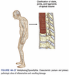
What is and what causes Ankylosing spondylitis?
- It is a chronic inflammatory joint disease characterized by stiffening and fusion (ankylosis) of the spine and sacroiliac joints
- Due to a HLA-B27 histocompatibility, there is an immune response to the cartilage → inflammation of fibrocartilage in the joints → fibroblasts proliferate and repair damaged cartilaginous structures → uncontrolled bone formation → fibrosis, ossification, and fusion of joints (primarily the sacroiliac joints and vertebral column)

What is and what causes osteomalacia?
- It is a metabolic disease characterized by inadequate and delayed mineralization of osteoid in mature and compact spongy bone.
- During bone remodeling replaced bone consists of soft osteoid instead of rigid bone
- Vitamin D deficiency is the most common cause of osteomalacia → A lack of vitamin D leads to low plasma calcium levels → PTH is secreted to ↑ Ca lvls → ↓’d phosphate levels → mineralization cannot proceed normally.


