7.1 Renal and Urologic Flashcards
(31 cards)
What is overactive bladder syndrome (OAB)?
- OAB is a chronic syndrome of detrusor overactivity in the absence of infection. Early, forceful detrusor contractions before the bladder is filled to capacity create a sensation of urgency and cause frequent voiding. Urethral resistance may be overcome, resulting in incontinence.
- Symptoms: incontinence, urgency, frequency, dysuria, and/or nocturia.

What is the Neurogenic Theory of an overactive bladder
- Neurologic damage to central inhibitory pathways or sensitization of peripheral afferent terminals in the bladder unmasks primitive voiding reflexes causing bladder overactivity. This may occur in association with neurologic diseases like stroke, multiple sclerosis, or spinal cord injury.

What is the Myogenic Theory of an overactive bladder?
- Increased intravesical pressure partially destroys peripheral efferent nerve terminals. Partial denervation of the detrusor muscle results in increased excitability of individual cells and spontaneous depolarization. Electrical activity spreads rapidly and there is an uncontrolled increase in intravesical pressure.

What is Nephrotic Syndrome?
- Nephrotic Syndrom is the excretion of 3.5 or more grams or more of protein per in the urine per day, especially albumin which leads to hypoalbuminemia (less than 3.0 g/dl), and peripheral edema.
- A characteristic of glomerular injury
- Nephrotic syndrome is a glomerular disorder that is more common in children.

What is the pathophysiology of nephrotic syndrome?
- Metabolic, biochemical or physiochemical disturbances damage glomerular basement membrane and podocytes, increasing its permeability to protein.
- Occurs secondary to disease in adults. The damage may be from glomerulonephritis, other kidney diseases, diabetes mellitus (most common), drugs, malignancies or vascular disorders.
- Albumin is lost in the greatest quantity because of its high plasma concentration and low molecular weight.
- Decreased dietary protein intake (malnutrition, anorexia) or liver disease (can’t synthesize enough albumin)complicates the disease by reducing the body’s ability to produce albumin.

What are the clinical manifestations of nephrotic syndrome?
- Proteinuria up to 10 grams/24.hours. Many clinical manifestations are related to loss of serum proteins and retention of sodium.
-
Edema, especially in areas of low tissue pressure (e.g. around eyes).
- Water accumulates in tissues because without adequate proteins in the serum, there is inadequate oncotic pressure to draw it back into the venous side of capillaries after hydrostatic pressure has pushed it out of the arterial side.
- Decreased plasma volume activates renin-angiotensin-aldosterone system and antidiuretic hormone. ADH causes sodium and water retention.
- The damaged kidney is unresponsive to physiologic mechanisms to reduce water retention
- Hyperlipidemia and lipiduria (fat bodies may be present in urine)
- Vitamin D deficiency
- Lipid casts or free fat droplets leak across glomerular capillary walls and are excreted in urine

What is Vesicoureteral Reflux (VUR)?
Vesicoureteral reflux (VUR) is the retrograde flow of bladder urine into the kidney ureters or both. Reflux perpetuates infection by preventing complete emptying of the bladder as refluxed urine drains back into the bladder at the end of each void.
- Normal vesicourethral junction doesn’t permit reflux by functioning as a one-way valve
- Trigone muscle contracts just prior to voiding. The increased pressure causes ureter to collapse
- The ratio of the length of ureteral segment in muscle to diameter of ureter determines the effectiveness of valve. Normal ratio is 5:1
- Reflux occurs secondary to short, wide, malpositioned ureter that doesn’t provide an adequate tunnel for valve to work
- Renal papillae are designed not to let refluxed urine in if reflux does occur

What is Acute Kidney Injury (AKI)?
- AKI is a sudden decline in kidney function with a decrease in glomerular filtration and accumulation of nitrogenous waste products in the blood as demonstrated by an elevation in plasma creatinine and blood urea nitrogen levels.
- Three categories: Prerenal, intrarenal or intrinsic, postrenal
- all cause an inflammatory response inside the kidney tissue
- Usually reversible if underlying cause is diagnosed and treated early

What causes Prerenal AKI?
- Reduced effective arterial blood volume causes renal hypoperfusion that occurs rapidly over a period of hours with elevation of BUN and Cr
- Poor perfusion can result from renal artery thrombosis, hypotension related to hypovolemia (dehydration, diarrhea, fluid shifts) or hemorrhage. Sepsis/septic shock and cardiogenic shock following cardiac surgery are the most common causes of AKI in the ICU
- sudden stress imposed on already marginally functioning kidneys may precipitate this

What causes Intrarenal AKI?
Intrarenal AKI may result from:
- dysfunction within the kidney
- ischemic acute tubular necrosis, nephrotoxic ATN (i.e. exposure to radiocontrast media or antibiotics), glomerulonephritis, vascular disease (malignant HTN, disseminated intravascular coagulation, and renal vasculitis), allograft rejection, or interstitial disease (drug allergy, infection, tumor growth)

What is Acute Tubular Necrosis (ATN)?
- Epithelial cells that line the tubule necrose/die via ischemia or nephrotoxins.
-
ATN caused by ischemia is the most common cause of intrarenal AKI
- Loss of blood supply (hypovolemia, hypotension) = loss of oxygen –> low ATP –> Free Radicals–> Necrosis
- Epithelial death also occurs via nephrotoxins like antibiotics especially aminoglycosides (gentamicin, tobramycin), heavy metals (mercury, arsenic), myoglobin, and radiocontrast media.
- Whatever the cause of cell death, when those cells die they slough off into the tubule and plug it up. The plug generates a higher pressure in the tubules which means fluids don’t flow as well from the high pressured arterioles and the GFR lowers. With less blood being filtered, less urine is produced (oliguria) and less Urea and Cr get filtered out ( azotemia).
- Transport of sodium and other molecules is disrupted
- The dead cells that make up the plug in the tubule form a brown tubular granular cast and eventually get secreted in the urine
- Damage primarily to the proximal tubular epithelium but may be distributed along any part of the nephron tubules.

What causes Post Renal AKI?
- Postrenal acute kidney injury is rare and usually occurs with urinary tract obstruction that affects the kidneys bilaterally
- (e.g., bilateral ureteral obstruction, bladder outlet obstruction–
prostatic hypertrophy, tumors or neurogenic bladder,
and urethral obstruction, calculi or renal vein thrombosis).
- (e.g., bilateral ureteral obstruction, bladder outlet obstruction–
- The obstruction causes an increase in intraluminal pressure upstream from the site of obstruction (can lead to nephrosis) with a gradual decrease in GFR

List the three mechanisms of Oliguria?
- Oliguria (less than 400 ml of urine output per day) can occur in AKI, and three mechanisms have been proposed to account for the decrease in urine output. All three mechanisms probably contribute to oliguria in varying combinations and degrees throughout the course of the disease
- Alterations in renal blood flow.
- Tubular obstruction.
- Backleak

Describe the three mechanisms of Oliguria
- Alterations in renal blood flow. Efferent arteriolar vasoconstriction may be produced by intrarenal release of angiotensin II or by redistribution of blood flow from the cortex to the medulla. Autoregulation of blood flow may be impaired, resulting in decreased GFR. Changes in glomerular permeability and decreased GFR also may result from the ischemia.
2. Tubular obstruction. Advanced injury with necrosis of the tubules causes sloughing of cells, cast formation, or ischemic edema that results in tubular obstruction, which in
turn causes a retrograde increase in pressure and reduces the GFR. Renal failure can occur within 24 hours.
3. Backleak. Glomerular filtration remains normal, but tubular reabsorption or “leak” of filtrate is accelerated as a result of permeability caused by ischemia and increased tubular pressure from obstruction.

What is Chronic Kidney Disease (CKD)?
- CKD is the progressive and irreversible loss of renal function or loss of nephrons that will eventually lead to kidney failure. The disease is classified by stages 1-5 that is based on the glomerular filtration rate (GFR).
- Kidney damage less than 60 ml/min for 3 months or more, irrespective of cause.
- Risk factors:
- Diabetes and hypertension cause more than 60% of chronic renal failure.
- The elderly are at higher risk because of normal age-related decline in renal function added to higher risk for other diseases.

Describe CKD Stage 1
- Normal GFR
- Kidney damage but usually no symptoms
- GFR greater than or equal to 90
Describe CKD Stage 2
- Mildly decreased GFR (60-89)
- Subtle HTN

Describe CDK stage 3
- Moderately reduced kidney function, Stage 3 GFR 30-59
- Stage 3A GFR 45-59
- Stage 3B GFR 30-44
- Erythropoietin deficiency, anemia
- Symptoms: mild hypertension, fatigue, fluid retention, changes in urine

Describe CKD Stage 4
- Severe Kidney Damage
- GFR 15-29
- Symptoms related to complications of kidney disease (osteodystrophy and anemia)
- symptoms: moderate htn, hyperphosphatemia, anemia

Describe CDK Stage 5
- End Stage Renal Disease
- GFR < 15
- Symptoms: Severe hypertension, hyperphosphatemia, anemia
- Dialysis

Briefly, explain how osteodystrophy develops in patients with CKD.

- Kidneys play an important role in balancing Calcium levels. Normally the kidney helps activate Vitamin D which increases absorption of Calcium from the diet.
- In CKD, kidneys are not able to activate as much Vitamin D so less Calcium is absorbed into the blood resulting in hypocalcemia.
- When Calcium levels fall, parathyroid hormone (PTH) is released which causes the bones to loose Calcium.
- Over time this resorption of Calcium from the bones leaves them weak and brittle.
- Leading to Renal Osteodystrophy
- Hypocalcemia leads to secondary hyperparathyroidism, GFR falls, and progressive hyperphosphatemia, hypocalcemia, and dissolution of the bone result.

Briefly, explain how Anemia develops in patients with CKD.
- Kidneys secrete a hormone called Erythropoietin which stimulates the production of RBC from the bone marrow.
- In CKD, Erythropoietin levels fall, leading to a lowered production of RBC and ultimately Anemia.
- Uremic toxins shorten red blood cell survival and alter platelet function
- Lethargy, dizziness, and low hematocrit are common.

What is Acute Glomerulonephritis?
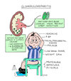
- Acute glomerulonephritis includes renal diseases in which glomerular inflammation is caused by immune mechanisms that damage the glomerular capillary filtration membrane including the endothelium, basement membrane (GBM), and epithelium (podocytes)
- The classic symptoms include sudden onset of hematuria including red blood cell casts and proteinuria, and in more severe cases, these symptoms are also accompanied by edema, hypertension, and impaired renal function.

What causes Acute Glomerulonephritis?
- Acute glomerulonephritis commonly results from inflammatory damage to the glomerulus as a consequence of immune reactions including deposition of circulating immune complexes, antibodies reacting in-situ to planted antigens, and antibodies directed against the glomerular basement membrane. The urine sediment may contain large amounts of protein (nephrotic sediment) or have red and white blood cells and protein (nephritic sediment).


