Midterm I - Miscellaneous Photos & Info Flashcards

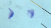
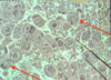
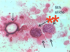
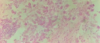
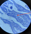
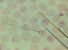
Trypanosoma sp.: Trypomastigotes
Blood smear

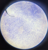
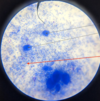
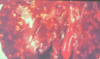
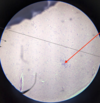
Trypanosoma equiperdum: Trypomastigotes

Trypomastigote in the blood

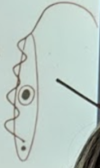
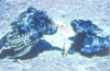
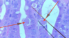
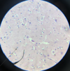
Trypanosoma sp.: Trypomastigote
Fish blood
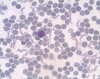
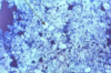
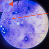
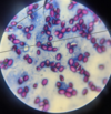
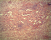
Leishmania sp.: Promastigotes

- Leishmania sp.*: Promastigotes
- Extracellular forms*
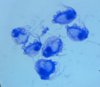
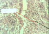
Leishmania sp.: Amastigotes
Divided & intracellular - Only in macrophages
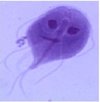
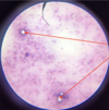
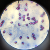
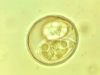
Giardia sp.: Trophozoite
Containing 2 nuclei

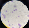
Giardia sp.: Trophozoites
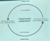
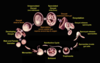
AfricanTrypanosoma sp.: life cycle

Trypanosoma sp.: Form type in the vertebrate host tissue
Amastigotes

Trypanosoma sp.: Form type(s) in insects
- Promastigote
- Epimastigote

Trypanosoma sp.: Form type in the vertebrate host’s blood
Trypomastigote
“Metacyclic form”
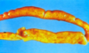
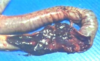
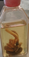
Trypanosoma sp.: Symptoms
- Genital & abdominal oedema
- Cachexia
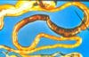
African Trypanosoma sp. “Salivaria”: Vector
Tsetse fly
Males & females
Trypanosoma equiperdum: Life cycle
Trypanosoma cruzi: Life cycle

Leishmania sp.: Life cycle
Passed on by the saliva (not faeces)

Leishmania sp.: Form type In vertebrates
Amastigotes
Leishmania sp.: Form type In insects
Promastigotes
Leishmania sp.: Vector
Female sand fly
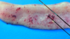
Leishmania tropica: Pathological form
Cutaneous form (skin)
Leishmania braziliensis: Pathological form
Mucocutaneous form (oral & nasal cavity)
Leishmania donovani: Pathological form
Visceral form (liver, spleen etc.)
Leishmania infantum: Pathological form
Visceral & cutaneous form
Leishmania chagasi: Pathological form
Visceral & cutaneous form
Giardia sp.: Life cycle
Spreading by cysts


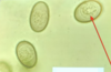
Giardia sp.: Cysts
Giemsa stain not good - Cysts appear empty

Giardia sp.: Cysts
Faecal smear, Poor staining - Nuclei of cysts cannot be seen
Giardia sp.: Cysts
Faecal smear, poor staining - Nuclei of cysts cannot be seen

Giardia sp.: Cysts
Floatation method - not good, empty cysts
Trichomonadida sp.: Life cycle

Trichomonas species are grouped by…
The number of anterior flagella they have

- Trichomonas foetus*
- In cattle*

- Trichomonas gallinae*
- In poultry*
Trichomonas sp.: Trophozoites
Trichomonas sp.: Trophozoites

Trichomonas sp.: Trophozoite

Trichomonas sp.: Trophozoites
Broth culture

Trichomonas gallinae: Necropsy specimen

Trichomonas gallinae: Liver necrosis
Histomonas sp.: Life cycle

Histomonas sp.: Susceptible species
- Turkey
- Partridge
- Quail
- Guinea fowl
- (Chicken)
Histomonas sp.: Forms

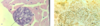
Histomonas sp.: Necropsy findings


- Histomonas meleagridis*: Trophozoite
- Liver, PAS Stain*

Histomonas sp.: Trophozoite
Histomonas meleagirdis: Black head disease
Caused by cyanosis, only becomes black post mortem

Generalised histomosis

Histomonas meleagirdis: Infection of the caeca of a turkey
Eimeria sp.: Life cycle
Eimeria sp.: Summarise the zoites
Sporozoites & merozoites
- Unicellular forms
- Asexual form in all apicomplexan parasites
- Lunar shaped

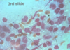
Apicomplexan sporozoites & merozoites
Giemsa stain

Apicomplexan parasite: Sporozoites & merozoites

Apicomplexan parasite: Sporozoites & merozoites

Apicomplexan parasite: Sporozoites & merozoites

Apicomplexan parasite: Sporozoites & merozoites

Eimeria sp.: Trophozoite

Schizonts
Filled with trophozoites or merozoites

Intracellular schizont
Schizonts: Filled with trophozoites or merozoites

Schizonts: Full of trophozoites or merozoites

Schizonts filled with trophozoites or merozoites
Notice the nucleus pressed to the side

Schizont with other staining
Zoites aren’t visible due to staining

Eimeria sp. infection

- Eimeria sp.*: Schizont
- Zoites can’t be seen inside*
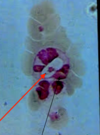
Eimeria sp.: Merozoites

Eimeria sp.: Microgamonts (male)
Eimeria sp.: Macrogamonts (female)

Schizogony

Gametogony

Gametogony

Schizogony

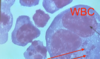
Eimeria sp.: Gamonts
Amongst chicken RBCs
Eimeria sp.: Macrogamonts (female)
Purple appearance
Eimeria sp. infection: Normal chicken RBC
Eimeria sp.: Empty oocysts
Eimeria sp.: Empty oocyst

Eimeria sp.: Macrogamonts
Eimeria sp.: Ripened oocyst
Gamonts

Nuclei of nurse cells
Eimeria sp.: Oocysts shedding into the lumen

Eimeria sp.: Oocysts enter the gut lumen
What is significant about the staining of unsporolated oocysts?
They cannot be stained with standard histological stains
Eimeria sp.: Unsporolated oocysts
Contains a zygote

Eimeria sp.: Unsporolated oocysts
Contains a zygote

Eimeria sp.: Unsporulated oocysts
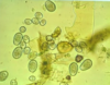
Eimeria sp.: Oocysts
Giemsa stain

Eimeria sp.: Oocysts
Iodine stain

Eimeria sp.: Oocysts
Iodine stain
Eimeria sp.: Oocysts
Faecal smear, Kinyoun stain
Eimeria sp.: Oocysts
Faecal smear, Kinyoun stain

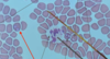
Eimeria sp.: Sporulated oocyst
Each contains 4 sporocysts (with 2 sporozoites each)

Eimeria sp.: Sporulated oocyst
Each contains 4 sporocysts (with 2 sporozoites each)

Eimeria sp.: Sporulated oocyst
Each contains 4 sporocysts (with 2 sporozoites each)

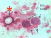
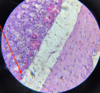
Avian coccidiosis: Trophozoites
Note the epithelium is intact: Development stage is therefore trophozoite
Avian coccidiosis: Trophozoites becoming schizonts
Note the epithelium being destroyed
Avian coccidiosis: Schizogony & new generations of trophozoites
Note the epithelium being destroyed

Avian coccidiosis: Gametogony

Avian coccidiosis: Younger oocysts
Note the empty space around them

Eimeria acervulina: Necropsy specimen
Characterised by white foci in the duodenum & jejunum
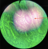
- Eimeria acervulina*
- Epithelial cells infected with gamonts*
Eimeria tenella

Eimeria necatrix

Eimeria maxima
Eimeria acervulina
Eimeria mitis

Eimeria acervulina
Epithelial cells infected with gamonts → Loss of epithelial cells

Eimeria acervulina
Epithelial cells infected with gamonts → Loss of epithelial cells
Eimeria acervulina
Macrogamonts & oocysts in the destroyed epithelium of duodenum
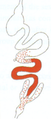
- Eimeria maxima*
- Characterised by “Salt and pepper” appearance in the intestine*
- Eimeria maxima*
- Characterised by “Salt and pepper” appearance in the intestine*

Eimeria maxima: Gamont

Eimeria maxima: Schizonts
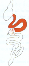
- Eimeria necatrix*
- Haemorrage of the middle third of the intestine
- Eimeria necatrix*
- Haemorrage of the middle third of the intestine

Eimeria necatrix: Gamonts

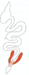
- Eimeria tenella*
- Enlarged, bloody caecum

- Eimeria tenella*
- Enlarged, bloody caecum

- Eimeria tenella*
- Enlarged, bloody caecum

- Eimeria tenella* infection
- Enlarged, bloody caecum

Eimeria tenella infection: Schizogony
- Eimeria tenella* infection
- Enlarged, bloody caecum

- Eimeria brunetti* infection
- Large intestine - Bloody content

- Eimeria anseris* infection
- Blood, ulceration, dark dots & inflammation in the gut

- Eimeria anseris* infection
- Blood, ulceration, dark dots & inflammation in the gut

Eimeria truncata infection
- White lines on kidneys
- Red spots inside tubules

Eimeria truncata infection
- White lines on kidneys
- Red spots inside tubules

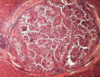
Eimeria truncata infection: Gamonts
Note the renal glomerulus seen in the kidney sample

Eimeria truncata infection: Gamonts
Note the renal tubules seen in the kidney sample
Which two types of development are possible in the life cycle of bovine coccidiosis?
- With small schizonts (meronts)
- With large macroschizonts (globidia) (usually the first generation)


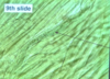
Sheep coccidiosis: Globidia in the intestine

Sheep coccidiosis: Globidia inside the intestine

Goat coccidiosis
Globidia inside the intestine

Large intestine: Ruminant
Gamonts seen visible with the naked eye (white)
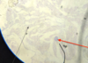
Coccidiosis of calf: Oocyst in the jejunum
Surrounded by RBCs (difference to chicken)

- Eimeria stiedai* infection (liver)
- Oocyst

- Eimeria stiedai* infection (liver)
- Gamonts

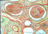
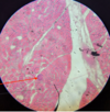
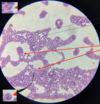
Eimeria stiedai: Biliary hyperplasia
Proliferation is characteristic of this parasite

Eimeria stiedai (biliary)
- Gamonts
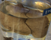
Biliary coccidiosis of liver (E. stiedai)

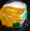
Eimeria stiedai infection: Rabbit liver
with small nodules

Eimeria coccidiosis: Intestine of a rabbit
Eimeria stiedai infection: Liver
Eimeria stiedai infection: Oocysts
Eimeria stiedai infection
Intestinal coccidiosis in rabbit gut
Darker than the surrounding RBCs
Isosporosis: Summarise sporulated oocysts
Each contains 2 sporocysts (containing 4 sporozoites)

Isospora canis infection: Oocyst
ZN stain

Prolapse of cattle rectum
- Detection of unsporulated oocyst by floatation
- E. bovis
Oocysts of E. bovis (larger) & E. zuernii (smaller)
Cryptosporidiosis: Life cycle


Cryptosporidiosis infection
Note the parasite isn’t inside the cell

Cryptosporidium baileyi infection: 2 oocyst types
Red = Thick wall; White = Thin wall
Kinyoun & ZN staining
Cryptosporidium baileyi infection: 2 oocyst types
Red = Thick wall; White = Thin wall
Kinyoun & ZN staining

Cryptosporidium destroyed part of the jejunum (calf)
Toxoplasmosis: Life cycle

Toxoplasmosis: In cells, trophozoites & cysts can be identified by their…
Lack of wall

Toxoplasma cyst (in brain)

Toxoplasma cyst (brain(

Toxoplasma cyst (Brain)
Besnoitiosis (Besnoitia besnoiti): Clinical signs
- Subcut. connective tissue → Elephant skin
- Lesions on the sclera


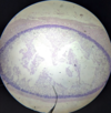
Besnoitiosis: Cysts surrounded by connective tissue
Note the cysts have an ‘empty’ border
Besnoitiosis: Cysts surrounded by connective tissue
Note the cysts have an ‘empty’ border

Besnoitiosis: Cysts surrounded by connective tissue
Note the cysts have an ‘empty’ border
Sarcocystiosis: Life cycle
Note that Sarcocystis species use host macrophages for their lifecycle


Sarcocystosis of a bird
White cysts on the muscle
Sarcocystis suihominis: Cyst
Sarcocystosis: Cyst
Sarcocystosis: Cyst cross section (with a wall)
In skeletal muscle

Sarcocystosis: Cyst cross section (with a wall)

Sarcocystosis: Cyst cross section (with a wall)
In skeletal muscle
Sarcocystosis: Cyst cross section (with a wall)
In skeletal muscle
Sarcocystis tenella infection: Cyst
- Oesophagus of sheep*
- Really large*

- Sarcocystis tenella* infection: Cyst
- Oesophagus of sheep*
- The free zoites can be seen*

- Sarcocystis rileyi* infection
- Cysts in mallard muscle

Sarcocystis rileyi infection: Muscle cysts

Sarcocystis tenella infection: Cysts in oesophagus (sheep)

Sarcocystis tenella infection: Cysts in oesophagus (sheep)
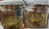
Sarcosporidium sp.: Mallard (intermediate host)
Hepatozoonosis of dog: Gamont inside neutrophil
Hepatozoonosis of dog: Life cycle

Hepatozoonosis of dog: Gamont
Spleen smear
Hepatozoonosis of dog: Gamont
Spleen smear

Hepatozoonosis of dog: Gamonts
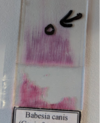
When sampling babesia, where in the sample would they be found in:
- Smear
- Centrifuged blood sample
- Smear: At the end of the smear
- Blood sample: Buffy coat layer
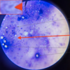
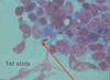
Babesia in peripheral blood
Unconfirmed as there is only one white speck in each
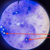
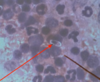
Babesia in peripheral blood: 2 merozoites in RBCs
Confirmed: As there are 2 white specks present

Babesiosis
Confirmed: As there are 2 specks

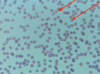
Babesia in a RBC
Babesiosis: RBCs with 2 merozoites
Confirmed: because there are 2 white specks
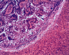
Babesiosis in the spleen
Brown appearance

Babesia canis
Theileriosis: Koch’s bodies (Macroschizonts)
Theleriosis: Merozoites in RBCs

Theleriosis: Merozoites in RBCs

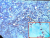
Encephalitozoon cuniculi infection: Tissue reactions caused by Microsporidia
Brain tissue
Encephalitozoonosis: Meronts & schizonts
Brain tissue
Encephalitozoonosis: Meronts & schizonts
Brain tissue
Encephalitozoon cunuculi infection: Nephritis & CT proliferation
Kidney tissue
