Microbe Library Flashcards
(62 cards)

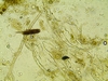
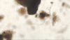
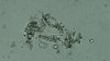
actinobacteria
An actinobacteria filament is coming out of the aggregate, but the strands in the upper left are too out of focus to ID

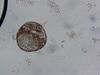
Testate Amoebae
Shape, OM plug, amoeba
The organism has some organic matter inside which indicates it is alive

actinobacteria
1 micrometer in diameter
The actinobacteria is growing off plant material. It is out of focus - when in perfect focus, it should look like a single pencil line. It is actinobacteria because the filament is evenly 1um in diameter, smooth curve. Another small segment of actinobacteria is more blurry. Measurements: 1 um diameter / 0.2 units lenght

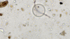
Testate Amoebae
shape
There is an amoeba in the clear bottom. The materials above are organic matter that the amoeba pulled in. Material looks like organic matter in the surrounding area, which was pulled in by the amoeba when the tube was shaken (otherwise material would not be inside.)

Tardigrade
shape
Look for the stumpy “feet” and claws

Arcella
shape and color
In the upper left corner there is a fungal spore. Look at the pattern of 2 septated cells and then a narrow point. This is a stalk of spores.
round naked amoeba
shape/bacterial density inside
Under the microscope, this organism’s internal contents were moving. Single membrane
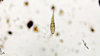
rotifer, tardigrade/nematodes
shape of organisms
Rotifer is attached to OM in the top left of photo. Tardigrade is in the center, you can see its feet and claws. Nematodes to the right

Testate Amoeba/ fungi
shape
Chain of spores in front of the testate amoebae.
Inside of the testate you can see that it is either dividing or has very apparent vacuoles. Video needed to confirm.
The upper section of the fungal hypha is out of focus therefore the position of the focal plane relative to the curvature of the cell wall creates the appearance of a line in the middle of the hyphae. Measurements: 0.58 Lenght / 4.5-5 Diameter

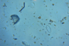
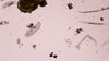
Flagellate, cyst
Flagellate is very clear but hard to find. Possible flagellate cyst on left side, single wall
Several mineral pieces in this sample with sharp edges and clear reflective surfaces. There is a spore to the right of center in this image. Find center of photo and move about a inch to the left you will find the flagellate. Potential cyst on left. Would need focus control to confirm it is a cyst and not an air bubble.

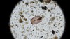
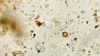
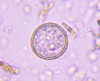
Testate Amoeba

Arcella
color, shape
Dark spot is the opening to the test. Several fungal spores in this image. This sample should of been diluted more so that all of the organisms could of been easily identified.

Naked Amoebae
shape

Bacterial Omnivore Nematode
Mouth
Wide open mouth, no tooth, rectangular chamber. This Bacterial Omnivore does not have any bulbs or the bulbs are not in focus.

Testate Amoebae
Shape, OM plug, amoeba in test
Top right one could be mistaken for empty, but is different from background. It looks like it is expelling its OM plug. There are two testate amoeba in the lower left corner that are possibly eating the same food source. Actinobacteria in the upper right corner are slightly out of focus.

Ciliate
cilia
Actinobacteria to the right in the center. The filamentous organism to the right of center is plant material because it is shredded and frayed along the length of it.
Amoebae Cyst
double walled

Ciliate
Shape, stalk

Testate Amoebae
Shape
Several fungal spores in image

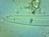
Fungi (beneficial)
even septa,color, uniform diameter
This is a mature fungal hyphae with thick cell walls. Oxalate crystals on the surface of the hypha. Measurements: .95 units Length 19-20 um Diameter
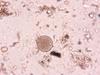
Arcella
color, shape
you can see the amoeba in the bottom left for the test. The opening in the top middle is not perfectly round, organism looks clearish inside test.

Fungi (beneficial)
even septa,color, uniform diameter
Fungal hypha, Actinobacteria strand below it. Measurements of fungal hypha: 1.05 units Lenght / 2.5-3 um Diameter

Fungi (beneficial)
even septa,color, uniform diameter
This particular species of fungi has dominated the organic material. It will be considered an outlier if it lands in a FoV unless every fov has whefts of fungi. Massage the sample, shake again, make a new drop to make sure this is common to the soil. Hyphae in focus is 3.5-4 um
testate amoebae
Shape
Not empty, note difference from background.