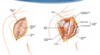
MEMORIZE Approaches Flashcards
Deltopectoral
Interval: Deltoid (axillary n.) & pec major (med & lat pectoral n.)
Incision: Coracoid to deltoid insertion, Coracoid to axillary fold
- cephalic vein is landmark
Approach: Split deltoid and pec, incise clavipec fascia, come down on subscapularis then capsule
Dangers:
- cephalic vein,
- axillary n.,
- musculocutaneous n.
- Biceps tendon,
- anterior circumflex vessels
Deltoid Split
Interval: none
Approach: Split fibres of deltoid
Dangers: Axillary n.. Crosses humerus approximately 5-7cm distal from tip of acromion

Judet Approach Shoulder
Interval: Teres Minor (Axillary n.) and Infraspinatus (Suprascapular N.)
Incision: From posterolateral corner acromion medial along spine of scapula, then 90 degrees and distal to inferior pole
Approach: Split deltoid or elevate from scapular spine. Fat stripe between teres minor and infraspinatus. Brings you down on capsule.
Dangers:
- Axillary N with aggressive retraction of Deltoid or Teres Minor.
- Suprascapular nerve and artery, at superolateral border of interval: which goes from supra to infraspinous fossa and can be damaged by retracting too medially;
- Vessels: Posterior Humeral Circumflex Artery: in the quad space with the axillary nerve

Anterolateral Approach Humerus
Interval:
- Proximal: Deltoid (Axillary N.) & Biceps (Musculocutaneous)
- Distal: Brachialis Split (Medial - Median n. lateral- radial N.)
Approach:
- Incision- coracoid to deltoid tuberosity then along lateral boarder of biceps.
- Establish deltopecotral interval, and separate between biceps and deltoid.
- As brachialis emerges, develelop split. - Can be extended into a henry approach of the volar forearm.
Dangers:
- Musculocutaneous N. Deep to biceps, superficial to brachialis.
- Radial N. Between brachialis and brachioradialis laterally and in spiral groove.
- LABCN between brachialis and brachioradialis
Paratricipital Approach
Interval: Lateral head of triceps (radial n.) and lateral intramuscular septum (no true plane)
**Incision: **Midline posterior from tip of olecranon to 8cm distal to acromion
Approach:
- split superficial fascia in line with incision
- identify and protect radial n.
- btw lateral head triceps (Retract lateral) and long head triceps (retract medial)
- Either identify LABCN (and trace to radial nerve proper) or radial nerve as it plunges into the intramuscular septum (~10cm proximal to the lateral epicondyle. Once radial nerve proper identified protect, peel triceps off of posterior humerus.
Dangers:
- Radial nerve – Identify if needing to go proximally,
- Ulnar nerve – deep to the medial head, stay subperiosteal medially,
- Profunda brachii artery – lies with the radial nerve in the spiral groove – protect it with the radial nerve
* can do medial or lateral. Medially, radial nerve enters spiral groove ~14cm proximal to medial epicondyle. Ulnar nerve to be identified deep to brachioradialis.

posterior transolecranon approach to elbow (Olecranon Osteotomy)
Interval: n/a
** Incision:** midline posterior over olecranon
**Intervals: **
- Proximal: Distal to radial nerve innervation of triceps, between Lateral head Triceps and BR lateral.
- Identify and isolate ulnar nerve medial.
- Distal: Anconeus and ECU lateral, FCU medial.
Olecranon Osteotomy: Predrill Olecranon, Chevron with point distal into bare area of sigmoid notch, take piece with triceps superior
**Dangers: **
- Radial n.– can’t extend past the dist 1/3 of humerus
- Ulnar n.– identify and protect it.
- Cartilage damage to olecranon.

Boyd Approach
Interval: Between both anconeus (radial n.) and ECU (PIN), and subcutaneous boarder of ulna/ FCU (ulnar n.)
Approach: Develop interval between both anconeus and ECU, and lift both anteriorly. Release supinator subperiosteally.
Dangers: Increased risk of synostosis.

Kocher Approach
Best for LUCL repair
Interval: Anconeus (radial n.) & ECU (PIN)
Approach: Look for fat stripe between the two. Anconeus fibres will run obliquely. Will need to elevate some of supinator to reveal distal insertion of LUCL on crestor supinatore.
Dangers:
PIN – limit dissection proximal to annular ligament with arm pronated, LUCL instability
* can extend proximally by detatching anconeus from its origin on the distal humerus, and triceps from lateral intramuscular septum.

EDC Split
Interval: Split EDC Tendon
Approach: It is the “shiny” tendon on the lateral aspect of the elbow. Split 50/50. Gives more access to anterior structures of the elbow (ie coranoid).
Dangers: PIN, LUCL.

Kaplan
Interval: EDC (PIN), ECRB (PIN)
Approach: Split interval above. Proximal interval of the Thompson approach to the forearm.
Dangers: PIN
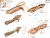
*pictured: kocher vs kaplan

Hotchkiss Medial Over-the-top
good for antermedial - access to top of coronoid process and anterior elbow joint
Interval:
* Distally: Through flexor pronator mass. FCU (ulnar n.) & FDS/Palmaris Longus (Median n.)
Approach:
- Unroof, identify and mobilize the ulnar n.
- Split flexor pronator mass, and elevate anteriorly. Care to be taken for MUCL
Dangers:
- Ulnar N.
- MUCL,
- Median N.
- Brachial A.,
- MABCN (found on fascia anterior to septum)

FCU Split
Interval: None, between two heads of FCU (Ulnar N.)
Approach:
- Identify, unroof and protect median n. - Split two heads of FCU and elevate anteriorly. Care to be taken not to injure MUCL
Dangers: MUCL, Ulnar N. Median N. Brachial A.

Modified Taylor and Scham
Interval: ECU (PIN), FCU (Ulnar N.)
Approach:
- identify and protect ulnar n.
- Dissect down to subcutaneous boarder of ulan and lift everything anteriorly. Akin to the boyd but on the medial side.
- Gives access to base of coranoid/sublime tubercle
Dangers: Ulnar N., MUCL
*1 = Hotchkiss, 2= FCU Split, 3=Taylor Scham

Approach to Ulnar Shaft
Interval: ECU (PIN), FCU (Ulnar N.)
Approach: Dissect onto subcutaneous boarder of ulna. Lift ECU and FCU subperiosteally to expose.
Dangers: Ulnar N (under FCU, ontop of FDP), Dorsal cutaneous branch of ulna distally, Ulnar A (runs with Ulnar N, radial to ulnar N)

Henry Approach Volar Forearm
Approach:
- Landmark incision from lateral aspect of biceps tendon to radial styloid.
* Superficial
* Proximally brachioradialis (radial n.) & FCR (median n.)
* ligate the arterial branch of lateral side of radial a. to mobize radial a. medially
* careful of superfical radial n under BR and retract laterally
* Distally brachioradialis (radial n.) & radial a.*
* Deep
* Proximal 1/3 - BR and PT, supinate forearm to protect PIN, incise supinator on medial edge and elevate
* middle 1/3 - pronate to expose lateral border of PT and can detach some of it
* distal 1/3 - elevate FPL, and PQ ulnarly
Dangers:
* Superficial radial N. (Deep to brachioradialis)
* PIN - radial neck under supinator
* Radial A. - under BR
*FCR approach, the interval is between FCR and radial artery (radial artery goes radially). In Henry, the radial artery comes medially.

Thompson Approach Dorsal Forearm
Interval:
- Superficial: EDC (PIN), ECRL/ECRB (Radial n., PIN)
- Deep: Supinator (PIN) and Pronator Teres (median n.)
Approach:
- incision: anterior to lateral epicondyle to just ulnar to Lister’s tubercle
Intervals:
- Superficial:
- Proximal: ECRB (radial nerve) & EDC (PIN); (Kaplan)
- Distal: ECRB (radial nerve) & EPL (PIN);
- Deep:
- Proximal: Supinator – dissect PIN, then supinate forearm & subperiosteally dissect supinator;
- Middle: APL & EPB covers radius, elevate these;
- Distal: between ECRB (2nd compartment) and EPL (3rd compartment), on bone
Dangers:
Lateral antebrachial cutaneous, PIN, superficial radial nerve

Modified Henry FCR Approach to the Distal Radius
Interval: FCR (Median N.) and Radial a. (brachioradialis- radial n.)
Approach:
- Sharp incision over FCR tendon, sharply through skin, subcutaenous tissue and FCR sheath. Retract FCR tendon radially and incise through FCR subsheath.
- Retract FPL tendon ulnarly
- Incise PQ along distal and radial boarder and peel off subperiosteally to reveal distal radius
Dangers: Radial A., Median N., palmar cutaneous branch of median n.

Volar Ulnar Approach at the Wrist
Interval: Flexor Tendons [(FDS) - Median N.)] & Ulnar A.
Approach: Develop interval between FDS and FCU. Work proximally to identify ulnar a. and ulnar n. Develop interval between ulnar a. and flexor tendons. Identify PQ, lift PQ radially to reveal ulnar aspect of distal radius. Can follow ulnar n. and relase guyons canal, can release carpal tunnel through this approach as well.
Dangers: Ulnar A. Ulnar N.

Dorsal Approach to the Wrist
Interval: 3rd & 4th extensor compartments (both PIN).
Position: Supine, hand table
**Incision: **Just ulnar to Listers, in line with 3rd metacarpal.
**Interval: **
1st Layer:
- extensor retinaculum between 3rd compartment - EPL (PIN) radial and 4th compartment - EDC (PIN) ulnar
- use retinaculum to retract tendons.
2nd layer:
- Ligament Sparing: V-Shaped, with tip at triquetrum, Proximal limb in line with DRC fibers and Distal limb in line with DIC fibers, elevated radially with attachment left at radial styloid.
- Non-Ligament Sparing: In line dorsal capsulotomy, subperiosteally dissect under RSC radially and DRC ulnarly.
- Scaphoid: T-Shaped capsulotomy, along border of radius and distal to DIC fibers. Must flex wrist. Allows visualization of proximal scaphoid, SL ligament.
Dangers: PIN purely sensory at the wrist (can be ablated for pain control), Dorsal sensory branch of radial nerve, Dorsal carpal artery (supplies scaphoid)
Approach:
- Incision centered over Lister’s tubercle
- Elevate skin flap, excise extensor retinaculum.
- Identify 3rd and 4th compartment and split the two.
- Identify dorsal wrist capsule, dorsal intercarpal ligament, and dorsal radiocarpal ligament. (Ligament sparing capsulotomy)
Dangers: PIN purely sensory at the wrist under 4th compartment (can be ablated for pain control). SL, LT ligaments.

Russe Approach (Volar Approach to Scaphoid)
Interval: Radial a. and FCR (Median N.)
Approach:
- wagner iIncision centred over the scaphoid tubercle
- Glabrous border of thumb between radial artery and FCR.
- Identify FCR tendon, then cut through subsheath , retract FCR ulnarly
- Split thenar muscles distally
- : Incise RSC and long Radiolunate in line.
- Incise capsule over scaphoid and expose scaphoid up to ST joint
- Must extend wrist and often excise part of trapezium to obtain start point.
Dangers: Dorsal sensory branch of radial nerve, radial artery, volar scaphoid branch of radial artery.

Subvastus/Lateral Approach to Femur
Interval: Lateral Intramuscular Septum & Vastus Lateralis (femoral n.). Technically none.
Approach:
- Incise IT Band
- Incise Vastus Lateralis fascia.
- Lift Vastus lateralis from septum and femur, taking care to cauterize perforator branches.
- Can go either through vastus (direct lateral) or under vastus (posterolateral)
Dangers: Perforating branches of Profunda Femoral A.

Anteromedial Approach to the Distal Femur
Interval: Vastus Medialis (femoral n.) & rectus femoris (femoral n.)
Approach:
- Incision from anteromedial thigh to medial patella
- Develop plane between vastus medialis and rectus femoris
- Split vastus intermedius deep
- Need to repair any detatched vastus medialis from quadriceps tendon insertion to patella
Dangers:
- Medial geniculate artery (ligate)

Medial Approach to Distal Femur
Interval:
- Superficial: Adductor Longus (Obturator n.) & Sartorius (Femoral N.)
- Deep: Adductor Longus (Obturator n.)& Vastus Medialis (Femoral N.)
Approach:
- Incision over adductor tubercle and extend proximally
- Develop plane between adductor longus and sartorisu, then deep vastus medialis.
- Cannot go distal to vastoadductor membrane (9cm proximal to jont. This is the membrane that covers hunters canal deep to sartorius.
Dangers:
- Saphenous n. (identify over adductor tendon)
- Femoral A. (crosses ant to post 13cm bove joint)

Smith-Peterson (Anterior) Approach to the Hip
Interval: *only approach to the hip with TRUE internervous plane
- Superificial: TFL (Superior Gluteal N.) & Sartorius (Femoral N.)
- Deep: Rectus Femoris (Femoral N.) & Gluteus Medius (Superior Gluteal N.)
Approach:
* Incision vertically from ASIS towards lateral side of patella (8-10 cm)
* Incise TFL fascia, retract muscle laterally, and incise deep subfascia to protect LFCN.
* Bluntly develop plate between Rectus & Gluteus medius. Take care to identify and ligate/cauterize branches of lateral femoral circumflex a. in this interval.
* Can detatch reflected head of rectus from acetabulum to better expose capsule
* Come directly ontop of hip capsule and make capsulotomy
Dangers:
- Lateral Femoral Cutaneous N. (branches over sartorius 2.5cm below ASIS)
- Ascending branches of Lateral Femoral Circumflex A.


