Management cards (Slide exam) Flashcards

1
Q

Name, mx (3)

A

Conjunctival papilloma

Patient reassurance, optional surgery for cosmesis, general ddx (check FAT, feeder blood vessels, surround tissue invasion, biopsy)

2
Q

.Name, mx (2)

A

CIN (conjunctival intraepithelial neoplasia):

Mx: surgery for removal, general ddx

3
Q

Name, mx (1)

A

SCN:

Mx: surgery for removal, ddx

4
Q

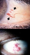
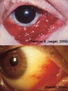
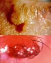
How should you manage any unusual/unepected growth on the eye (e.g. the ones in the image) or adnexa? (4)

A
  1. Check for fast development/spread (e.g. with FAT)
  2. Check amount of feeder blood vessels
  3. Check for surrounding tissue invasion
  4. Refer for biopsy as needed
5
Q

.Name, mx (1)

A

Naevus: Benign ocular pigmented lesion

Mx: refer for biopsy if suspicious

6
Q

Name, mx (1)

A

Congenital melanocytosis

Mx: (if malignant) refer for excision + biopsy

7
Q

Name, When is a biopsy indicated? (2)

A

Primary Acquired Melanosis (PAM):

Biopsy indicated if: - diffuse lesion found at limbus + corneal involvement; - expansive/overgrowth of diffuse lesion in one eye (which shows its pleomorphic)

8
Q

Name, Biopsy appearance (1), mx (1)

A

Melanoma

biopsy: invasive lesion with pleomorphism and anaplasia

Mx: surgical removal

9
Q

Name, mx (2)

A

Telangiectasia:

Mx: surgery for cosmesis, tx any assoc. systemic conditions (e.g. sturge-weber, HHT)

10
Q

Name, mx (7)

A

Kaposi’s sarcoma:

Mx: refer for diagnostic workup if AIDS dx unknown, or for therapy (e.g. HAART - highly active anti retroviral therapy) if AIDS present.;

Tx often palliative (i.e. relieve symptoms/pain), surgery, focal radiation, anti-mitotics, AIDS protease inhibitors

11
Q

Name, mx (4)

A

Sturge-Weber Syndrome

Mx: consider referral for ax of neurological status; follow up appt. in 3-12 months to check for glaucoma; if choroidal hemangioma present = check for signs of retinal detachment. If choroidal hemangioma grows into central vision, leaks or causes detachment: tx it by laser or radiation

12
Q

.Name, define, mx (1)

A

Cavernous sinus fistula:

.- Break in the cavernous sinus- i.e. “fistula” = break in wall of an artery –> causing higher arterial blood to enter venous space—-

Mx: refer for closure of fistula + mx of IOP

13
Q

.Name, mx (4)

A

Cyst of Moll/sweat glands

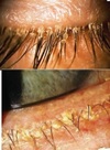
Excision for cosmesis
• Pouching of small hydrocystoma
• Refer for excision
• Lidnocaine as skin anaesthetic before pouching

14
Q

Name, mx (3)

A

Cyst of Zeis Gland - Opaque lesion

Mx: pouching incision, curettage, refer for excision

(note: curretage = Removal of tissue with a curette from the wall of a cavity or another surface)

15
Q

Name, mx (4)

A

Stye/Hordeolum

Mx: Warm compress, Most resolve 2-3 days, Topical broad spectrum antibiotic (chlorsig) if persistent, Can lead to cellulitis (indicated by fever and V.A) –> that requires antibiotics tx

16
Q

Name, mx (5)

A

Chalazion

Mx: spontaneous resolution in few months (when fluid resorbed), Tx = heat compress (qid), lid massage (to help glands open + soften oil), incision w/curettage + steroid injection (long lasting), doxycline if recurrent (40-100mg bid) or tetracycline (500mg bid)

17
Q

Name, mx (3)

A

Sebaceous gland carcinoma: malignant tumour of sebaceous gland

Mx: careful examination, review with referral of suspicious cases, 6-12 monthly reviews

18
Q

Name, mx (1)

A

Viral Warts (Verruca)

Mx: refer for excision + cautery

19
Q

Name, Ax (2), Mx (3)

A

Molluscum contagiosum

Ax: check eyebrows and evert; ensure no lymphadenopathy, check lymph nodes

Mx: counsel about hygeine, spread skin-skin contact; self-limiting, non-scarring; refer for excision + cauterisation

20
Q

name, ax (1), mx (3)

A

Keratoacanthoma

Ax: biopsy of keratin core

Mx: refer for excision + biopsy; exclude SCC; spontaneous involuted by one year

21
Q

Name, mx (4)

A

Xanthelasma

refer to GP to manage systemic overlay
check /control lipid levels (TGs, cholesterol),
systemic work up (fasting blood lipids)
Consider excision for cosmesis(recurrent) + refer for biopsy (to make sure it’s benign)

22
Q

Name, Ax (1), Mx (1)

A

Seborrheic Keratosis (SK):

Ax: referral for excision and biopsy (if suspicious)

Mx: progressive, suspicious or cosmesis excise excluding BCC, SCN

23
Q

Solar or Actinic Keratosis. How do you manage it? (3)

A

malignant potential
photos to monitor
refer for biopsy & excision (exclude BCC, SCC)

24
Q

Epithelial Basement Membrane Dystrophy (EBMD): Mx (4)

A

Lubricants; Mx of any co-existing surface disease if there are any symptoms; mx of any pre-existing erosion;

any co-existing surface disease will increase the risk of recurrent corneal erosions.

25
Q

Meesman’s dystrophy: Mx (3)

A

Mx is conservative; Surface lubrication to minimise discomfort; Bandage CLs

26
Q

How can you manage Reis-Buckler’s Dystrophy? (3)

A

Treat RCEs (recurrent corneal erosions) as required; Must monitor px for decreased vision; Difficult to mx because of reoccurence; surgical intervention often required for patients over 50

27
Q

How can you manage Thiel-Behnke corneal dystrophy? (3)

A

Same as Reis-Bucklers. So: Treat RCEs (recurrent corneal erosions) as required; Must monitor px for decreased vision; Difficult to mx because of reoccurence; surgical intervention usually for over 50yo

28
Q

How do you manage Lattice Dystrophy? (4)

A

Mx of any surface discomfort; Mx of RCEs; May refer to GP to determine if amyloidosis is systemic; surgical intervention (refer for PTK if vision significantly reduced)

29
Q

How can you manage Granular Dystrophy (4.5)

A

Early tx involves treating symptoms (use lubricants); Try to reduce occurrence of RCEs (reccurent corneal erosions); Soft bandage CLs; surgical (superficial keratectomy or PTK, lamellar keratoplasty, penetrating keratoplasty)

30
Q

Name, mx (1)

A

Avenillino Dystrophy:

tx same as for other stromal dystrophies

31
Q

Name, mx (3)

A

Macular Dystrophy

Mx: Vision usually significantly affected by 30s; referral for PK; Recurrence in graft is uncommon

32
Q

Name, mx (2)

A

Schnyder’s Crystalline Dystrophy:

Mx: Refer for workup of blood lipids as systemic disease may be life threatening; Refer for PTK to remove superficial crystals in px with severe glare

33
Q

How can you manage Fuchs Endothelial Dystrophy? (4)

A

6mth review recommended (work up patchy, specular microscopy, IOP); Hypertonic saline, warm hairdryer upon waking to reduce corneal oedema; Ocular lubricants/bandage CL (for bullae); Eventual referral for PK

34
Q

Name, mx (3)

A

Posterior polymorphous corneal dystrophy

Mx: Most don’t require tx for corneal changes; check IOP at each visit (b/c increased incidence of glaucoma with epithelial overgrowth); if tx required = as per Fuch’s

35
Q

Name, mx (6)

A

Filamentary Keratopathy

Mx: Tx underlying conditions to prevent reoccurence; Remove filaments under topical anaesthesia w/sterile forceps; Removal will cause epithelial defect (warn px it’ll feel worse before it gets better); Prescribe prophylactic antibiotics and ocular lubricants (for comfort); Topical steroids if filaments persistent (except for HSV); Bandage CL

36
Q

Name, mx (5)

A

Superficial punctate keratopathy (SPK)

Mx: Tx underlying cause; Will typically repair within 24 hours; Ocular lubricant to promote repair (non-preserved); If severe - may require prophylactic topical antibiotics cover or steroids if underlying condition is inflammatory; Discontinue any CL wear

37
Q

What’s this? How can you manage it? (3)

A

Subepithelial Infiltrates

Mx: If symptomatic - ocular lubricants; topical corticosteroids w/slow taper; antibiotics if significant epithelial breakdown

38
Q

Name, mx (6)

A

Neurotrophic keratopathy

Mx: Non-preserved ocular lubricants; Withdrawal of topical medicatiosn; Risk of microbial keratitis due to incomplete epithelium (consider prophylactic tx with chlorsig); Non-preserved topical corticosteroids (?); Bandage CL (?); Refer for surgery - tarsorrhaphy

39
Q

Name, mx (4)

A

Bullous Keratopathy:

Mx: Non-preserved ocular lubricants; Hypertonic saline (5%) to aid epithelium; Bandage CL for symptoms; Refer for surgery - graft for PTK

40
Q

Name, Mx (3)

A

Interstitial Keratitis

Mx: Refer for tx of any assoc. systemic disease; Cogan’s = life threatening (urgent referral); Tx corneal inflammation with potent, frequent topical corticosteroids

41
Q

Name, Mx (6)

A

Marginal Keratitis

Mx: Tx of underlying lid disease/blepharitis; Topical steroid for inflammation; Prophylactic topical antibiotic for epithelial defect and to reduce bacterial load; Monitor closely for improvement; If no improvement - consider oral doxycycline; Stop CL wear

42
Q

Name, mx (3)

A

Phlyctenulosis

Mx: If concerned for TB - refer for chest X-ray, mantoux and sputum culture; Manage staph lid disease; Monitor closely for improvement

43
Q

Name, Mx (4)

A

Mooren’s ulcer

Mx: Refer to establish diagnosis (to exclude ddx); Systemic steroids + other immunosuppressants; Topical steroids, prophylactic antibiotics can also be used; Conjunctival resection surgery

44
Q

How can you manage Thygeson’s Superficial Punctate Keratitis? (4)

A

Ocular lubricants for discomfort; Mainstay = moderate strength topical steroid during exacerbation (followed by careful taper); Bandage CL use occassionally to reduce symptoms; Monitor Px IOP regularly if using steroids

45
Q

Name, How would you manage the condition related this?

A

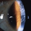
Vogt’s Striae (it’s me!)

It’s from keratoconus, do topo regularly check progression, RGPs, etc. You name it, it’s my condition so I know this

46
Q

How can you manage Pellucid Marginal Degeneration? (4)

A

Similar to KC but specs are better; CL fiting more complex (need larger RGP or mini-scleral); CXL(?); Low success rate for corneal grafts

47
Q

What’s this and how can you manage it? (4)

A

Keratoglobus: .- Globular protrusion of entire cornea—–

Mx: Difficult to mx; Specs improve vision + provide protection; Penetrating Keratoplasty (PK) = poor results; RGP = difficult to fit

48
Q

What’s this? How can you manage? (7)

A

Band keratopathy

Mx: Identify underlying cause (Hx, ocular examination, referral); If mild - artificial tears or bandage CLs for comfort or vision; If poor comfort/VA affected: chelating agent (manual Ca removal), superficial keratectomy, PTK (laser removal of epi); Px counselling that reccurence likely if underlying cause remains

49
Q

Name, Mx (3)

A

Salzmann’s Nodular Degeneration

Mx: Usually tear supplements for comfort; Occasionally manual removal or PTK; At worst - lamellar graft or penetrating graft

50
Q

Name, Mx (4)

A

Terriens Marginal Degeneration

Mx: Corneal topo; Pachymetry; Astig correction; Occasionally keratoplasty (if visual status poor enough)

51
Q

Name, Mx (4)

A

Spheroidal degeneration

Mx: Advice on UV protection; Initially ocular lubricants to enhance comfort; Referral for epithelial debridement, superficial keratectomy; Occasionally PK (penetrating keratoplasty)

52
Q

Name, Mx (1)

A

Polymorphic Amyloid Degeneration

No treatment required

53
Q

How can you manage a corneal arcus? (1)

A

No tx required, unless referral warranted to check blood lipids

54
Q

Name, how can you manage? (1)

A

Lipid Keratopathy

Mx: Topical corticosteroids - reduce inflammatory response + regress vessel –> reduce lipid deposition over time

55
Q

What’s this? How can you manage it? (1)

A

Vogt’s Limbal Girdle

Mx: No tx required, it’s totally benign

56
Q

What’s this? How can you manage it? (2)

A

Vortex Keratopathy

Mx: Tx any associated systemic conditions (often linked to systemic conditions e.g. Fabry’s) or drug induced; Refer any younger px for 2nd opinion on systemic conditions

57
Q

Can ocular siderosis affect mx of a FB? How?

A

Location affects mx of a FB

58
Q

How can you manage Kayser-Fleischer rings? (3)

A

Referral for diagnosis + mx of systemic disease; Copper chelating agents; Corneal ring will disappear with systemic therapy for copper levels

59
Q

Name, How can you manage? (1)

A

Crocodile Shagreen

No mx required

60
Q

What’s this called? How can you manage? (1)

A

Corneal Farinata

No mx required

61
Q

What is this? How can you manage it? (1)

A

Corneal Guttata.

Mx: Check for Fuch’s endothelial dystrophy and manage that (central guttata is associated with Fuch’s)

62
Q

What is this condition? How can you manage it? (5)

A

Pingueculum

Mx: Vasoconstrictors to reduce redness (?); Ocular lubricants, cold compress if FB sensation; NSAIDs (topical or oral) or topical steroids for severe inflammation; Refer for cosmetic surgery if required (or if dellen persists); Follow up at normal review or px requirest

63
Q

What is this condition called? How can you manage it? (5)

A

Pterygium

Mx: Do NOT use vasoconstrictors (chronic use can result in rebound –> vasodilation); Ocular lubricants for ocular irritation + corneal signs; Topical steroids or NSAIDs for severe irritation; Follow up dependent on rate of progression (stable = 1-2 yr, progressive/new px = 3-6M); Advise UV protection; Refer for surgery if advancing rapidly

64
Q

What’s this? How can you manage? (7)

A

Concretions: are deposits on palpebral conj.

Mx: Monitor if asymptomatic; Tx if symptomatic; Review CL care + environmental factors; Ocular lubricants; Remove using topical anaesthesia (may result in bleeding); Prophylactic antibiotic after removal; Follow up (if not removed = at px request, if removed = 3-5 days after)

65
Q

What is this called? How can you manage it? (4)

A

Amyloidosis

Mx: Check if amyloid present elsewhere in eye (lids, AC, ON, cornea, iris, lacrimal glands); Removal if FB sensation or cosmesis; Biopsy for ddx and to rule out systemic amyloidosis; Early review if uncertain diagnosis, or observed change in size or symptoms

66
Q

Name, mx (5), When follow up (1)

A

Conjunctival cyst/lymphangiectasia

Mx: Monitor if aysmptomatic; Lance cyst with needle + anaesthesia + massage closed lid to drian; Will usually re-occur; Prophylactic antibiotics (?); Refer for surgical removal from base if required; Follow up to monitor if lanced and antibiotic prescribed or at px request

67
Q

What is this? How can you manage? (4) When follow up? (2)

A

Ecchymosis

Mx: If traumatic, mx as appropriate; Reassure px; Cold packs to stop bleeding in first 1-2 days followed by hot packs to assist with haemolysis; May take 1-3 days to resolve; Do NOT use vasoconstrictors (b/c you need WBCs to come in to mop it up)

Follow up: review at 1 week if concerned or no resolution; if more than 2 recurrences in 1 year then refer to determine if systemic cause or cauterisation

68
Q

What’s this? How can you manage? (5)

A

Bitot’s spot

Mx: Refer for blood test to confirm VitA deficiency; Will normally disappear with high dose VitA therapy; May regress within weeks; If long standing - may have permanent epithelial metaplasia and will remain; Mx dry eye symptoms as appropriate

69
Q

What’s this condition called? How can you manage it? (8)

A

Superior limbic keratoconjunctivits

Mx: Refer to evaluate thyroid function; Evaluate CLs; Tx to reduce inflammation; Topical steroid (Flarex/Predforte iTDS to iQiD and taper); Ocular lubricant (punctal plug?); Topical vasoconstrictors (?); Soft bandage CL; Surgery to cauterise bulbar conj or fuse conj and episclera

70
Q

What’s this? How can we manage it? (6)

A

Floppy Eyelid Syndrome

Mx: Refer to investigate sleep apnoea; mx apnoea; weight loss program; ocular lubricant ointment before sleep; taping lid shut during sleep; surgical horizontal tightening of upper lid

71
Q

What’s this? How to manage? (1)

A

Conjunctival Papillae

Mx underlying cause

72
Q

What’s this? How to manage? (1)

A

Conjunctival Follicles

Mx underlying cause

73
Q

What’s this? How to manage? (1) Why must you do this? (1) Will the conj epithelium remain intact upon doing so?

A

Pseudomembrane

Must remove (peel off with forceps or cotton bud) or else you’ll get scarring, blepharon, or entropion.

Conj epithelium will remain intact when you peel off the pseudomembrane

74
Q

What’s this? How can you manage? How does this differ from the management of pseudomembranes? List 4 potential causes of this condition

A

True membrane

Remove/peel off just like pseudomembrane. Note: more bleeding will occur because of how it’s attached and it can rip off the corneal epithelium but it’s still perfectly safe to do.

Causes: e.g. strep, gonococcal, autoimmune, diptheria (corynebacterium)

75
Q

What’s this? How can you manage? (4)

A

Bacterial conjunctivitis

Mx: Self limiting –> monitor until resolution (resolves quicker with antibiotic); If little/no corneal involvement –> may be treated with NaCl solution bathing; Broad spectrum antibiotic drops w/without ointment at night for 7-10 days (chloramphenicol or aminoglycoside); Follow up (3-5 days then 7-10 days, if no resolution then review diagnosis)

76
Q

What’s this? How can you manage? (2)

A

Bacterial Conjunctivitis (gonococcal)

Mx: Refer - often systemic –> IM or IV antibiotics (cephalosporin and/or oral FQ); Consider co-existing chlamydia

77
Q

What’s this? How can you manage it? (8)

A

Pharynogoconjunctival fever (PCF)

Mx: Gloves during exam/clean equipment; Educate px; GP referral for time off (contagious ~1wk); Povidone-iodine therapy (?); No antiviral agent proven effective; Supportive therapy (cold compress, artificial tears, fever relief,), Steroid if severe inflammation (Flarex BD to QiD with slow taper); Resolution within 7-14 days (check for corneal involvement in this time)

78
Q

What’s all this? How can you manage? (7)

A

Trachoma

Mx: Try and find source of exposure to help confirm diagnosis; Refer for lab test for diagnosis; Treat active infection with oral macrolides (e.g. azithromycin), tetracyclines (e.g. doxycycline); Refer for surgery; Corneal grafts rarely succesful; Inactive scarring requires topical lubrication; Improve hygeine + education for prevention

79
Q

Name, how can manage? (5.5)

A

Adult inclusion conjunctivitis (AIC)

Mx: Refer Px + sexual partner for lab tests to confirm diagnosis; Tx systemic macrolide or tetracycline; Identify + tx co-existing infections; Mx ocular signs + symptoms with topical tetracycline; Oral medication critical (either azithromycin 1gm PO or Erythryomycin 250mg QiD 2-6 wks)

80
Q

Name, How can you manage this? (2)

A

Anterior blepharitis

Mx: Lid therapy = mainstay tx (scrub lid margins up to 4x a day, hot compress + lid massage for coexisting posterior bleph, follow up to see success); Tear supplements (for ocular surface problems)

81
Q

What’s this? How can you manage this? (5)

A

Posterior blepharitis

Mx: Oral tetracyclines = mainstay of tx; Lid therapy for anterior bleph but hot compresses and lid massage essential to improve gland function; Tear supplements; Antibiotic ointment (for anterior); Mild topical steroids to reduce inflammation along lid margin

82
Q

What is this called? How can you manage this? (6)

A

Ocular rosacea

Mx: Topical antibiotic (microbial assoc.); Metronidazole gel for skin rash; Oral tetracyclines (doxcycline) or macrolides = mainstay; Lid therapy (for bleph); Tear supplements; Mild topical steroids

83
Q

How can you manage demodex? (4)

A

Consider removal if px has demodex +1 ocular problem; TTO (tea tree oil) kills demodex but also toxic to cornea/conjucntiva; Weekly application of 50% TTO combined with daily use of TTO shampoo or lid wipes; Ivermectin 3mg one tablet PO

84
Q

What MUST you do to confirm if someone has an adenovirus infection? (1)

A

Lymphadenopathy