Lower limb random questions Flashcards

1
Q

To prevent the joint space from being obscured by the magnified shadow of the medial femoral condyle in the lateral projection of the knee, the radiographer should: a. Angle the central ray 5 degrees cephalad b. Place the knee joint in 90 degree flexion c. Rotate the knee so that the patella forms a 45 degree angle to the film d. Fully extend the patients lower leg

A

a. Angle the central ray 5 degrees cephalad

2
Q

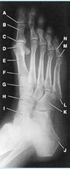
The bone identified in the figure attached is the:

a. tibia
b. cuboid
c. talus
d. navicular

A

Talus

3
Q

To perform this position, the central ray is angled:

a. 5-7 degrees cephalic
b. 5-7 degrees caudad
c. angulation depends on the patient
d. There is no angulation in this position

A

5-7 degrees cephalic

4
Q

The tibial plateaus slope:

a. anteriorly 10-20 degrees
b. posteriorly 10-20 degrees
c. laterally 10-20 degrees
d. medially 10-20 degrees

A

posteriorly 10-20

5
Q

In which of the following positions can the sesamoid bones of the foot be demonstrated free of superimposition with the metatarsals or phalanges?

a. dorsoplantar metatarsals/toes
b. tangential metatarsals/toes
c. 30 degrees medial oblique foot
d. 30 degrees lateral oblique foot

A

tangential metatarsals/toes

6
Q

Where does the CR enter the knee for a lateral projection of the patella?

a. posterior margin of the medial epicondyle
b. anterior margin of the medial epicondyle
c. through the patellofemoral joint space
d. directly to the lateral aspect of the patella

A

Through the patellofemoral joint space

7
Q

For a lateral projection of the ankle, the CR must enter the:

a. navicular
b. tibiofibular joint
c. medial malleolus
d. lateral malleolus

A

medial malleolus

8
Q

The bone part identified in the figure attached is the:

a. lateral condyle
b. medial condyle
c. lateral malleolus
d. medial malleolus

A

lateral malleolus

9
Q

The talus articulates with how many bones:

a. 1
b. 2
c. 3
d. 4

A

4

10
Q

Which of the following projections of the calcaneous is obtained with the leg extended, the plantar surface of the foot vertical and perpendicular to the IR, and the central ray directed 40 degrees caudad?

a. Axial plantodorsal projection
b. Axial dorsoplantar projection
c. Lateral projection
d. Weight-bearing lateral projection

A

Axial dorsoplantar projection

11
Q

How far should the IR extend below the knee for a lateral projection of the femur?

a. 1 in
b. 2 in
c. 3 in
d. 4 in

A

2 in

12
Q

What is the CR angle for an AP projection of the leg?

a. 0
b. 5 degrees caudad
c. 7 degrees caudad
d. 5-7 degrees cephalad

A

0 degrees

13
Q

What is the CR angulation for the axial (plantodorsal) projection of the calcaneous?

a. 25 degrees
b. 30 degrees
c. 35 degrees
d. 40 degrees

A

40 degrees

14
Q

The best projection to demonstrate the articular surfaces of the femoropatellar articulation is the:

a. AP knee
b. PA knee
c. tangential (“sunrise”) projection
d. “tunnel” view

A

tangential (“sunrise”) projection

15
Q

In the lateral projection of the ankle, the:

  1. talotibial joint is visualized
  2. talofigular joint is visualized
  3. tibia and fibula are superimposed
A

1 and 3 only

16
Q

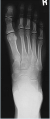
In the attached image, the central ray is directed to:

a. The PIP joint of the third digit
b. The head of the third metatarsal
c. The base of the third metatarsal
d. The PIP joint of the digit of interest

A

The head of the third metatarsal

17
Q

Which of the following bones participate in the formation of the acetabulum?

  1. Illium
  2. Ischium
  3. Pubis
A

1, 2 and 3

18
Q

Which of the following is recommended to better demonstrate the tarsometatarsal joints in the dorsoplantar projection of the foot?

a. Invert the foot
b. Evert the foot
c. Angle the central ray 10 degrees posteriorly
d. Angle the central ray 10 degrees anteriorly

A

Angle the central ray 10 degrees posteriorly

19
Q

How many tarsal bones are there in the foot?

a. 4
b. 5
c. 6
d. 7

A

7

20
Q

The most commonly performed oblique projection of the foot is the:

a. AP oblique in medial rotation
b. AP oblique in lateral rotation
c. PA oblique in medial rotation
d. PA oblique Grashey method

A

AP oblique in medial rotation

21
Q

For an AP oblique projection of the knee, the limb is rotated:

a. 25 degrees
b. 30 degrees
c. 45 degrees
d. 30-40 degrees

A

45 degrees

22
Q

For a plantodorsal projection of the toes, the central ray should be directed to enter the anatomy at the:

a. 1st metatarsophalangeal joint
b. 3rd metatarsophalangeal joint
c. 2nd metatarsophalangeal joint
d. navicular bone

A

3rd metatarsophalangeal joint

23
Q

In which of the following projection is the talofibular joint best demonstrated:

a. AP
b. Lateral oblique
c. Medial oblique
d. Lateral

A

Medial oblique

24
Q

All of the following bones area associated with condyles except the:

a. femur
b. tibia
c. fibula
d. madible

A

fibula

25
Which of the following must be rotated for all oblique projection of the ankle? 1. pelvis 2. leg 3. foot
2 and 3
26
The medial epicondyle of the femur is the structure lettered: a. B b. C c. I d. J
I
27
The central ray in this projection is angled: a. 0 degrees b. To match the angle of the femur c. 40 degree cephalic d. 40 degrees caudad
0 degrees
28
The CR angle for AP oblique projections of the knee is: a. 0 degrees b. 5 degrees caudad c. 7 degrees cephalad d. variable, depending on the ASIS/tabletop distance
variable, depending on the ASIS/tabletop distance
29
When the malleoli of the ankle are positioned parallel with the IR, the ankle is in position for which projection: a. AP b. AP oblique, 45 degree lateral rotation c. AP oblique, 45 degree medial rotation d. AP oblique., 15-20 degree medial rotation for the ankle mortise
AP oblique., 15-20 degree medial rotation for the ankle mortise
30
For this position, the sole of the foot should form what angle with the plane of the cassette: a. 10-20 degrees b. 30-40 degrees c. 40-50 degrees d. 50-60 degrees
30-40 degrees
31
Which of the following bones participates in the formation of the knee joint? 1. Femur 2. Tibia 3. Patella
1 and 2 only
32
The superior portion of the calcaneous contains a groove called the calcaneal sulcus. The inferior potion of the talus contains a matching groove called the sulvus tali. Collectively, the two sulci form the: a. trochlea b. ankle mortise c. sinus tarsi d. sustentaculum tali
sinus tarsi
33
How many degrees of angulation are required to open the interphalangeal joint spaces of the toes on an AP projection? a. 0 degrees b. 10 degrees c. 15 degrees d. 20 degrees
15 degrees
34
in the lateral projection of the foot, the 1. plantar surface should be perpendicular to the IR 2. metatarsals are superimposed 3. talofibular joint should be visualized
1 and 2 only
35
In order to place the patella parallel with the plane of the IR for a PA projection, the heel must be rotated: a. 5-10 degrees laterally b. 5-10 degrees medially c. 10-15 degrees laterally d. 10-15 degrees medially
5-10 degrees laterally
36
How much is the knee joint flexed for tha PA axial projection (Homblad method) of the intercondylar fossa? a. 20 degrees b. 45 degrees c. 50 degrees d. 70 degrees
70 degrees
37
The ankle is rotated how many degrees for this projection: a. 0 degrees b. 30 degrees c. 45 degrees d. 90 degrees
45 degrees
38
For this position, the sole of the foot should form what angle with the plane of the cassette: a. 10-20 degrees b. 30-40 degrees c. 40-50 degrees d. 50-60 degrees
30-40 degrees
39
Which projection would you use to demonstrate the space between the first and second metatarsal joints and the intertarsal joints between the first and second cuneiforms? a. Tangential projection of the instep b. Lateral oblique projection of the foot c. Medial oblique projection of the foot d. Mediolateral projection of the
Lateral oblique projection of the foot
40
How many metatarsal bones are there in the foot? a. 4 b. 5 c. 6 d. 7
5
41
The cuboid bone in the structure lettered: a. A b. B c. C d. D
D
42
The CR angulation for a lateral projection of the knee is: a. 0 degrees b. 5 degrees cephalad c. 7 degrees cephalad d. 5-7 degrees cephalad
5-7 degrees cephalad
43
Which projection is demonstrated in the attached figure? a. PA foot b. AP axial foot c. axial calcaneous (plantodorsal) d. axial calcaneous (dorsoplantar)
axial calcaneous **(plantodorsal)**
44
For an AP oblique projection of the foot in either medial or lateral rotation, the plantar surface of the foot should form an angle of: a. 15 degrees b. 30 degrees c. 45 degrees d. 15-30 degrees
30 degrees
45
Which angulation is used for this position if the patient measures 18 cm at the ASIS: a. No angulation b. 5 degrees cephalic c. 5 degrees caudad d. Depends on the patient
5 degrees caudad
46
Where is the CR directed for an AP projection of the knee? a. 1/2 inch above the patellar base b. 1/2 inch above the patellar apex c. 1/2 inch below the patellar base d. 1/2 inch below the patellar apex
d. 1/2 inch below the patellar apex
47
Which lateral projection of the foot is the most commonly performed? a. lateromedial (lateral recumbent position) b. mediolateral (lateral recumbent position) c. lateromedial (standing weight-bearing) d. mediolateral (standing weigh-bearing)
mediolateral (lateral recumbent position)
48
Which of the following is NOT clearly demonstrated on an AP projection of the ankle? a. tibiotalar b. lateral malleoli c. ankle mortise d. tibiofibular overlapping
ankle mortise
49
This position is used to demonstrate the: a. Intercondyloid fossa b. Medial tibial Plateau c. Tibial tuberosity d. Intercondyloid Eminence
Intercondyloid fossa
50
AP stress studies of the ankle may be performed: 1. to demonstrate fractures of the distal tibia and fibula 2. following inversion or eversion injuries 3. to demonstrate a ligament tear
2 and 3 only
51
Which of the following articulate(s) with the bases of the metatarsals? 1. The heads of the first row of phalanges 2. The cuboid 3. The cuneiforms
2 and 3 only
52
The inferior aspect of the foot is termed the: a. posterior surface b. caudal surface c. dorsal surface d. plantar surface
plantar surface
53
Which projection of the patella will aid in the diagnosis of a vertical fracture? a. AP projection b. AP axial projection c. Lateral projection d. Tangential projection of the profile
Tangential projection of the profile
54
Which projection of the foot will best demonstrate the longitudinal arch? a. Mediolateral b. Lateromedial c. Lateral weigh-bearing d. 30 degree medial oblique
Lateral weight-bearing
55
The patient position and CR method demonstrated in the attached figure is the: a. Homblad (intercondylar fossa) b. Camp-Coventry (intercondylar fossa) c. Settegast (patellofemoral joint) d. Huston (patellofemoral joint)
Camp-Coventry (intercondylar fossa)
56
How far should the knee be flexed for the tangential projection (Settegast method) of the patella when done in the prone position? 1. 50-60 degrees from the table 2. as much as possible 3. until the patella is perpendicular
2 and 3
57
With the patient and the x-ray tube positioned as illustrated in the attached figure, which of the following will be visualized? 1. Intercondyloid fossa 2. Patellofemoral articulation 3. Tangential patella
2 and 3 only
58
The proximal end of the tibia presents two prominent processes called the: a. tubercles b. condyles c. malleoli d. tuberosities
condyles
59
To prevent lateral rotation, how should the foot be positioned for a lateral projection of the ankle? a. In dosiflexion b. In plantar flexion c. on a 10 degree angle wedge d. on a 15 degree angle wedge
in dorsiflexion
60
When the knee is properly positioned for an AP projection, the patella will lie: a. directly in the center of the limb b. slightly to the medial side c. slightly to the lateral side d. parallel with the tabletop
slightly to the medial side
61
Which bones articulate distally with the tarsal navicular? a. Phalanges b. Metatarsals c. Talus and calcaneus d. Cuneiforms
Cuneiforms
62
In the lateral projection of the knee, the 1. femoral condyles are superimposed 2. patellofemoral joint is visualized 3. knee is flexed about 20 to 30 degrees
1,2 and 3
63
Which of the following projections of the ankle would best demonstrate the distal tibiofibular joint? a. Medial oblique 15-20 degrees b. Lateral oblique 15-20 degrees c. Medial oblique 45 degrees d. Lateral oblique 45 degrees
Medial oblique 45 degrees
64
The femur forms an angle of ____ degrees with the table top for this projection: a. 40 b. 50 c. 70 d. 90
70
65
How is the patient placed for a lateral projection of the great toe and second toe? a. supine, with the affected leg turned out b. supine, with the affected leg turned in c. lateral, on the affected side d. recumbent, on the unaffected side.
recumbent, on the unaffected side
66
Which of the following can be used to demonstrate the intercondyloid fossa? 1. Patient PA, knee flexed 40 degrees, central ray directed caudad 40 degrees to the popliteal fossa 2. Patient AP, cassette under flexed knee, central ray directed cephalad to knee, perpendicualr to tibia. 3. Patient PA, patella parallel to IR, heel rotated 5-10 degrees lateral, central ray perpendicular to knee joint.
1 and 2 only
67
The CR angulation for the AP ankle projection is: a. 0 degrees b. 5 degrees caudad c. 10 degrees caudad d. 15 to 20 degrees caudad
0 degrees
68
Which of the following is (are) located on the posterior aspect of the femur? 1. intercondyloid fossa 2. Intertrochanteric crest 3. Intertubercular groove
1 and 2 only
69
Tangential axial projections of the patella can be obtained in which of the following positions? 1. supine flexion 45 degrees (Merchant) 2. prone flexion 90 degrees (Settegast) 3. Prone flexion 55 degrees (Hughston)
1, 2, and 3
70
The description that best describes this position is: a. AP Knee b. Lateral knee c. AP oblique knee (Lateral rotation) d. AP oblique knee (medial rotation)
AP oblique knee (medial rotation)
71
The navicular is the structure lettered: a. A b. B. c. C d. D
A
72
On the anterior surface of the tibia is a prominent process called the: a. body b. anterior border c. tibial tuberosity d. intercondylar eminence
tibial tuberosity
73
For a lateral projection of the foot, the CR is directed to the: a. head of the third metatarsal b. base of the third metatarsal c. tibiotalar joint d. navicular
base of the third metatarsal
74
A lateral projection of the tibia/fibular is commonly performed in which position? a. Lateromedial b. Anterolateral c. Mediolateral d. Lateroposterior
Mediolateral
75
To demonstrate the ankle mortise, the leg and foot should be rotated medially how many degrees? a. 15 degrees b. 20 degrees c. 45 degrees d. 15-20 degrees
15-20 degrees
76
In the AP projection of the ankle, the 1. plantar surface of the foot is vertical 2. fibular projects more distally than the tibia 3. calcaneus is well visualized
1 and 2 only
77
Which of the following projection should be used to demonstrate a transverse fracture of the patella? 1. Lateral 2. PA 3. Tangential (Settegast method)
1 and 2 only
78
To correctly adjust the foot for medial oblique position, the sole must: a. form an angle of 15 degrees to the image receptor b. form an angle of 30 degrees to the image receptor c. form an angle of 45 degrees to the image receptor d. form an angle of 60 degrees to the image receptor
form an angle of 30 degrees to the image receptor
79
The radiograph seen in the attached figure can be produced with the: 1. long axis of the plantar surface perpendicular to the image recorder 2. CR 40 degrees cephalad to the base of the third metatarsal 3. CR 20 degrees cephalad to the talotibial joint
1 and 2 only
80
which of the following articulates with the base of the first metatarsal? a. First cuneiform b. third cuneiform c. Navicular d. Cuboid
First cuneiform
81
The two flat-like superior surfaces of the tibia are called the: a. tubercles b. melleoli c. condyles d. tibial plateaus
tibial plateaus
82
What is the CR angle for a PA projection of the patella? a. 0 degrees b. 5 degrees cephalad c. 10 degrees cephalad d. 5-15 degrees cephalad
0 degrees
83
In which projection of the foot are the sinus tarsi, cuboid, and tuberosity of the fifth metatarsal best demonstrated? a. Lateral oblique foot b. Medial oblique foot c. Lateral foot d. Weight-bearing foot
Medial oblique foot
84
How much is the knee flexed for a lateral projection of the patella? a. 0 degrees b. 5 degrees c. 10 degrees d. 5-10 degrees
5-10 degrees
85
Which of the following projections is used to demonstrate vertical patellar fractures and the patellofemoral articulation? a. AP knee b. Lateral Knee c. Tangential patella d. "Tunnel" view
Tangential patella
86
When the femur is vertical, the medial condyle is lower than the lateral condyle. How many degrees difference is there between the two? a. 3 degrees b. 5 degrees c. 5-7 degrees d. 8-10 degrees
5-7 degrees
87
The CR angle for an AP, bilateral weight-bearing knee is: a. 0 degrees b. 5 degrees cephalad c. 5 degrees caudad d. 5-7 degrees cephalad
0 degrees
88
In which of the following positions of the knee are the proximal fibula and tibia superimposed? a. AP b. Lateral oblique c. Lateral d. Medial Oblique
Lateral oblique
89
Where should the patella be demonstrated on the medial AP oblique projection of the knee? a. over the medial condyle of the femur b. over the lateral condyle of the femur c. superimpsed over the tibiofibular articulation d. centered between the tibial condyles
over the medial condyle of the femur
90
Which of the labeled bones in the attached figure identifies the tarsal navicular? a. Number 2 b. Number 3 c. Number 6 d. Number 7
Number 6
91
In the attached image, the Navicular is: a. G b. H c. J d. K
H
92
For this position, the foot should be rotated no more than: a. 45 degrees laterally b. 45 degrees medially c. 25 degrees laterally d. 25 degrees medially
45 degrees medially
93
To perform this position, the central ray is angled: a. 5-7 degrees b. 30 degrees c. 40 degrees d. To make the angulation of the femur
40 degrees
94
In which projection of the foot are the sinus tarsi, cuboid, and the tuberosity of the fifth metatarsal best demonstrated? a. Lateral oblique foot b. Medial oblique foot c. Lateral foot d. Weight-bearing foot
Medial oblique foot
95
Which of the following articulations participate(s) in formation of the ankle mortise? 1. Talotibial 2. Talocalcaneal 3. Talofibular
1 and 3 only
96
Which of the following is/are proximal to the tibial plateau? 1. Femoral condyles 2. Tibial condyles 3. Tibial Tuberosity
1 only
97
Which of the following articulate(s) with the bases of the metatarsals? 1. The heads of the first row of phalanges 2. The cuboid 3. The cuneiforms
2 and 3 only
98
Which of the following projections of the ankle would best demonstrate the distal tibiofibular joint? a. Medial oblique 15 to 20 degrees b. Lateral oblique 15 to 20 degrees c. Medial oblique 45 degrees d. Lateral Oblique 45 degrees
Medial oblique 45 degrees
99
In the attached image, item "A" is the: a. Cuboid b. Navicular c. 1st cuneiform d. Lateral cuneiform
Cuboid
100
The centering for the attached image is: a. The medial malleolus b. 1" inferior to the medial malleolus c. The lateral malleolus d. 1" inferior to the lateral malleolus
1" inferior to the medial malleolus
101
For this projection, the central ray is angled: a. 0 degrees b. 5-7 degrees cephalic c. 5-7 degrees caudad d. Can be any of the above, it depends on the patient
0 degrees
102
The tube is angled _____ for this projection. a. 10 degrees b. 15 degrees c. 20 degrees d. 30 degrees
10 degrees
103
The best projection to demonstrate the articular surfaces of the femoropatellar articulation is the: a. AP knee b. PA knee c. tangential ("sunrise") projection d. "tunnel" view
tangential ("sunrise") projection
104
In the attached image the central ray may be directed ____ degrees. a. 10 b. 15 c. 20 d. 30
15
105
The CR angulation for a lateral projection of the calcaneous is: a. 0 degrees b. 5 degrees cephalad c. 10 degrees cephalad d. 5-10 cephalad
0 degrees
106
In the AP projection of an asthenic patient whose knee measures less than 19 cm from ASIS to tabletop, the central ray should be directed a. perpendicularly b. 5 degrees medially c. 5 degrees cephalad d. 5 degrees caudad
5 degrees caudad
107
Improper support of a patient's fractured lower leg (tibia/fibula) while performing radiography could result in 1. movement of fracture fragments 2. tearing of soft tissue, nerves, and blood vessels 3. initiation of muscle spasm
1,2, and 3
108
The second largest tarsal bone, and the one that occupies the highest position in the foot is the: a. talus b. navicular c. calcaneous d. medial cuneiform
talus
109
For an axial projection of the calcaneous the ankle should be dosiflexed so the plantar surface of the foot is: a. parallel with the CR b. perpendicular to the CR c. 70 degrees from the plane of the IR d. 90 degrees from the plane of the IR
90 degrees from the plane of the IR
110
With the patient positioned as illustrated in the attached figure, which of the following structures is best demonstrated? a. Patella b. Patellofemoral articulation c. Intercondyloid fossa d. Tibial tuberosity
Patellofemoral articulation
111
Which of the following should be clearly demonstrated on a lateral projection of the leg? 1. knee joint 2. ankle joint 3. entire femoral condyles
1 and 2
112
Which of the following positions can be used to perform the tangential projection (settegast method) of the patella? 1. seated 2. supine 3. prone
1 and 3
113
The CR for an AP oblique projection of the foot is: a. 0 b. 5 degree posteriorly c. 10 degree posteriorly d. 15 degree posteriorly
0 degrees
114
which of the following objects should be available in the radiography room for performing radiographs of the lower limb? 1. angle sponges 2. sandbags 3. pull straps
1, 2 and 3
115
For an AP projection of the toes, the CR is directed to the a. 1st metatarsophalangeal joint b. 2nd metatarsophalangeal joint c. 3rd metatarsophalangeal joint d. 4th metatarsophalangeal joint
3rd metatarsophalangeal joint
116
The CR angulation for an AP oblique projection of the ankle is: a. 0 degrees b. 5 degrees caudad c. 7 degrees caudad d. 15 degrees caudad
0 degrees
117
what is the position of the femoral condyles when the leg is properly positioned for an AP projection? a. perpendicular to the IR b. parallel to the IR c. at a 15-20 degree oblique position (laterally) d. at a 15-20 degree oblique position (medially)
Parallel to the IR
118
To perform the attached position, the bottom of the foot will form an angle of ___ with the cassette. a. 0 degrees b. 30 degrees c. 45 degrees d. 90 degrees
30 degrees
119
Where will the fibula be located on a properly positioned lateral radiograph of the ankle? a. directly over the tibia b. behind the tibia c. over the anterior half of the tibia d. over the posterior half of the tibia
over the posterior half of the tibia
120
The preferred method of positioning the patient for the tangential projection (settegast method) of the patella is: a. prone b. supine c. seated on the table d. seated at the end of the table
prone
121
How is the CR directed for the PA axial projection (Homblad method) of the intercondylar fossa? a. perpendicular to the lower leg b. perpendicular to the femur c. 40-50 degrees to lower leg d. 40-50 degrees to the femur
perpendicular to the lower leg
122
Which of the following best describes how the attached image was made? a. Dorso-plantar foot with 10 degree angulation b. Dorso-plantar foot with 15 degree angulation c. Two exposures on one film. One with a posterior angle and one with an anterior angle. d. 40 degree cephalic angle to the base of the third metatarsal
Two exposures on one film. One with a posterior angle and one with an anterior angle.
123
Which of the following positions is used to demonstrate verticla patellar fractures and the patellofemoral articulation? a. AP knee b. Lateral Knee c. Tangential patella d. "Tunnel" view
Tangential patella
124
Item "D" is the a. Lateral tibial plateau b. Medial tibial plateau c. Tibial tuberosity d. Intercondyloid eminence
Medial tibial plateau
125
126
What angulation is used for this position if the patient measures 20 cm at the ASIS: a. No angulation b. 5 degrees cephalic c. 5 degrees caudad d. Depends on the patient
No angulation
127
To better demonstrate the interphalangeal joints of the toes, which of the following procedures may be employed? 1. Angle the CR 15 degrees caudad 2. Angle the CR 15 degrees cephalad 3. Place a sponge wedge under the foot with toes elevated 15 degrees
2 and 3 only
128
Which of the following projections will best demonstrate the tarsal navicular free of superimposition? a. AP oblique, medial rotation b. AP oblique, lateal rotation c. Mediolateral d. Lateral weight-bearing
AP oblique, medial rotation
129
For this position, the tibia and fibula should form an angle of no more than ____ with the femur: a. 10 degrees b. 30 degrees c. 45 degrees d. 90 degrees
10 degrees
130
In which of the following projections is the talofibular joint best demonstrated? a. AP b. Lateral oblique c. Medial Oblique d. Lateral
Medial oblique
131
In the lateral projection of the knee, the central ray is angled 5 degrees cephalad to prevent superimposition of which of the following structures on the joint space? a. Lateral femoral condyle b. Medial femoral Condyle c. Patella c. Tibial eminence
Medial femoral condyle
132
To better visualize the joint space in the radiograph in the attached figure, the radiograph should: a. flex the knee more acutely b. flex the knee less acutely c. angle the CR 5 to 7 degrees cephalad d. angle the CR 5 to 7 degrees caudad
angle the CR 5 to 7 degrees cephalad
133
The central ray for this projection should be centered to pass through: a. the ankle joint b. the base of the third metatarsal c. the third metatarsophalangeal joint d. the posterior talus
the base of the third metatarsal
134
To center the part to the film for this projection, the cassette is placed: a. with the bottom of the cassette 2 inches below the knee joint b. with the top of the cassette 2 inches avove the pubic symphysis c. with the top of the cassette at the level of the ASIS d. with the top of the cassette at the level of the illiac crest
with the top of the cassette at the level of the ASIS
135
Which of the following is (are) accurate posistioning or evaluation criteria for an anteroposterior projection of the normal knee? 1. Femorotibial interspaces equal bilaterally 2. Patella superimposed on distal tibia 3. CR enters 1/2 inch distal to base of patella
1 only
136
For this position, the central ray is angled: a. 0 degrees b. 5-7 degrees cephalic c. 5-7 degrees caudad d. can be any of the above, it depends on the patient
0 degrees
137
The radiograph shown in the attached figure demonstrates the articulation between the 1. talus and the calcaneus 2. calcaneus and the cuboid 3. talus and the navicular
2 and 3 only
138
With the patient positioned as illustrated in Figure A, how should the central ray be directed to best demonstrate the intercondyloid fossa? a. Perpendicular to the popliteal depression b. 40 degrees caudad to the popliteal depression c. Perpendicular to the long axis of the femur d. 40 degrees cephalad to the popliteal depression
40 degrees caudad to the popliteal depression
139
Posterior displacement of a tibial fracture would be best demonstrated in the a. AP projection b. lateral projection c. medial oblique projection d. lateral oblique projection
lateral projection
140
Which of the following positions would best demonstrate the proximal tibiofibular articulation? a. AP b. 90 degrees mediolateral c. 45 degrees internal rotation d. 45 degrees external roation
45 degrees internal rotation
141
To perform this position, the tibia and fibula are placed at what angle in relation to the femur: a. 5-7 degrees b. 20-30 degrees c. 45 degrees d Whatever is comfortable for the patient
20-30 degrees
142
The bone part identified in the figure attached is the: a. medial epicondyle b. . tibial tuberosity c. medial malleolus d. medial condyle
medial condyle
143
How many bones are there in the foot? a. 14 b. 26 c. 27 d. 29
26
144
Which of the following is clearly demonstrated on an AP oblique projection of the knee in medial rotation? a. distal fibula b. tibiotalar articulation c. patellofemoral joint space d. tibiofibular articuation
tibiofibular articuation
145
What is the patient position for a lateral projection of the third, fourth, or fifth toes? a. supne, with the leg and foot laterally rotated b. 45 degree oblique, with the leg and foot laterally rotated c. lateral recumbent, on the affected side d. lateral recumbent, on the unaffected side
lateral recumbent, on the affected side
146
Where should the central ray be directed for the lateral projection of the foot? a. to the center of the longitudinal arch b. to the midpoint of the calcaneus c. to the base of the third metatarsal d. to the head of the fifth metatarsal
to the base of the third metatarsal
147
The bone part identified in the figure attached is the: a. lateral condyle b. lateral malleolus c. medial condyle d. medial epicondyle
medial condyle
148
For the lateral projection of the knee, the knee should be flexed how many degrees? a. 30 degrees b. 45 degrees c. 35 degrees d. 90 degrees
30 degrees
149
In which of the following positions/projections will the talocalcaneal joint be visualized? a. Dorsoplantar projection of the foot b. Plantodorsal projection of the os calcis c. Medial oblique position of the foot d. Lateral foot
Plantodorsal projection of the os calcis
150
How many phalanges are there in the great toe? a. 0 b. 1 c. 2 d. 3
2
151
The bone of the foot indicated in the figure attached is the: a. navicular b. talus c. calcaneous d. medial cuneiform
talus
152
Which of the following projection of the knee best demonstrates the narrowing of a joint space? a. AP b. AP oblique c. lateral d. AP of both knees with weight-bearing
AP of both knees with weight-bearing
153
Which of the following will ensure that the knee is in proper position for a lateral projection? 1. epicondyles are perpendicular to the IR 2. patella is perpendicular to the IR 3. leg is flexed 20-30 degrees
1,2 and 3
154
When the ASIS to tabletop measurement is greater than 24 cm, the CR angulation for an AP knee is? a. 0 degrees b. 5 cephalad c. 5 caudad d. 7 cephalad
5 cephalad
155
How many degrees should the limb be internally rotated for an AP projection of the femur? a. 10 degrees b. 15 degrees c. 18 degrees d. 10-20 degrees
15 degrees
156
Which of the following is the essential method of demonstrating the patella in the tangential projection? a. Merchant b. Settegast c. Hughston d. Kuchendorf
Settegast
157
In which of the following positions was the radiograph in the attached figure made? a. AP with perpendicular plantar surface b. 45 degree lateral oblique c. 20 degree medial oblique d. 45 degree medial oblique
20 degree medial oblique
158
The central ray is directed to: a. The metatarsophalangeal joint of the third digit b. The base of the third metatarsal c. The cuneiforms d. The PIP joint of the third digit
The base of the third metatarsal
159
The projection of the patella demonstrated in the attached figure is the: a. Hughston b. Merchant c. Settegast d. Holmblad
Settegast
160
AP dorsoplantar and lateral foot radiographs were requested to rule out fracture. A fracture through the base of the fifth metatarsal was suggested on the lateral radiograph, but bony superimposition interfered. Which of the following positions would result in demonstration of the fifth metatarsal base without superimposition? a. AP (dorsoplantar) medial oblique b. AP (dorsoplanter) lateral oblique c. PA (plantodorsal) medial oblique d. PA (plantodorsal) with 15 degree cephlad angle
AP (dorsoplantar) medial oblique
161
Which projection will clearly demonstrate the joint spaces between the talus and both malleoli? a. AP b. AP oblique, 15 to 20 degree internal rotation c. AP olbique, 45 degree internal rotation d. AP oblique, 45 degree external rotation
AP oblique, 15 to 20 degree internal rotation
162
In order to better demonstrate the tarsometatatarsal joint spaces of the foot, a posterior angulation of how many degrees is required? a. 0 degrees b. 10 degrees c. 15 degrees d. 20 degrees
10 degrees
163
What is the preferred term when referring to the AP projection of the foot? a. PA b. Dorsoplantar c. Tangential d. Plantodorsal
Dorsoplantar
164
When the ASIS to tabletop measurement is less than 19 cm, the CR angulation for an AP knee is: a. 0 degree b. 5 degrees cephalad c. 5 degrees caudad d. 7 degrees caudad
5 degrees caudad
165
Which position is the patient placed in for a PA projection of the patella? a. supine b. prone c. lateral d. upright
prone
166
The essential projections of the calcaneous are: 1. axial (plantodorsal) 2. axial (dorsoplantar) 3. lateral (mediolateral)
1 and 3
167
How far should the IR extend beyond the ankle or knee joint for an AP projection of the leg? a. 1/2 in b. 1 in c. 1 1/2 in d. 2 in
1 1/2 in
168
How much should the leg be flexed for a lateral projection of the knee? a. 20 degrees b. 30 degrees c. 10-20 degrees d. 20-30 degrees
20-30 degrees
169
The superior surface of the foot is termed the: a. dorsal surface b. plantar surface c. anterior surface d. superior surface
dorsal surface
170
Which of the following is a necessary criterion for demonstrating the intercondyloid fossa utilizing the Camp-Coventry position? a. the knee should be flexed 90 degrees b. the central ray should be directed perpendicular to the knee joint c. the patients lower leg should be rotated medially 45 degrees d. the central ray should be directed perpendicular to the lower leg
the central ray should be directed perpendicular to the lower leg
171
Where is the fovea capitis located? a. Occipital bone b. Proximal femur c. Distal tibia d. Innominate bone
Proximal femur
172
For an AP projection of the ankle, the CR must enter the: a. Talus b. Subtalar joint c. talofibular joint d. ankle joint, midway between the malleoli
ankle joint, midway between the malleoli
173
The largest and strongest bone in the body is the: a. tibia b. femur c. hip d. skull
femur
174
Which of the following projections will demonstrate the tibiofibular articulations open and free of superimposition? a. AP projection of the knee b. AP tibia/fibula c. Medial oblique of the knee d. Lateral oblique of the knee
Medial oblique of the knee
175
What may be demonstrated on a tangential projection of the patella (Settegast method)? 1. Transverse fractures of the patella 2. Patellofemoral articulation 3. Longitudinal fractures of the patella
2 and 3 only
176
Failure to dorsiflex the patients ankle during an AP medial oblique projection of the ankle will result in: a. inaccurate demonstration of the distal tibiofibular ariculation b. inaccurate demonstration of the lateral malleolus c. superimposition of the malleoli d. superimposition of the phalanges and the tarsal bones
inaccurate demonstration of the distal tibiofibular ariculation
177
Where is the CR directed for the tangential projection (Settegast method) of the patella? a. through the patellofemoral joint space b. to the anterior aspect of the patella c. at the level of the femoral condyles d. to the apex of the patella
through the patellofemoral joint space
178
How many degrees are the lower leg and foot rotated for the AP oblique projection of the toes in medial rotation? a. 25 degrees b. 30 degrees c. 45 degrees d. 30-45 degrees
30-45 degrees
179
The CR is directed to which of the following for an AP or AP axial projection of the foot? a. head of the second metatarsal b. head of the third metatarsal c. base of the third metatarsal d. base of the fourth metatarsal
base of the third metatarsal
180
Which of the following are the essential oblique projections of the ankle? 1. AP Oblique, 45 Degree medial rotation 2. AP Oblique, 45 degree lateral roation 3. AP Oblique, 15-20 degree medial rotation for the ankle mortise
1 and 3
181
What is the degree of angulation for the tangential projection of the patella (settegast method)? a. 30 degrees b. 45 degrees c. perpendicular to the long axis of the femur d. variable depending on the degree of knee flexion
variable depending on the degree of knee flexion
182
What is the CR angle for an AP projection of the femur? a. 0 degrees b. 5 degrees c. 7 degrees d. variable depending on body habitus
0 degrees
183
What is the correct obliquity required to demonstrate the mortise joint of the ankle? a. 15 degrees b. 45 degrees c. 30 degrees d. 60 degrees
15 degrees
184
The largest and strongest tarsal bone is the: a. calcaneous b. navicular c. medial cuneiform d. Lateral cuneiform
185