Lower Extremity Flashcards
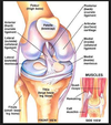
LCL (fibular collateral ligament), MCL (tibial collateral ligament)

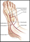
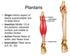
muscles of the foot (1st layer - most superficial)
origen: calcaneous
- abductor hallucis, i: base of proximal phalanx of big toe, flexes/abducts big toe, braces medial longitudinal arch
- flexor digitorum brevis, i: tendons to 4 lateral toes (middle phalanx), flexes lateral 4 toes, braces medial and lateral longitudinal arch
- abductor digiti minimi, i: proximal phalanx of fifth toe, flexes and abducts fifth toe, braces lateral longitudinal arch

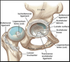
ligament of head of the femur
aka ligamentum teres
flexor digitorum (longus)

common fibular nerve
aka peroneal nerve

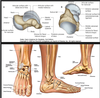
bones of the foot

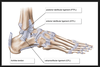
deltoid ligament at ankle

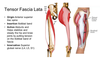
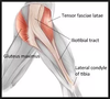
tensor fascia lata
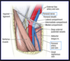
tibial nerve - posterior
tibial nerve, vein, artery from lateral to medial
lateral ligament at ankle
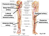
femoral artery, popliteal artery, anterior tibial artery, posterior tibial artery
plantar calcaneo-navicular ligament (spring)
- medial view

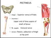
pectineus
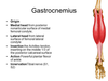
gastrocnemius
flexor hallucis longus

fibular artery

plantar fascia

muscles of the foot (3rd layer - second deepest)
- flexor hallucis brevis, o: cuboid, lateral cuniform i: lateral and medial side of base of proximal pahalnx of big toe, a: flexes big toe, supports medial lognitudinal arch
- adductor hallucis, o: base of 2-4 metatarsals (oblique head), plantar ligaments (lateral head), i: lateral side of base of proximal phalynx of big toe, a: flexes bid toe and holds together metatarsals
- flexor digiti minimi brevis, o: base of 5th metatarsal, i: lateral side of base of proximal phalynx of little toe, a: flexes little toe
tibia

tibialis anterior

dorsalis pedis artery
muscles of the foot (2nd layer - 3rd deepest)
- 4 lumbricals, o: tendons of flexor digitorum longus, i: base of proximal phalanges of lateral four toes, extends toes

soleus
- under gastrocnemius

sciatic nerve

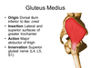
gluteus medius
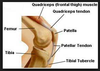
quadriceps and attachments

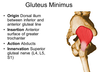
superior and inferior gluteal nerves
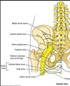
superior and inferior gemelli
superior: origen = spine of ischium, insertion = greater trochanter of femur, action = lateral rotation of thigh at hip joint
inferior: origen = ischial tuberosity, insertion = greater trochanter of femur, action = lateral rotation of thigh at hip joint

piriformis
- posterior view
femoral triangle
gluteus minimus
obturator externus

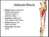
adductor brevis
ligaments of the knee
sacroiliac joint

medial and lateral plantar nerves

pes anserine
sartorius, gracilis, semitendinosus - goose foot
- inserts onto anteromedial tibia

obturator internus
posterior view

meadial view of ankle
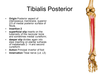
tibialis posterior, flexor digitorum longus, posterior tibial artery, tibial nerve, flexor hallucis longus

popliteus muscle
extensor digitorum brevis
- also note the extensor hallucis brevis

patella
femoral nerve

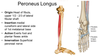
fibularis longus
tibialis posterior
plantaris
Superior and inferior pubic rami

iliopsoas
origin for iliacus = iliac fossa

extensor digitorum (longus)

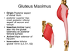
gluteus maximus
extensor hallucis longus

Ischial tuberosity
sartorius

tibial plateau (medial and lateral)

fibularis tertius

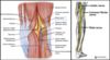
posterior leg muscles

talus (head, body, neck)
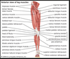
anterior leg muscles
iliotibial band
hamstrings and attachments

adductor magnus

gracilis

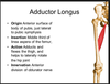
adductor longus
quadratus femoris

fibularis brevis

inferior tibiofibular joint and ligaments

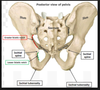
ilium, pubis, and ischium

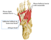
lateral ankle: fibularis longus and brevis

superior tibiofibular joint and ligaments

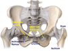
pelvis - body of ilium, iliac crest, Anterior Superior Iliac spine (ASIS), Anterior Inferior Iliac spine (AIIS), Posterior Superior Iliac spine (PSIS), pubic symphysis, obturator foramen, acetabulum

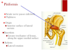
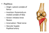
3 Hip Ligaments

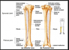
fibula
ACL, PCL, medial and lateral meniscus

muscles of the foot (4th layer - deepest)
- 3 plantar interossei, o: inferior surface of 3-5 metatarsal, i: medial side of bases of proximal phalanges of lateral 3 toes, a: adduction of toes, flexes metatarsophalangeal joint, extends interphalangeal joints
- 4 dorsal interossei, o:adjacent sides of metatarsal bones, i: base of proximal phalanges, a: abduction of toes, flexes metatarsophalangeal joint, extends interphalangeal joints

Ischiopubic ramus
femur

