Liver conditions Flashcards
Describe the architecture of a liver lobule
Histologically, the liver is composed of hepatocytes, bile canaliculi, and hepatic sinusoids:
- Hepatocytes are arranged in chords, with blood flowing inbetween in sinusoids from the portal triads → central vein
- Endothelial cells form space of Disse between hepatocytes and sinusoids

Describe the roles of the several vessels entering or leaving the liver
- Portal vein bringing food-rich blood from the gut.
- Hepatic artery bringing oxygenated arterial blood. Liver has dual blood supply.
- Heptic veins taking away processed blood into the vena cava.
- Lymphatics taking away some lymph and some by-products.
- Hepatic ducts removing bile to the gallbladder & gut (biliary system).
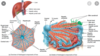
Describe the general anatomical features of the liver
- Large, lobated exocrine/blood-processing gland, with vessels/ducts entering/leaving at the porta
- Enclosed by a thin collagen tissue capsule, mostly covered by mesothelium.
- Collagen tissue of the branching vascular system provides gross support.
- Parenchymal cells are supported by fine reticular fibres.
Describe the hepatic lobular flow
Blood flows:
- From branches of the portal vein and hepatic artery; from the periphery towards the centre;
- in the sinusoids, between the cell plates (blood flows slowly from portal triad to central vein, allowing exchange of nutrients for the enrichment of blood)
- Blood collected in central veins → sublobular veins → collecting veins → hepatic veins leaving the liver.

Describe the liver acinus
The territory of an acinus (as its axis) has one final branch
of the portal vein, and is subdivided into:
- Periportal zone – roughly spheroid, isolated from periportal zones of adjacent acini
- Intermediate zone
- Perivenous zones – near the central vein

What does this slide show?

Normal liver

No fatty infiltration
No inflammation
No fibrosis or cell plate thickening
What types of cells are found within the sinusoids of the liver?
Kuppfer (stellate) cells- resident liver macrophages
Under physiological conditions, they are the first innate immune cells and protect the liver from bacterial infections
Roughly speaking, what do the 3 main LFTs measure?
LFTs measure liver enzymes which can leak from damaged liver cells- so they reflect liver injury
- ALT + AST = liver parenchymal damage (hepatocellular)
- ALP = obstructive cause (cholecystatic)
Gamma-GT = mirrors ALP (unlike ALP, only produced by liver so good way of discerning hepatic pathology)

State the sources of ALT, AST, ALP
ALT = hepatocytes ONLY
AST = liver, heart, skeletal muscle, kidneys, pancreas
ALP = biliary ducts, bone, placenta, small intestine, kidneys
What is the correct way of assessing ALT and AST levels? Explain which pathologies would affect assessment of AST:ALT
must look at AST:ALT ratio
High AST:ALT >2.5 = alcoholic damage/cirrhosis, metastases
Low AST:ALT <1 = paracetamol OD (toxin-induced hepatitis), viral hepatitis (hep A/B/E, EBV, CMV), hepatic obstruction
SALT

Compare the likely diagnoses for a:
- Marked
- Moderate
- Mild
increase in ALT
Marked increase (1000s):
- Toxin/drug induced hepatitis e.g. paracetamol
- Acute viral hepatitis e.g. hep A/B/E, EBV, CMV
Moderate increase (300-500):
- chronic/alcoholic/autoimmune hepatitis
- biliary obstruction
MIld increase (<300):
- Cirrhosis
- Haemachromotosis
- hepatocellular carcinoma
- Wilson’s
- NASH
State some non-hepatic causes for raised ALP
- placenta: pregnancy
- small intestine: fatty meals
- kidney: CKD
- bone: paget’s, bony metastases, fractures, osteomalacia, renal bone disease
Which marker can be measured to determine whether ALP is hepatic in origin?
Gamma-glutamyl transferase (Gamma-GT)
Mirrors ALP but specific to liver
And isolated rise in Gamma-GT is suggestive of what?
alcoholic liver disease
What is a high ALP with high gamma-GT suggestive of?
bile duct disease
liver metastases
Summarise the production of bilirubin
reticuloendothelial cells metabolise Hb into haem and globin
haem → iron and bilviderin
bilviderin → unconjugated bilirubin (binds to albumin and travels to liver)
conjugated bilirubin excreted into bile
in colon: conjugated bilirubin → urobiligen (20% absorbed via enterophepatic circulation)
80% urobiligen → stercobilin

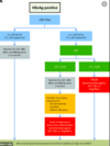
How can jaundice be categorised? For each, state the underlying cause and the type of bilirubin that is in excess
Pre-hepatic = increased haemolysis = ^UNCONJUGATED
Heptic =liver impairment = ^UNCONJUGATED+CONJUGATED
Post-hepatic = biliary tree obstruction = ^CONJUGATED

EXPLAIN the causes of pre hepatic jaundice
Caused by increased haemolysis.
This results in the increased presence of unconjugated bilirubin in the blood as the liver is unable to conjugate it all at the same rate.
This is caused by:
- Tropical disease, e.g. malaria, yellow fever
- Genetic disorders, e.g. sickle-cell anaemia, Gilbert’s syndrome
- Haemolytic anaemias

EXPLAIN the causes of hepatic jaundice
Hepatic jaundice is caused by liver impairment.
This causes the decreased ability of the liver to conjugate bilirubin, resulting in the presence of conjugated and unconjugated bilirubin in the blood. Liver damage can result from:
- Viral hepatitis
- Hepatotoxic drugs, e.g. paracetamol overdose, alcohol abuse

EXPLAIN the causes of post hepatic jaundice
Post-hepatic jaundice is caused by the blockage of bile ducts.
This results in backflow of conjugated bilirubin into the blood as it cannot move past the obstruction.
Bile duct obstruction can be caused by:
- Gallstones
- Hepatic tumours
- Pancreatic tumours

What is the key liver protein and what is a specific outcome of not having enough
Albumin
When albumin is low, there is reduced oncotic pressure so water leaks out of cells contributing to ascites.
May be low due to low production or increased loss (nephrotic syndrome)
State some pathologies that cause:
- Low albumin + low protein
- Low albumin + normal protein
- Low albumin + high protein
-
Low albumin + low protein =
- advanced cirrhosis
- protein malnutrition
- chronic inflammation
-
Low albumin + normal protein =
- infection
-
Low albumin + high protein =
- myeloma
State some causes of raised INR
- liver disease with impaired function
- vit K deficiency
- consumptive coagulopathy (DIC)
- overdose of oral anticoagulants (VKAs)
- Coagulation factors deficiency (fibrinogen and factors II, V, VII, or X, or a combined deficiency) in the extrinsic pathway
Define Liver Abscess and Liver Cyst
Localised infection in the liver parenchyma that may be bacterial, fungal, or parasitic in origin…
resulting in a walled off collection of …
- abscess = pus
- cyst = cystic fluid