Biliary tract conditions Flashcards

1
Q

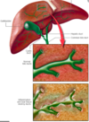
Label the biliary tree

A

Hepatocytes line bile cannaliculi → form ductules that drain into portal tracts

portal tracts form L and R hepatic ducts (L/R lobe)

these merge into the common hepatic duct, joining the cystic duct to form the common bile duct

common bile duct travels through the head of pancreas and merges with the pancreatic duct at the sphincter of Oddi

Exits in second part of duodenum

2
Q

What is the makeup of bile?

A

water, bile acids, cholesterol and bilirubin

3
Q

Which hormone stimulates bile release from the gallbladder?

A

Cholecystokinin

bile is concentrated and stored in the gallbladder

4
Q

State the terminology for:

  • gallstones
  • gallbladder inflammation
  • inflammation/infection of the biliary tract
  • gallstone occlusion of the the cystic/common bile duct
  • slowing of bile flow through biliary tree
A
  • gallstones (uncomplicated)
    • cholelithiasis
  • gallbladder inflammation
    • (usually associated N+V)
    • cholecystitis
  • inflammation/infection of the biliary tract
    • (2/2 choledocholithiasis)
    • cholangitis
  • gallstone occlusion of the the cystic/common bile duct
    • (associated with deranged LFTs)
    • choledocholithiasis
  • slowing/stopping of bile flow through biliary tree
    • cholestasis
5
Q

ERCP versus MRCP

A

Endoscopic retrograde cholangeopancreatography (ERCP)

  • contrast injected through endoscope, allowing radiographic visualisation of biliary tree + pancreas

Magnetic resonance cholangeopancreatography (MRCP)

  • uses MRI to visualise biliary and pancreatic ducts in a non-invasive manner
6
Q

Define biliary colic

A

steady, severe pain (intensity >5 on a scale of 1-10) in the RUQ lasting >15-30 minutes.

An attack of simple biliary colic commonly requires an analgesic but should resolve within 8 hours.

caused by contraction of biliary tree to remove obstruction

7
Q

Define acute cholecystitis

A

Biliary colic lasting >8 hours, accompanied by features of inflammation:

  • fever
  • marked RUQ tenderness (Murphy’s sign)
  • leukocytosis

caused by acute gallbladder inflammation

8
Q

What are the 3 primary types of gallstones?

A

MIXED = 75%

  • cholesterol, calcium bilirubinate, protein, phosphate
  • imbalance between bile salts, cholesterol, phospholipids + gallbladder immotility

PURE CHOLESTEROL = 20%

  • classic patient is a fair, fat, fertile female of forty (the five Fs)

PURE BILE PIGMENT= 5%

  • black stones made of calcium bilirubinate
  • bile pigments are Hb breakdown products
9
Q

State the risk factors for gallstones

A
  • 6 Fs
    • Fair (Caucasian)
    • Fat
    • Fertile- high oestrogens increases risk
    • Forty
    • Female
    • Family history
  • Diabetes mellitus
  • Drugs (OCP, octreotide)
  • Interruption of the enterohepatic recirculation of bile salts (e.g. Crohn’s), terminal ileal resection
  • Bile pigment stones- haemolytic disorders:
    • hereditary spherocystosis
    • sickle cell disease
    • G6PD deficiency
  • TPN
10
Q

Summarise the epidemiology of gall stones

A
  • Very COMMON (developed countries)
  • UK prevalence of gallstone disease = 10-15%
  • 3 x more common in FEMALES in younger population
  • More common with increasing age
11
Q

Describe the typical pain experienced by someone presenting with gallstones using SOCRATES

A

S- RUQ/epigastric

O- sudden onset

C- colicky, constant, increasing in intensity

R- radiates to right scapula

A- nausea , dyspepsia, heartburn, flatulence, and bloating

T- lasting >30mins

E- often 1hr onset after fatty meal, relieved by simple analgesics

S- severe

12
Q

What 2 conditions can cholelithiasis develop into? State key symptoms of each

A

ASCENDING (ACUTE) CHOLANGITIS

  • Charcot’s triad- RUQ pain, rigors, jaundice

CHOLECYSTITIS

  • fever
  • prolonged pain >8hrs referred to R shoulder tip (diaphragmatic irritation)
  • Murphy’s sign- (respiratory arrest upon deep inspiration on palpation of the biliary fossa)
13
Q

Explain the key difference between symptomatic cholelithiasis (biliary colic) and cholecystitis

A

inflammatory component

Tachycardia

Pyrexia

Local peritonism

RUQ pain or epigastric tenderness

⇒ May be guarding and/or rebound tenderness

Murphy’s sign positive

NOTE: this is only positive if the same test in the LUQ does not cause pain

14
Q

Identify appropriate management of gallstones

A
  1. If found incidentally + asymptomatic = observation
    • recommend low fat diet
  2. Symptomatic cholethiasis
    • analgesia
      • paracetamol, diclofenac/other NSAIDs
    • antispasmodic
      • hyoscene
    • cholecystectomy
      • elective laproscopy to prevent reccurrence
  3. Choledocholethiasis
    • ​​removal of stones via ERCP and biliary sphincterectomy (then cholecystectomy)
    • or laproscopically alongside cholecystectomy
    • temporary biliary stenting if the abpve to options unsuccessful
15
Q

Identify some common complications of gallstones

A

STONES IN GALLBLADDER

  • Biliary colic
  • Acute cholecystitis
  • cholecystoduodenal fistula →
    • Gallstone ileus- stone passes through and obstructs terminal ileum
    • Bouveret syndrome- fistula obstructs duodenum
  • Gallbladder empyema
  • Gallbladder cancer (RARE)

STONES IN DUCTS

  • acute ascending cholangitis (charcot’s triad)
  • biliary fistula- erosion of stone from cystic duct to common hepatic

PANCREATITIS

  • acute biliary pancreatitis- pancreatic bile outflow obstruction
  • Following ERCP - indometacin prophylactically
16
Q

State some common complications of cholecystectomy

A
  • Bleeding
  • Infection
  • Bile leak
  • Fat intolerance due to inability to secrete a large amount of bile into the intestine as pt no longer has a gall bladder
  • Post-cholecystectomy syndrome – abdominal symptoms e.g. dyspepsia, nausea/vomiting, RUQ pain
  • Port-site hernia
17
Q

How is acute biliary pancreatitis managed?

A
  • IV hydration
  • IV analgesia
  • ERCP with sphincterotomy and stone extraction within 72 hours of admission

Mild acute pancreatitis requires only fluids and supportive care (+ elective cholecystectomy when condition improves)

18
Q

Define cholecystitis

A

acute gallbladder inflammation, and one of the major complications of cholelithiasis

19
Q

What is the aetiology of cholecystitis?

A

90% = GALLSTONE causing cystic duct/gallbladder neck obstruction

5% = ACALCULOUS- bile inspissation (due to dehydration) or bile stasis (due to trauma or severe systemic illness) blocks the cystic duct

20
Q

How does cholecystitis present?

A

RUQ pain + tenderness

  • severe and steady, radiates to back + right shoulder
  • Murphy’s sign

Signs and symptoms of inflammation

  • fever and raised inflammatory markers
  • systemically unwell- tachycardia

Palpable gallbladder

N+V, anorexia

21
Q

How would you investigation cholecystitis/cholelithiasis?

A

Bloods

  • FBC
    • in inflammation (cholecystitis):
    • high WCC, CRP, ESR
  • LFTs
    • amylase normal unless pancreatitis
    • high ALT + GGT in ascending cholangitis
  • Blood cultures- if suspected sepsis

Imaging

  • no sepsis- abdominal USS confirms diagnosis
    • see thickened wall, distended biliary tree/bladder, stones, pericholecystic fluid
  • sepsis- abdo MRI/CT contrast
    • see abscess, perforation, increased density of fatty tissue around gallbladder
22
Q

Define cholangiocarcinoma and its subdivisions

A

Carcinomas arising from the bile duct epithelium

>95% are adenocarcinomas

can be intrahepatic or extrahepatic (perihilar/distal) depending on location in biliary tree

23
Q

What are the risk factors for cholangiocarcinoma?

A
  • Cholangitis- primary sclerosing and acute
  • Cholecysto- / choledo- lithiasis (gallstones in gallbladder or common bile duct)
  • Alcoholic liver disease- intrahepatic
  • Ulcerative cholitis
  • Cirrhosis
  • Hepatitis
  • HIV
24
Q

Summarise the epidemiology of cholangiocarcinoma

A
  • VERY RARE
  • 50-70yrs
  • More common in the developing world due to the increased prevalence of parasitic infections
25
What are the presenting symptoms of cholangiocarcinoma?
90% present with **_painless jaundice_** SYMPTOMS OF OBSTRUCTIVE JAUNDICE * **Yellow sclera** * **Pale stools** * **Dark urine** * **Pruritis** May present with abdo pain and weight loss
26
Identify appropriate investigations for cholangiocarcinoma
_Bloods:_ * LFTs * **bilirubin- high** * **ALP- high** (AST/ALT- minimally raised) * **Gamma GT- high** * Clotting * **increased PTT**- decreased fat soluble vitamins ADEK) * Tumour markers * **CA 19-9** - elevated in cholangiocarcinomona, pancreatic or gastric malignancy * **CA-125** _Imaging:_ * **Abdominal USS**- shows dilated intrahepatic ducts + mass * **CT/MRI** - differentiate between solid and cystic biliary contents * ERCP/MRCP/PTC * **MRCP** is diagnostic only not therapeutic * **ERCP** is invasive * **PTC-** when stricture completely occludes duct so ERCP/MRCP won't work
27
# Define acute (ascending) cholangitis
**Infection of the biliary tree**, most commonly caused by obstruction
28
Explain the aetiology of acute cholangitis
1. Most commonly caused by **cholelithiasis** → **choledolithiasis** 2. → biliary **obstruction** 3. → **cholestasis** 4. → **bacterial seeding** from duodenum 5. **Sludge** can form = growth medium 6. If left untreated, extravasation of bacteria into bloodstream → **sepsis**
29
What are the present symptoms of acute cholangitis?
_CHARCOT'S TRIAD_ * **RUQ pain** (may refer to right shoulder tip) * diffuse- Murphy's NEGATIVE * **Jaundice** * **Fever w/rigors** Result of biliary obstruction and bacterial growth in the biliary tree. Other symptoms: * **pale stools** * **pruritis** * severe- **hypotension + mental status changes**
30
Identify appropriate investigations for acute cholangitis
Bloods * FBC- **raised WCC** * LFTs: typical pattern of obstructive jaundice (**raised ALP + GGT**) * U+Es - **urea + creatine raised** in severe disease * ABG- in sepsis, **metabolic (lactic) acidosis** * Inflammatory markers- **raised CRP, ESR** * **Culture**- usually gram negative * Clotting- **raised PTT** in sepsis Imaging: * **Abdo USS**- dilated ducts, stones * **ERCP**- find and remove stones. best first intervention * **CTKUB contrast -** is abdo USS negative, diagnostic
31
Generate a management plan for acute cholangitis
* **A-E approach** * IV fluids, electrolytes * **sepsis 6** * **IV broad spectrum Abx** * until blood and bile culture results are obtained * **Biliary decompression and drainage** * if refractory to Abx * by ERCP +/- sphincterotomy and placement of a drainage stent * **Opiod analgesics** * morphine, fentanyl
32
Identify the possible complications of acute cholangitis
* **Acute pancreatitis-** via obstruciton of the pancreatic duct * **Inadequate biliary drainage** following ERCP/surgical intervention. Leads to worsening of condition * **Hepatic abscess** * **Sepsis** → organ dysfunction
33
Summarise the prognosis for patients with acute cholangitis
Most patients experience rapid clinical improvement once **adequate biliary drainage is achieved** **Mortality high** if comorbidities/develop **sepsis**
34
Define Primary Biliary Cirrhosis/cholangitis (PBC)
**Chronic inflammatory disease** of the **intrahepatic bile ducts** causing **cholestasis**, progressive **bile duct damage** and eventual cirrhosis Thought to have an **autoimmune** component
35
What are the risk factors/aetiology of PBC?
**Autoimmune-** presence of **antimitochondrial antibodies** in 95% Therefore increased risk if other autoimmune conditions: * **Sjogren's syndrome** * **Coeliac disease** * **Scleroderma** Genetic + environmental factors- **smoking, UTI** can be triggers
36
Describe the pathophysiology
Damage to + progressive **destruction of the biliary epithelial cells** lining the small intrahepatic bile ducts. due to portal tract **granulomatous autoimmune inflamation**
37
Summarise the epidemiology of primary biliary cirrhosis
Mainly affects **middle-aged women** (9:1 female: male)
38
What are the presenting symptoms/signs of primary biliary cirrhosis?
Findings may be incidental. Insidious onset of: * **Fatigue** (due to sleep disturbance) * **Pruritis** * Smooth, non-tender **heptomegaly** * **Xanthelasama (**due to hypercholesteraemia) May present with a **complication of liver decompensation** (e.g. jaundice, ascites, variceal haemorrhage) May present with **symptoms of associated conditions** (e.g. Sjogren's syndrome, arthritis, Raynaud's phenomenon)
39
What investigations would you do for PBC?
_Bloods_ * **High ALP and gamma GT**- evidence of cholestasis * **ALT and AST** are normal initially but will increase as the disease progresses and cirrhosis develops * **Low albumin** * **High PTT** _Immunology_ * **ANTIMITOCHONDRIAL ANTIBODIES** * **antinuclear** antibodies * EILSA and immunofluorescence * Raised **IgM** and **IgG** _Other_ * **Liver biopsy** * bile duct lesions and granulomata formation * late stages- loss of ducts + biliary fibrosis * **Upper GI endoscopy** * Check for oesophageal varices if abnormal LFTs/biopsy
40
Discerne between Primary Sclerosing Cholangitis (PSC) and Primary BiIliary Cholangitis (PBC)
_PSC_ * Affects **large intrahepatic and extrahepatic ducts** * Diagnostic on **USS + MRCP** * **Increased risk of colon cancer + cholangiocarcinoma** * associated with **IBD (80% patients)** * **60%** men * mostly affected **40yrs/any age** * neg association with smoking _PBC_ * Affects **only small intrahepatic ducts** * Not visible on USS or MRCP * **Diagnostic = liver biopsy and AMA serology** * Not associated with IBD * **90% women** * mostly affected **\>45yrs** * associated with **smoking**
41
Define Primary Sclerosing Cholangitis (PSC)
A **chronic cholestatic** liver disease characterised by progressive **inflammatory fibrosis** and obliteration of **_large_** **intrahepatic and extrahepatic bile ducts** Results in diffuse multi-focal **stricture** formation
42
In what sense is PSc not a classic autoimmune disorder?
* More common in men * Doesn't respond to immunosuppressive treatment
43
Explain the pathogenesis of PSC
**Injury and inflammation of medium-large hepatic ducts → fibrosis and stricturing → cholestasis** _Biliary strictures of PSC contribute to:_ * **jaundice** * **pruritus** * episodes of **bacterial cholangitis** * progression to **biliary cirrhosis** _Ongoing injury and continued destruction:_ * secondary **parenchymal damage** * **fibrosis, cirrhosis, end-stage liver disease**
44
Explain the risk factors of PSC
UNKNOWN Possible **immune and genetic predisposition** with environmental triggers **_Close association with inflammatory bowel disease_** (especially ulcerative colitis) - 90% UC
45
Summarise the epidemiology of primary sclerosing cholangitis
Usually presents between 25-40 years of age Relatively rare
46
What are the presenting symptoms of PSC?
* Asymptomatic, persistant high ALP * **RUQ pain** * **fatigue** * **pruritis** * **weight loss** * **jaundice** * May present with episodes of **fever and rigors** caused by acute cholangitis * late stage- symptoms of **cirrhosis** (ascites, splenomegaly, encephalopathy, steatorrhoea) **HISTORY OF UC**
47
What investigations would you do for PSC?
Bloods: * **LFTs- high ALP + GGT** (indicate bile duct injury/obstructive). * high bilirubin + low albumin in late stages * **FBC- late stage thrombocytopaenia ± anaemia** * Serology- * Anti-mitochondrial antibodies (AMA) ABSENT * **No specific autoimmune antibodies** for PSC, **ASMA** and **ANCA** present in 30% * **MRCP/ERCP** * MRCP preferred, non-invasive + specific * See strictures, and dilations ± dominant biliary stricture * ERCP if intervention is needed
48