Lecture 4-Development of the nervous system Flashcards
What does the embryo look like about 2 weeks after fertilisation?
-at that stage the embryo is indistinguishable from a flat disk of cells -sits in this layer between two spaces that are like bubbles -the embryo is in between, no organs, no recognisable parts -trilaminar embryo

What are the three layers of the trilaminar embryo called?
-ectoderm -mesoderm -endoderm
What is the process of neuralation?
-patch of tissue at the tip of the ectoderm (the neural plate) starts to specialise and becomes neuroepithelium (=, the stem cells of the nervous system, basic type of tissue, associated with the skin and gut, this is as the nervous system in evolutionary terms is modified patch of skin -reflects the fact that in the past skin used to be the endophase between the organism and the outside

What does the neural plate develop into?
-neural groove and then neural tube
How does the neural tube form?
-at about 2-3 weeks of age, the first signs of structure -tissue made of three layers, inwards fold of the ectoderm into the mesoderm and endoderm -this forms a groove and the sides start to bend towards each other -then forms a tube (derived from ectoderm) -the tube becomes free from ectoderm and floats beneath it amongst the mesoderm and endoderm -this tube= neural tube and it will become the CNS -only a cell thick
What is the process of neural tube formation along the lengths of the embryo?
-the invagination runs from the head end towards the tail end, so the head end is a bit older in terms of development -rostral to caudal gradient in formation of nervous system -rostral is older

What are the 5 stages of the neural fold closure?
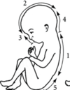
-happens in stages 1.the back and goes in both directions 2. the head 3. face 4. nape of the neck 5. the lower part of the back

What is spina bifida?
-lesion in the lower back of the baby where the neural tube failed to close -brings problem in spinal development, problems with motor skills -failure in stage 5
What is anecephaly?
-failure in stage 2 of neural fold closure -no upper part of the brain and head, no higher centers -stillborn
What happens in segmentation of the neural tube?
-the rostral end starts swelling (vessiculation) -forms 3 distinct vesicles 1.Prosencephalon (forebrain) 2. Mesencephalon (midbrain) 3. Rhombencephalon (hindbrain) -what is left of the tube will form the spinal cord

What happens in further segmentation of the neural tube?
-the three vesicles divide further -the Prosencephalon splits into= telencephalon and diencephalon -Rombencelphalon- divides into 7 segments -rets is spinal cord -looks like a worm and it points to the evolutionary history

What is the third stage in segmentation?
-what was just straight folds and double backs on itself -rombencephelon splits into metencephalon and myelencephalon these will be pons and medulla respectivelly -telencephalon is at the front will become cortex -diencephalon= deep cortex -mesencephalon= midbrain -in diencephalon there are two outgrows= will become retinae, (optic vesicles) (eyes are also part of the brain) -telencephalon= already forming two hemispheres

What does the Prosencephalon (forebrain) develop into?
Five vesicle stage= Telencephalon and Diencephalon -Mature brain= 1. Telencephalon=cortex,basal ganglia, hippocampus 2.Diencephalon=Thalamus and hypothalamus
What does the Mesencephalon (midbrain) develop into?
-five vesicle stage: Mesencephalon -mature brain= midbrain
What does the Rhombencephalon (hindbrain) develop into?
-five vesicle stage: Metencephalon, Myelencephalon -mature brain 1. Metencephalon= Pons and cerebellum 2. Myelencephalon= Medulla
What does the caudal neural tube develop into?
-five vesicle stage: caudal neural tube -mature: spinal cord
What is the brain like after the segmentation process?
-series of thin-walled bubbles
What is the neural crest?
-cells at the top of neural tube form neural crest -at around the time the tube is being sealed -migrate away from neural tube to form a wide range of structures

What are the neural crest derivatives?
-PNS- dorsal root ganglia, sympathetic and parasympathetic ganglia, enteric ganglia, Schwann cells -Melanocytes (pigment cells) -Muscle cartilage and bone of skull, jaws, face and pharynx -Dentine cells (teeth- partly)
How do the neural crest cells migrate?
-from neural crest 1.laterally under the skin(melanocytes) 2. to site of dorsal root ganglia 3-Through somite to sympathetic ganglia -the migration occurs from top of the neural tube -neural crest cells follow specific paths through the embryo
What happens if the neural crest cells fail to migrate?
-born without a face (everything on it is formed from those cells) -milder version= cleft palate, some failure, the neural crest cells didn’t finish the job
What can you see forming in the second embryo?
-big change of size in a day -green= neural crest cells -older embryo can see the migrated cells and how they form what will be the PNS

How does the enteric system arise?
-neural crest cell migration from what will be the hindbrain down what will be the vagus nerve into the gut -the longest migration in the body and goes on for the longest period of time -9m long gut on avg.
How does the neuroepithelium add layers to generate the cortex?
-the brain is still mostly empty space, needs to be filled with neurons -cortex is a structure with different cells from top to bottom= formed: - single cell layer, cells dividing quickly, the spin off daughter cells that start to differentiate and turn into neurons -first neurons that are born are right at the bottom of the neuroepithelium -then they migrate up to form multiple layers in the cortex or else -cell bodies sink to the lower level -inside out process = building of the cortex
Where are all new neurons born?
-at the ventricular surface (zone) which contains stem cells
What is a stem cell?
-self renewing indefinitely and undifferentiated cells -daughter cells capable of differentiation into specialised cells -have to produce at least one daughter cell and one stem cell
What are radial glia?
–specialised cells= no matter how thick the cortex the radial glia have one processs attached to the bottom and one at the top -guide the migrating neurons -have leading process and trailing process -bipolar-shaped cells that span the width of the cortex in the developing central nervous system

What is the process of induction?
-so far only have generic neurons, but need several types -signals pass between structures or tissues, involves ligands and receptors -signal induces a particular response (next step in development)
What are somites?
developing muscle and bone
What is the notochord?
-forms centre of vertebral body, - associated with the neural tube, one of the first things to form, goes down the midline of the embryo -degenerates later after the vertebrae form -initially this is what the major structural elemnet of the embryo is
What is the neural tube?
-developing spinal cord
What is the organisation of the spinal cord?
-core of grey matter surrounded by white matter
Where are the motor neurons in the spinal cord?
-ventral horn -driven by interneurons

What are the distal muscles innervated by?
-lateral cells (neurons)

What are the proximal muscles innervated by?PIC12
-more medial cells (neurons)
Where in the spinal cord is the sensory processing?
-dorsal horn

What are the circuits of neurons in the spinal cord like?
-sensory axons (afferent) going to the spinal cord and going into interneurons (in the spinal cord) and then the motor axons (efferent)

How does induction work?
-the notochord is critical because it releases chemicals that set up gradients -much of induction happens by reading the presence of gradients -you have a source of signaling chemical and as you move further away the chemical concentration drops= cells can detect these changes and use this to determine their phenotype- set up different parts of the nervous system as needed -wide range of signaling molecules important, set up gradients that define topography
How does the spinal cord develop (induction)?
-the notochord releases sonic hedgehog -it is at the highest concentration at the bottom of the neural tube and influences formation of cells called= floor plate -the floor plate cells then release sonic hedgehog as well broadcasting it into the environment -then the gradient is set up and motor neurons develop in the ventral part (because of the gradient there)
What is the sonic hedgehog?
-retinoic acid, noggin and chordin in floorplate and notochord -signalling chemical in induction
Where is the roof plate and floor plate in the neural tube?
-

How can you prove the role of notochord in the induction process?
-in a chicken embryo you can remove it and see that the motor neurons don’t develop -you can also get an extra one and see that the motor neurons develop depending on where you place the notochord around the neural tube
What does the floor plate do?
-induces ventral horn motor neurons
How does the induction of interneurons work?
-hierarchy of gradients occurs -have notochord releasing sonic hedgehog that forms the floor plate -the floor plate releases sonic hedgehog and motor neurons form -the motor neurons release the motor neuron factor and that sets up the gradient for the interneurons to form -interneurons appear just dorsal to motor neurons -depends on this chain of inductive events starting with the notochord

Are axons present when the neurons first form?
-no -it is a key feature of mature neurons -axons have to grow to meet their target cell
How do the first axons form?
e.g. frog embryo= the first axon forms about 16hrs after fertilisation, forms in the midbrain, grows to the spinal cord and then others appear in the medulla and forebrain -the first axons that grow can be used like scaffolding for the others thus they are the most important ones -the axons extend over millimeters in an embryo
What is the role of the growth cone?
-at the tips of the growing axons -the cone is the growing tip -axons grow because the cone is pulling itself like a dog on a chain through the tissue -all that the neuron cell body does is adding raw materials and makes the chain longer -the growth is directed by the growth cone -if you cut off the growth cone= it would crawl itself (self-powered attractor) -axon is towed into position by growth cone
What is the growth cone made of?
-dynamic structure of cytoskeleton -has actin, microtubules and they also overlay
How is the movement of axons directed?
-by chemicals developed in the nervous system= the growth cone responds to the chemical gradient, tries to get to the point of highest concentration -and signals on top of the cells they have to grow over (can have yes or no signal= this creates no go zones)
What is the brain like when you’re born? (neonatal brain
-plastic -lot of functions not working yet -development not finished= continues for a significant amount of time (21 yrs) -connections in the brain must be refined -activity in neural circuits is a key to plasticity
How does the visual system develop after birth?
-it performs badly at birth (can tell dark or light at most) -the brain has to integrate vision from both sides -at first the eyes can’t focus properly -it takes 10 years for sight to completely mature (critical period) -can prove by the kitten experiment= cover one eye with an eye patch (when newborn) and take off after three months, then no functional vision in that eye despite the neural circuitry being intact -visual cortex now completely devoted to the uncovered eye -it is caused by the lack of input from the eye= cortically blind!
What are the critical periods?
-periods when you need input so the neural circuits form properly -in many areas, language, hearing, social interactions… -due to changes in weight and presence of synaptic connections -without input the neural circuits won’t settle into adult behaviour
Are axons present when the neurons first form?
-no -it is a key feature of mature neurons -axons have to grow to meet their target cell
How do the first axons form?
e.g. frog embryo= the first axon forms about 16hrs after fertilisation, forms in the midbrain, grows to the spinal cord and then others appear in the medulla and forebrain -the first axons that grow can be used like scaffolding for the others thus they are the most important ones -the axons extend over millimeters in an embryo
What is the role of the growth cone?
-at the tips of the growing axons -the cone is the growing tip -axons grow because the cone is pulling itself like a dog on a chain through the tissue -all that the neuron cell body does is adding raw materials and makes the chain longer -the growth is directed by the growth cone -if you cut off the growth cone= it would crawl itself (self-powered attractor) -axon is towed into position by growth cone
What is the growth cone made of?
-dynamic structure of cytoskeleton -has actin, microtubules and they also overlay
How is the movement of axons directed?
-by chemicals developed in the nervous system= the growth cone responds to the chemical gradient, tries to get to the point of highest concentration -and signals on top of the cells they have to grow over (can have yes or no signal= this creates no go zones)
What is the brain like when you’re born? (neonatal brain
-plastic -lot of functions not working yet -development not finished= continues for a significant amount of time (21 yrs) -connections in the brain must be refined -activity in neural circuits is a key to plasticity
What are the critical periods?
-periods when you need input so the neural circuits form properly -in many areas, language, hearing, social interactions… -due to changes in weight and presence of synaptic connections -without input the neural circuits won’t settle into adult behaviour
What happens if something goes wrong in development?
-disorders of development -many neurological diseases have origin in developmental problems -including complex behavioural disorders such as schizophrenia and autism


