Lec 4- Adaptive immunity Flashcards
(27 cards)
Adaptive immunity is the 3rd level of defence
- Physical barriers
- Innate immunity
+instant- complement- cells- cytokines +hours-days
-Adaptive immunity +lymphocytes-
B&T cells
+Similar effector mechanisms to innate immunity
+unique system of recognition
Pathogen recognition- innate immunity
-A fixed repertoire of receptors and soluble molecules (PRR) -All components are inherited -New variants rarely arise -Overall strategy +Recognise pathogenic structures (PAMP’s) or +Detect alterations in infected or damaged cells
Pathogen recognition- adaptive immunity
-Each lymphocyte express just one molecule type of receptor
+B cells- BCR (membrane immunoglobulins
+T cells- TCR
- The receptors are made by gene rearrangement so we all have millions of different specificities 3 disadvantages
- Precise targeting
- Memory cells
- Recognise ‘new’ pathogens
Receptors of the adaptive immunity B cell
Surface immunoglobulins
- Antigen-binding site is on the light chain, this is a variable region
- Heavy chain- this is a constant region
- Transmembrane region Plasma cells
- Once a B cell starts producing (excreting) the immunoglobulins they become plasma cells and the immunoglobulins become antibodies T cells -a and b chain
- Consisting of antigen binding site in the variable region and then constant regions
Antibodies are highly specific
Anti-bodies made during infection with measles virus bind to the virus and prevent reinfection with measles virus
- Antibodies made during the infection with measles virus don’t bind to the influenza virus
- This is because the antibodies shape is only complementary to the configuration of 1 antigen
Receptor diversity- gene rearrangement
- Germline configuration of genes encoding B-cell and T cell receptors cannot be transcribed
- Gene rearrangement with nucleotide insertions at the joint produces a functional gene
- Different rearrangement and insertions occur in each lymphocytes
- Different combinations of randomly selected gene regions allow for a huge number of receptor permutations
- Genes are never cleaved in the same place it is random giving rise to a far greater number of different permutations
- V1 = variable -J1= joining -C= constant regions

Clonal selection
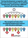
- During development, progenitor cells give rise to large number of circulating lymphocytes, each having a different form of cell surface receptors
- The receptors of only a few circulating lymphocytes interact with any give pathogen
- Lots of lymphocytes
+Different receptors
+receptor specificity generated through chance
-Small minority recognise Ag

Clonal expansion
- Pathogens-reactive lymphocytes are triggered to divide and proliferate
- Pathogen-activated lymphocytes differentiate into effector cells that eliminate the pathogens
- Ag recognition
+Survival signals- up-regulaiton
+division signals- up-regulation
-Effector cells generated- clear pathogens

Step 1 in adaptive immunity
- Dendritic cells carry Ag to lymph nodes
- Pathogens adhere to epithelium
- Skin wound allows pathogens to penetrate epithelium -
Local infection, innate immunity
- Dendritic cells take infection (breaks down microbe producing Ag) to lymph node and stimulate adaptive immunity
- Within the node it is the APC which educate the cells (B and T cells)
- Effector cells and molecules of adaptive immunity travel to the infected tissue -APC= antigen presenting cells
Step 2 in adaptive immunity
T cells recognise pathogen fragments
- Dendritic cells take up pathogen for degradations
- Pathogens is take apart inside the dendritic cells
- Pathogen proteins are unfolded and cut into small pieces
- Peptide bind to MHC molecules and the complexes go to the cell surfaces
- T cell receptor bind to peptide: MHC complexes on dendritic cell surface (the T cell can differentiate when the MHC contains our own peptides and not react)
- MHC= major histocompatibility complex

Antigen presentation in adaptive immunity
-T cells can only recognise small peptide fragments
+Ignore 3D structure of proteins
+Recognise linear elements
- Macromolecular structures are unfolded and cleaved into short pieces- ANTIGEN PROCESSING
- Antigens are then displayed on the cell surface in the context of MHC- Ag PRESENTATION
2 Types of MHC present antigen Why 2 types
- To deal with different pathogens (intracellular mainly virus /extracellular mainly bacteria )
- To interact with different T cells
- MHC class I is for intracellular peptides and has 1 anchor point
- MHC class II is for extracellular peptides and has 2 anchor point

MHC class I and II bind different effector T cells
- The interaction between MHC and TCR is incredibly weak and therefore we have another protein, otherwise the interaction would be so fleeting that it may not stimulate a reaction in the T cell
- Co-receptors that stabilise the cell interactions
- For MHC class I we have CD8 co-receptor, Tc (cytotoxic) use this co-receptor
- For MHC class II, we have CD4 co-receptor, TH (helper cell)
- The co-receptor defines the T. cell populations

Class I and II bind peptides from different cellular compartments
-Cells have 2 major compartments
+cytosol- peptides from intracellular pathogens
+Vesicular system- peptides from extracellular pathogens
MHC I presents Ag from intracellular infections
- Virus infects cell
- Viral proteins synthesised in cytoplasm
- Peptide fragments of viral proteins bound by MHC class I in ER
- Bound peptides transported by MHC class I to the cell surfaces
- Cytotoxic T cell recognises complex of viral peptide with MHC class I and kills infected cells

MHC II presents Ag from extracellular origin
- Macrophage engulfs and degrades bacterium, producing peptides -Bacterial peptides bound by MHC class II in vesicles
- Bound peptides transported by MHC class II to the cell surface
- Helper T cell recognises complex of peptide antigen with MHC class II and activates macrophages

B cells need help to
- Cell surface immunoglobulins of B cell binds to bacteria; the cell then engulfs and degrades them, producing peptides
- Bacterial peptides bound by MHC class II in endocytic vesicle
- Bound peptides transported by MHC class II to the cell surface and presents peptide
- Helper T cell recognises complex and activates B cells

Antibodies
- Antibodies are soluble effector molecules of adaptive immunity
- 5 classes (isotopes) of antibody
+IgA: IgG; IgM: IgE:IgD- these different types are based on there constant region
- Different locations and functions
- IgA, IgG and IgM- main antibodies in blood, lymph and tissue -IgM- first antibody made (primary immune response)
- IgA protects mucosal surfaces
Antibodies are multi-functional: Neutralisation
- This occurs when a bacteria release a toxin
- Cell with receptors for toxins can bind and cause damage
- If we release an Ig that is complementary they will start to bind the toxins together
- Because they have 2 arms they can cause aggregation and so steric hindrance of the toxin meaning it is no longer complementary to the shape of the receptor
- This leads to the ingestion and destruction of antibody and toxin
- Therefore doesn’t cause harm, this process is known as neutralisation

Antibodies are multi-functional: opsonisation
- The antibodies will bind to the bacteria
- This is opsonisation and will allow the macrophage to ingest and destroy the bacteria more easily
- This can also be done with the help of complement
Antibodies- how they change to improve response
- IgM is the 1st antibody made against infectious pathogen (primary response)
- Somatic hypermutation selects for antibodies that bind more tightly to the pathogen (improves response)
- Switching antibody isotope to IgG allows delivery of the pathogen to phagocyte
Memory cells make even better response
- Clonal expansion produces effector cells and memory cells
- Secondary adaptive immune response are faster and stronger
+Memory cells are more numerous
+Memory cells are more quickly activated
+Memory T cells patrol lymphoid tissue- detect infection early
+Memory B cells make better immunoglobulins- more robust and less likely to change
Lymphocytes are tolerant of self
- Lymphocyte receptors are generated by chance- so there’s a chance of SELF-reactivity
- We can induce tolerance towards self during lymphocyte development
- T cells undergo negative and positive selection in the thymus
T cell selection

- In the thymus, T cell progenitor give rise to billions of thymocytes, each with a different T cell receptor
- Thymocytes are positively selected by epithelial cells in the cortex of the thymus (is it self MHC reactive- if it can’t react with MHC it can’t kill pathogens)
- Positively selected thymocytes survive and divide
- Positively selected thymocyte clones are negatively selected in thymus medulla (do they react to self-peptide if yes then they will attack our own cells so must be destroyed)
- Clones surviving negative selection leave the thymus for the circulation
- Pathogens select upon less than 1% of T cells originating in the thymus


