learning objectives EXAM 1 Flashcards
biomechanics
study of forces acting on human body/body segments and the consequences of those forces related to posture and motion
kinematics
description of motion as a function of space and time, without regard to forces creating the movement
(no cause, just motion)
kinetics
the description of motion of a system in terms of forces acting on the system
(muscle activity)
linear motion
curvilinear and rectilinear

force
mechanical interaction between a system and its surroundings; a push or pull of one object or another
the base of kinetics
moment
the turning effect of a force, known as moment of force or torque
skeletal muscle cross-sectional area
anatomical, physiological
proportion to the muscle force that can be produced
PCS>ACS

absolute reference frame
based on the environment that movement occurs in

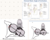
relative reference frame
moves with body segment
shows joint angle/ROM

velocity
change in position/change in time (s/t)
position/time, velocity/time, acceleration/time graphs
sign of velocity will be in direction of the change in position (if position slope is positive, then velocity value is negative)
peak/valley in position = 0 velocity

relationship between linear and angular motion of body segments
angular motion, theta = s/r
s=r(theta)
angular movements of a segment at the joint lead to linear movement of parts of segments
angular motion of the hip and knee lead to linear motion of the foot

kinematic graphs
area under a curve is the distance traveled
a change in position slope = 0 V
change in velocity slope = 0 acceleration
vector composition and resolution
make parallelogram for vector composition if vectors are coplanar but not collinear (if collinear, just add together)
resolution: split into X and Y components
- X to parallel to bony segment
- Y is perpendicular to bony segment
center of mass
COM is generally just anterior to S2
diarthroses (synovial joints)
“freely movable”
low-friction/frictionless
similarities in structure for all subtypes

difference between osteokinematics and arthrokinematics
osteokinematics: bone motion, physiologic motion (flexion, extension, abduction, adduction)
arthrokinematics: joint surface motion, accessory motion (roll, glide, spine) - necessary for physiologic motion
arthokinematic motions
- roll: series of points on one surface contacts a series of points on another
- glide/slide: a single point on one surface contacts a series of points on another
- spin: a single point on one surface rotates about a point on the other
convex on concave
- like femur on tibia
- roll and slide
- convex moves on stationary concave
- maximizes rotation and minimizes translation
concave on convex
- glide and roll
influence of articular structures on joint motion, beyond surface shape
ligaments, joint capsule, muscle-tendon units also influence
- frozen shoulder
stress-strain relationship for connective tissues
- stress: normalized force applied to deform a structure (tension, compression, shear)
- stress = force/area
- strain: quantification of object’s deformation (due to stress)
- no unit
- deformation - change in shape
- linear strain - change in length (from axial stress), tension or compression
- the more force is applied, the greater the deformation
- more strain = more stress
- change in stress/change in strain = stiffness

hysteresis
how water content affects stress-strain
loss of energy
when stress is removed, the tissue returns to normal but along a different path
less energy recovered

stress relaxation
also due to water content
with constant strain over time, stress decreases (stretching)

creep
also due to water content
increasing deformation under constant load
tendency of a material to move slowly or deform permanently under the influence of mechanical stresses
active muscle force generation
actin filaments overlapping
active force is greatest at intermediate length of muscle
passive muscle force generation
ability of whole muscle (active and passive components) to produce force, based on muscle length
total force = active force + passive tension
greatest total force of a maximally active muscle is at longest physiologic length of the muscle

describe active length-tension principles for 2-joint muscles
2-joint muscles can length and shorten across both
- hamstring
- knee ROM - 135
- hip ROM - 145
- total HS ROM is 280
manipulat posture and motion to create mechanical strength advantage during movement
- active can produce greatest force at an intermediate length
- intermediate length is approximately the resting length
- changing length can change muscle force

viscosity and influence on muscle force production
- viscosity: resistance of a fluid to flow
- rate dependent resistance - higher resistance with higher rate
- muscles do not behave ideally as elastic because of viscosity due to water content
- increases resistance to motion and lowers energy return of lengthening an elastic structure
determine muscle force capacity based on shortening/lengthening velocity
- lengthening velocity: eccentric
- faster lengthening increases resistance due to fluid
- more force due to crossbridge lengths and faster reattachments in lengthening rather than shortening
- shortening velocity: concentric
- limitation of time for crossbridges to form, detach, and reform limits force
- no velocity: isometric

how muscle line of pull, attachment sites, and joint axis of rotation determines muscle moment arm
- muscle moment production depends on
- cross-sectional area
- length (length-tension)
- velocity (force-velocity)
- muscle moment arm
- length of MMA directly proportional to the muscle moment created
- moment = force x moment arm

describe EMG signal and what it represents
- EMG - electromyographical
- represents sarcolemmal potential changes (APs)
- net change from all motor unit AP trains (MUAPT) in recording area
- interference signal is EMG result - individual motor units not distinguishable
4 ways in which EMG signal data are used clinically
- temporal muscle activity
- relative exertion level
- muscle fatigue
- biofeedback
type of muscle activity at whole muscle level

primary active muscles
- primary active muscle: muscle that acts directly to produce or control a desired movement at a given joint(s)
how to determine likely primary active muscle using muscle cross-sectional area, line of pull, length, and moment arm
- choose a functional task or exercise
- choose a joint moving during the task
- choose a specific phase of the task or exercise in which the joint is moving in one direction
- identify the primary active at the given joint, in the given phase
muscle synergist
a muscle working cooperatively to assist the performance of another muscle
- helping: muscle that acts loosely or peripherally to assist in producing or controlling the desired motion of a joint
- stabilizing: muscle that acts locally or peripherally to oppose undesired motion in plans other than the primary plan of motion or stabilize segments adjacent to the moving joint
muscle response to external moments
muscle respond in order to counter external demand moments
internal forces that can counter a given muscle moment
if internal forces = external forces, then the arm doesn’t move
net muscle moment (joint moment)
sum of all internal moments (force x moment arm) acting about a given axis for a given joint
accounts for muscle, tendon, ligament, joint capsule, and all other internal forces with lines of action that do not go through axis of motion
muscle action based on muscle line of pull and joint axes of motion
- muscle action: potential for a muscle to cause movement in a particular direction about an axis of rotation
- dictated by line of pull/action of the muscle
- dependent on starting position of the joint
muscle activation vs action
- muscle activation: neuromuscular process of muscle motor unit recruitment to create muscle tension
- muscle action: potential for a muscle to cause movement in a particular direction about an axis of rotation
variables related to mechanical work and power
- MW = (force)(displacement)
- quantification of force (or moment) applied and given displacement
- MP = (force)(displacement)/time = work/time = (force)(velocity)
- quantification of force applied and the given displacement over a given time period
- faster = greater power
- area under power-time curve = angular work
- power is concentric
- power is eccentric
relationship between positive/negative work and muscle activity
- net concentric activity - positive work (force and displacement are positive)
- bicep in elbow flexion
- net eccentric activity - negative work
- bicep in elbow extension
- isometric activity - no movement, no displacement, no work
mechanical energy
- mechanical energy: amount of mechanical energy is the capacity of a body to do work
- kinetic - 1/2(m)(v^2)
- potential - mgh
- rotational kinetic - 1/2(I)(w^2)
- total mechanical energy
- TE = KE(l) + PE + KE(r)
influence of kinetic and potential energy on mechanical work of an object (body or body segment)
U = change in ME (mechanical work = change in mechanical energy)
if holding a position: no ME (no velocity at COM), no change in PE, no change in KE
metabolic energy
- energy produced through anaerobic and aerobic metabolism
- aerobic metabolism: can be measured with O2 consumption (VO2)
- anaerobic metabolism: measured with CO2 production relative to consumption (VCO2/VO2)
- respiratory exchange ratio (glycolysis)
metabolic economy vs efficiency
- movement economy: metabolic energy usage (measured as rate of oxygen uptake) for a given amount of submaximal momvement
- amount of energy USED (O2 consumed)
- movement efficiency: work accomplished for a given energy consumed
- WORK/USE
mechanical efficiency
- effectiveness of a machine
- percentage of energy expended and converted to work (output/input)
anatomical differences between spine regions that influence movement
- CS: 45 facet orientation, lordotic curve (flexion in upper CS)
- uncovertebral joint C3-7
- TS: 60 facet orientation, natural kyphosis
- small disc to vertebral height ratio: limits rotation due to compression, provides stability
- LS: 90 facet joint, natural lordosis
explain how structure of joint in spine influence functional movement
- CS: has dens (axis) and atlas for increased rotation
- TS: disc to vertebral height ratio limits rotation and provides stability
- more ligaments, smaller ranges
- LS: mobility between thorax and palvis
- facets and ligaments stabilizes spine from excessive movement and shear forces

joint loads during functional activities
- larger vertebral body to accomodate greater load
- shock absorption
structure and variations of hip joint
- triaxial, increased ROM, promotes stability and movement
- ball in socket
- large convexity of acetabulum - increased weight-bearing and stability
- angle of inclincation: normal angle is 125
- coxa vara: decreased AoI, larger moment arm
- genu valgum (knock-knees)
- rearfoot inversion
- increase moment arm for hip abductors if (+)
- coxa valga: increased AoI (150), decreased moment arm
- genu varum ()
- coxa vara: decreased AoI, larger moment arm
- angle of torsion
- increased: excessive anteversion, toe in
- decreased: retroversion, toe-out

typical joint load at hip due to functional activity and influencing factors
- double limb standing: 35% BW
- SLS: 250-300% BW
- muscle forces on a joint
- stair climb: 300% BW
- walking: 400-700% BW
- running: 1000% BW
structure and variations of knee
- condyles: asymmetry between lateral and medial
- menisci: increase stability by deepening tibial plateaus
- decrease friction by 20%
- increase contact area by 70%
- absorb shock
joint positions influence functional movement
- IR and ER: in full extension, rotation is limited by interlocking femoral and tibial condyles
- rotational freedom is max at 90 degrees of flexion
- after 90 flexion, rotation decreases due to soft tissue limitations
typical joint loads for knee
- force at tibiofemoral joint is 2-4x BW with typical walking
- 50-100% of load is transmitted through menisci
- patellofemoral contact pressure is 0.5x BW with walking
- increases 2.5-3.5x BW on stairs
- up to 7x with running
- PF compression increases with increased angle and quad activation
- Q angle - formed by line from ASIS to mid patella and line from mid patella to tibial tuberosity
- genu varum: small or negative Q angle
- genu valgum: angle is greater than 17 degrees

ankle and foot positions and movement
primary movements are df and pf - correlated with tibiofibular joints
talocrural: lateral facet longer contact area than medial (larger fibular movement compared to tibia)
subtalar: talocalcaneal, 3 facets on talus and calcaneus (anterior, middle, and posterior)
* posterior takes the most weight

how to foot joint positions influence movement
- transverse tarsal
- supination: axes cross, lock joint, create rigid lever
- pronation: axes parallel, free motion, movement of mid and forefoot vary on terrain in WB, absorbs BW
- intertarsal: provide stability
- cuneonaviular, cubonavicular, cuneocuboid
- tarsometatarsal: primary df/pf, most motion at first ray
- arch: absorbs load to dampen impact, distribute BW across foot
- pes cavus: high
- pes planus: low

define inertia and how it is measured
- inertia: the resistance of an object to motion
- measured by mass - a person’s inertia is their mass
- larger mass = greater resistance to motion/change in momentum
explain how mass distribution of a body segment affects its rotational motion
- if mass distribution is closer to axis of rotation - lower moment of inertia (less resistance to motion)
- increased angular velocity
- if mass is further from axis of rotation - increased moment of inertia
- lower angular velocity

define momentum and how it is measured
- momentum: quantification of motion (mass x velocity)
- net force applied is proportional to the change in momentum over a given time

explain how the forces acting on a body/segment influence momentum
a change of motion/momentum is proportional to the forces impressed on the system, in direction of the straight line in which the net force is impressed
describe the effect of 2 objects interacting, based on mass and velocity of each object
- masses and velocities have to even out

explain how movement of one body segment effects adjacent segments
- movement of one segment affects another
- relative moments of inertia
- pitching in baseball
- UE extension moment
- torso moment opposes UE motion
- moment of inertia of arm is much smaller, so UE has greater momentum (angular velocity)
- walking
- muscles create a moment at the foot
- opposite moment created on the leg
apply the principle of mechanical input to human movement
- impulse = (force)(time)
- can change parts of impulse/momentum
- increase t, decrease F

estimate joint mechanical impulse based on visual analysis of moment versus time graph

identify forces on body segment from structures within and out of body
- always: JRF, net muscle, weight of segment
- can also have GRF (with foot on ground) or an extra weight (around ankle, in hand)
determine net effect of forces acting on a given body segment
- if forces are coplanar but not colinear, have to break it down to X and Y

describe how forces at one segment are translated to creates forces and moments on adjacent body segments
- can determine amount of force delivered to adjacent segment through JRF
- F = ma
- you could identify forces acting on segments and measure acceleration of segment, calculate JRF and how JRF moves up to adjacent segments

imaging
- common, position, and time data
- video is most common and most useful
electrogoniometers
joint position and time, one plane of motion
inertial measurement units
mostly accelerometer and gyroscope
- collect acceleration data
- used for joints and body segments
force plates-kinetics
- GRF data during movement in X, Y, and Z directions
muscle activity
EMG
- supplementary to joint kinematics, helps determine type of muscle activity
position, velocity, and acceleration graphs
