Lab1 Cell Injury Images Flashcards
(32 cards)
What type of adaptation has occured to the skeletal muscle fibers indicated by arrows? What possible cause?

atrophy
possbily caused by focal neurogenic denervation
What type of adaptation does this demonstrate?

Squamous metaplasia in trachea
Cells on right are nomral pseudostratified ciliated columnar
Cells on left are stratified squamous
What is the arrow pointing to? What two organs is it most commonly found in?

Lipofuscin: normal product of metabolism, wear and tear pigment
often in elderly/atrophic tissue
In liver or heart [this is liver]
Usually innocuous
What normal endogenous substance is shown accumulated in this liver?
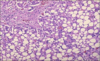
Triglycerides in liver of alcoholic
indicates reversible injury
What problem of liver is shown here? What type of endogenous substance might be accumulated here?

Cirrhosis of liver
May be due to hemosiderin accumulated in hereditary hemochromatosis
May cause irreversible injury
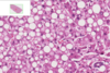
What disease is associated with the sphingomyelin accumulated in this image of liver? What mech?

Niemann-Pick disease
Inborn error due to failure in breakdown of sphingomyelin
What is the red arrow pointing to in this image of Niemann-Pick? What mech?

sphingomyelin accumulated in lysosome
Inborn storage disese
failure to breakdown
What are the arrows pointing to in the liver? Hint: accumulated substance because problem in folding protein

Alpha-1-antitrypsin
A protease inhibitor of neutrophil elastase
causes irreversible ER stress and thus cell death because abnormal a1-AT accumulates in ER
What is the blue line in this trichrome stain of an a1AT deficient patient’s liver? What other effect does this disease have on another organ in body?
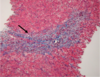
Fibrosis band from hepatic scarring
Due to hepatic injury from entrapment of alpha 1 antitrypsin in liver endoplasmic reticulum
pulmonary emphysema due to deficiency of a1AT in rest of body
What does this show that is common in smokers and city dwellers? What substance accumulated? What can be negative effect in large amounts?

This shows carbon duse [anthracotic pigment] accumulation in lung
Can cause pulmonary fibrosis in large amounts
What substance is accumulated in this hilar lymph node commonly found in smokers and city dwellers? What cell is it accumulated in?

Anthracotic pigment = carbon dust
Accumulated in macrophages
What disease does show in lungs? What substance accumulated? What do arrows point to?

Coal workers’ pneumoconiosis [clinically significant anthracosis]
Due to accumulation of carbon dust/anthracotic pigment
Left arrow = carbon dust
right arrows = scarring
Which is normal and which abnormal liver? What type of reversible cell injury? How can you tell?

Left is abnormal, right is normal
Hydropic change in hepatocytes
Indicated by swollen, clear cytoplasm
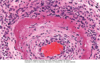
Which could have csuses this problem in aortic valve?

Dystrophic calcification
What injury are these arrows pointing to in this image of retinoblastoma?

Dystrophic calcification
What kind of calcification does this arrow point to in lung?
metastatic calcification
Which is normal? Which type of necrosis in heart? Etiology?

Right is normal
Left is coagulative necrosis
Due to irreversible ischemic injury
In heart after MI
What type of necrosis? What do arrows point to?

Myocardial coagulative necrosis
arrows point to degenerating neutrophils
What type of necrosis? Most common location and etiology?

Liquefactive necrosis
due to bacterial or fungal infection
most likely in lung or liver
What type of necrosis? Cause?

Fat necrosis
Due to release of digestive enzymes [lipase]
Occurs in pancreas
preserved architecture
What type of necrosis? Cause?
Fibrinoid necrosis
Often in blood vessels in response to immune-mediated or hypertensive injury
Preserved architecutre
get fibrin and neutrophils
What type of cell death? How can you tell?

Apoptosis
You can tell because of apoptotic bodies, lack of inflammatory cells, single + small cell
Describe the change in cardiac myocytes [right is normal]. What possible diagnosis?

large, hyperchromatic nuclei
hypertrophy of myocytes
likely left ventricular hypertrophy
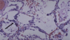
What is this cellular change in breast? Normal is right. Possible cause?

Hyperplasia [of glands]
Hormonal stimulation due to pregnancy and lactation