Intro to Anemia Flashcards
What is anemia?
- Anemia is decreased RBC mass. but we can’t measure that so we use hemoglobin concentration of blood as a surrogate
- Decreased O2 carring capacity of blood
- decreased O2 delivery to tissues (final physiologic consequence)

What are the 4 compensatory mechanisms of Anemia?
Increased RBC production
Increased 2,3-DPG (causes right shift)
Shunting of blood from non-vital to vital areas
increased cardic output
Increased pulmonary function
What 3 things cause right shift what 3 things cause left shift?
Right: increased temp, low DPG, low pH
Left: decreased temp, low DPG, high pH

A lot of the symptoms of anemai are caused by the compensatory mechanisms. What mechanisms lead to the following signs/symptoms:
weakness, malaise, easy fatigability
marrow expansion with potnetial bony abnormalities
pallor
tachycardia; cardiac ischemia in severe cases
dyspnea on exertion: increased pulmmonary function
weakness, malaise, easy fatigability: tissue hypoxia
marrow expansion with potnetial bony abnormalities: increased RBC production
pallor: shunting of blood from non-vital to vital areas
tachycardia; cardiac ischemia in severe cases: increased cardiac output
dyspnea on exertion: increased pulmonary function

Explain the disease process of anemia.
TRICK QUESTION!
Anemia is NOT a disease. It is a symptom of other diseases, and all anemias need to be explained
What are the 3 funcitonal classifications of anemai?
Blood loss
Decreased production
Accelerated destruction
What are the 3 morpholigc classifications of anemia?
Microcytic (MCV<80)
Normocytic (MCV=80-100)
Macrocytic (MCV>100)
What arer the two types of microcytic anemia? What are some things that can cause these!
- Normochromatic
- iron-deficiency-early
- thalassemis trait
- (anemia of chronic disease)
- some hemoglobinopathies eg Hemoglobin E
- Hypochromic
- iron deficiency
- thalassemis trait
- siderblastic anemia
- anemia of chronic disease
What are some things that case normochromic/normocytic anemia?
- anemia of chronic disease
- anemia renal failure
- marrow infiltration
- aplastic anemia
- blood loss***
- hemolysis**
What are some things that cause macrocytic anemia?
B12 and folate deficiency
liver disease
myelodysplastic syndromes
blood loss
hemolysis
some drugs
What are the 6 ways that we investigate anemia?
clinical history
physical exam
complete blood count (CBC)
Reticulocyte count
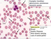
examination of peripheral blood smear
specific diagnostic tests (guided by above)
What things are shown on a CBC?
Hemoglobin concentration
Hematocrit
RBC count
Mean cellular corpuscular volume (MCV)
What does the measure of hemoglobin concentration tell us?
Hemoglobin in lysed sample reactedwith proprietary reagents
resulting complexes measured spectraphometrically
Most important parameter for assesment of O2 carrying capacity
(Hb; g/dL or g/L)
What is the most important parameter for assesment of O2 carrying capacity of blood?
Hemoglobin concentration!
What is the hematocrit?
(Hct; %)
- Packed cell colume (percentage of blood volume comprised by RBCs
- old method was centrifugation
- currentyl calculated as MCV* RBC
- usually 3X hemoglobin- does not add independent information in vast majority of cases (ie we do it for no reason)
What is Red blood cell count?
Direct measure of number of RBCs per unit volume
generally correlated well with Hb and hematocrit, adds little independent information
What is Mean cellular (corpuscular) volume (MCV; fL)
measured directly based on either electrical impedence or light scatter
very useful in the differential diagnosis of anemia (eg microcytic, normocytic, and macrocytic anemias)
What four things lead to a microcytic anemia?
iron deficiency
thalassemis
anemia of chronic disease
other (rare)
What 2 things lead to macrocytic anemia?
Megaloblastic
non-megaloblastic
What is megaloblastic anemia
imparied DNA synthesis
B12 and folate deficiency
some drugs
myelodysplastic syndromes
What is non megaloblastic anemia? What things cause it?
macrocytic anemia not caused by impaired DNA synthesis (Megaloblastic= impaired DNA synthesis)
reticulocytosis
liver disease
hypothyroidism
some drugs
What is mean corspuscular hemoglobin?
(MCH; pg)
calculated as Hb/RBC
measure of averae amoutn of hemoglobin per RBC
high correlation with MCV
What is mean corpuscular hemoglobin concentration?
(MCHC; g/dL)
measure of “chromicity” of RBCs
Calculated as Hb/(MCV*RBC)
decreased in hypochromic anemais
increased in a few “hyperchromic” states (eg. hereditary spherocytosis, hemoglobin CC disease)
What is Red cell distribution width?
(RDW)
Measure of variability of red cell volume
coefficient of variation of red cell volumes=standard deviation volume/MCV . Óvolume/MCV
useful for the separation of anisocytotic anemias (eg iron deficiency) from non-anisocytotic anemias (anemia of chronic disease)

What is poikilocytosis?
abnormal RBC shape
What poikilocytosis do these RBCs have? Describe their shape. What are 2 examples of things thaat cause this?

Spherocytes (spheres 3D)
round, small diameter, more densely staining, lack of central pallor
ex: hereditary spherocytosis, autoimmune hemolytic anemia

What poikilocytosis do these RBCs have? Describe their shape. What are 3 things taht cause this?

target cells
Ex: Liver dz, splenectomy, hemoglobinopathies

What poikilocytosis do these RBCs have? What are 4 things that cause this?

Elliptocytes (ovalocytes)
Ex: hereditary elliptocytosis, megaloblastic anemia, iron deficiency, myelofibrosis

What poikilocytosis do these RBCs have? What are 3 things that cause this?

Tear drop cells
megaloblastic anemia, myelofibrosis, extramedullary hematopoiesis

What is up with these RBCs?

sickle cells! from sickle cell disease
What poikilocytosis do these RBCs have? What are 3 things that cause this?

Fragments (schistocytes)
TTP, DIC, HUS, Malignant hypertension

What poikilocytosis do these RBCs have? What causes these?


What is anisocytosis?
red cell size variability- the the red cells aren’t the same size

What is polychromasia?
reticulocytes

What is in this picture?

Howell-Jolly Bodies (nuclear fragments)
ex: spelenectomy megaloblastic anemia

What is in this picture?

Pappenheimer bodies (iron granules)
ex: splenectomy, iron overload

What is in this picture?

Basophilic stippling (coarse)
thalassemias, MDS, lead poisoning
What is in this picture?

Hemoglobin C crystals
ex: Hb CC disease, Hb SC disease

What is the arangement of these RBCs called?

Rouleaux
decreased repulsive forced between RBCs
Ex: increased serum proteins (Ig, fibrinogen)

WHat RBC arrangement is shown in these pictures?

Agglutination
ex: IgM RBC antibodies (cold agglutinins)

What 8 things do we look at on a peripheral blood smear?
red cell shapes (poikilocytosis)
red cell variability (anisocytosis)
average red cell size (microcytosis, macrocytosis)
hemoglobinization (hypochromia, normochromia)
polychromasia (reticulocytes)
red cell inclusions
red cell arangements (Howell-Jolly Bodies, Pappenheimer bodies, Basophillic stippling)
White cell and platelet morphology
What hapens during acute anemia of blood loss?
initially no anemia by CBC parameters despite decrease in blooc volume
anemia develops as tissue fluid enters vascular space to restore blood volume, producing dilution of cellular elements
reticulocyte count increases after 2-3 days and peaks after 7-10 days
What happens during chronic anemia of blood loss?
no anemia initially because marrow is able to compensate
slight reticulocytosis
eventual development of iron deficicnecy with resultant iron deficiency anemia
Where are RBCs produced for an embryo? fetus (3 months after gestation until birth)? shortly after bibrth through adult life?
embryo: yolk sac
fetus: liver
shortly after birth through adult life: bone marrow
how is RBC production regulated?
decreased oxygen delivery induces production of erythropoietin by kidney
erythropoietin causes proliferation and differentiation of commmitted progenitor cells

How do normoblasts mature?

normoblasts (nucleated RBC precurors) obtain iron from plasma transferrrin for hemoglobin synthesis
up to 16 reticulocytes are produced from each pronormblast (earliest morphologically recognizabel erythroid precursor)
roughyl equal numbers of reticuloctes and normoblasts in marrow
As they mature they have more hemoglobin (more red) and less RNA (less blue)

Fill in the blanks


What are reticulocytes? What is in their cytoplasm? How long do they stay in marrow vs circulate?
earliest anucleate erythroid form
larger than mature RBCs
contain residual RNA which gives cytoplasm a blu tinge on routinely stained blood smears (polychromasia)
stay in marrow 1-2 days then circulate for ~1 day before losing residual ribosomes, mitochondria, and other organelles to becoe mature erythrocytes
WHat percentage of circulating RBCs are reticulocytes? How do we detect them? WHat is ti used as a measure of?
~1% of peripheral erythrocytes
can be detected using RNA stains to obtain a “reticulocyte count” which is expressed as % of total RBCs
used as a measure of RBC production-identifies anemias with decreased RBC production and those with adequate marrow response to blood loss or increased RBC destuction

WHat is the problem with a reticulocyte count? what is an average solution
what is a better solution?
prblem: reticuloycte % varies on total RBC count
solution: corrected reticulocyte percentage retic%*(patient HCT/45)
BETTER SOLUTION: Absolute reticulocyte count
What is the definition of ineffective erythropoiesis?
decreased red cell production despite increased RBC precursors in marrow
charcterized by defects in maturation
ex: iron deficiency (cytoplasmic maturation defect)m megaloblastic anemia (nuclear maturation defect), myelodysplastic disorders
WHat are 3 general features of ineffective erythropoiesis?
prominent morphologic abnormalities of erythrocytes due to disordered maturation
dysmaturation of erythroid precursors in marrow
decreased reticulocyte erythroid mass in marrow
What is the significance of decreased RBC precursors? WHat are 3 features of the progenitors
- proliferation defect
- characterized by an absolute decrease in the marrow mass of erythroid precursors
- decreased progenitors available or decreased proliferative capcaity of progenitors
- features
- normochromic/normocytic
- little anisopoikilocytosis (comapred to matration defects/ineffective erythropoiesis)
- decreased reticulocyte count
What are 2 possible results of stem cell defects with adequate erythropoietin? WHat are 3 congenital causes of this and 5 acquired causes?
red cell aplasia (pure) and pan-aplasia (aplastic anemia)
congenital: DIamons-Blackfan syndrome (pure red cell aplasia), Fanconi’s anemia (pan-aplasia)
acquired: idiopathic. drugs and toxins, autoimmune, infections, paraneoplastic
How do leukemias/lymphomas, metastatic carcinoma, fibrosis, storage disease impact the number of RBC precursors?
decrease RBC precuros! Bc they replace them in the bone marrow
How does anemia from renal failure impact number of RBC precursors?
decrease! Bc decrease Erythropoietin
How does anemia of chronic disease occur?
inflammatory block in erythropoiesis
mediated by IL-1, TNFa, IF gamma
probably multiple mechanisms
What is the size of a RBC? What is the volume? What does the special membrane structure provide?
7.5-9.7m in diamteter (can get through a 2.8m diameter capillary)
average volume of fluid is 90fl (can swell to 150fl)
special membrane structure which provides durability, flexibility and tensile strength. it springs back into shape after distortion (same)
How long do RBCs circulate? How does increased RBC destruction influence bone marrow production?
120 days!
Increased destrction results in increased marrow production (8X normal in ideal circumstances-enough iron, folate, good health)
What happens when rate of destruction exceeds bone marrow’s ability to compensate?
anemia develops, new steady state at a lower hemoglobin
you can have hemolysis wihtout anemia is bone marrow is able to compensate
What are the 3 kinds of hemolysis?
intravascular
extravascular
combination
What is extravascular hemolysis? When does it happen?
it predominates in most forms of hemolysis
final common pathway: decreased RBC deformability
- rigid, non-defomable cells have trouble traversing narrow slits between splenic cords and sinusoids
- cells are damaged further with prolnged exposure to splenic environment
- damaged cell phagocytized by cordal macrophages

What are the 7 general features of hemolysis?
reticulocytosis
increased indirect bili from heme metabolism
increased lDH released from destroyed RBCs
decreased hepatoglobin
morpholig abnormalities of red cells characteristic of specific disorder
splenomegaly in chronic cases
bony abnormalities in severe chronic hemolytic anemias