histology of skin Flashcards
19.07.30
what do the following characteristics of skin mean? explain why
- yellow
- blue-grey
- pale
- lack of pigment
- color of skin
- yellow= jaundice
- blue=cyanosis
- reflects a pathological condition of cardiovascular and or respiratory function.
- pale=anemia
- lack=albinism
- lack of enzyme tyrosinase
- involved in conversion of Y-> melanin
2.
- involved in conversion of Y-> melanin
- lack of enzyme tyrosinase
skin has seven unique functions.
- protection against physical, chemical and biological insult
- acts as a water barrier
- thermoregulation
- defense barrier to microorganism
- excretion of salts via sweat
- synthesis of Vit-D
- sensory organ
discuss the two types of skin and reference the differenciating features(junction).
What happens in the pronounced smoothing of the junction?

two classifications
- thick skin
- 5mm thick
- covers palms of the hands and soles of the feet
- has a thick epidermis and dermis
- thin skin
- 1-2mm thick
- covers everything, but palms of hands and soles of the feet
- each epidermal ridge corresponds to an underlying dermal papilla. tidges and papillae are permanent,
- finger print
- dermal papilla have a uniquepattern for each individual and thier impression creates a finger print
- dermal papillae are low in height and less in number
- finger print
dermal-epidermal junction
- primary epidermal ridge interlocks with a subsequent primary dermal ridge.
-
epidermal interpapillary peg
- projection downward from the primary dermal ridge, surrounded by smaller dermal pappilae.
- this arrangments is predominant in harless thick skin
pronounced smoothing of the epidermis-dermis junction
- results in decrease (50%) in dermal papilla
- less contact between dermis and epidermis

give 5 skin layers; reference to cell types and function of layer. note the 4 associated cell types; function and must know facts.
list the layers in thin vs thick skin
what disease affexcts the function of melanocytes?
what happens to the skin over aging process?
layers and features
- stratum basale
- cell types
- simple cuboidal - low columnar
- develop basement membrane
- melanocytes
-
merkel cells-mechanoreceptors
- sensitive to mechanical strain
- simple cuboidal - low columnar
- function
- mitotic capabilities
- cell types
- stratum spinosum
- cell types
- polyhedral cells
- Langerhans cells
- melanocytes
- function
- layers 1 +2 = stratum malpighi, responsible for cell renewal
- cell types
- stratum granulosum
- cell types
- squamous like cells
- function
- start granulation of keratohyaline
- cell types
- stratum lucidum
- cell types
- thin transparent membrane
- cells lack nuclei
- function
- cells contain eleidin, which is a tranfomation product of keratohyalin
- layer is absent in thin skin
- cell types
- stratum corneum
- cell types
- squames
- function
- squames are densly packed with keratin
- cell types
cell types
- keratinocytes
- most abundant of all the cell types.
- produce keratin= an intermediate filament protein
- melanocytes
- derived fron neural crest cells and responsible for melanin production.
- disease
- addisons-increased pigmentation
- albinism-hypopigmentation
- deplete with age
- skin becomes lighter and increase possibility of skin cancer
- langerhans cells
- act as APCs in skin
- merkel cells
- derived from neural crest cells
- involved in tacticle sensation
thin
- stratum basale
- stratum spinosum
- stratum corneum
thick
- stratum basale
- stratum spinosum
- stratum granulosum
- stratum lucidum
- stratum corneum

what type of skin is this, what is/isnt there?

thin skin
- lacks stratum granulosum
- the thickness of the other layers is also much less as compared to the thick skin
the dark pink layer is the epidermis, take note of the epidermal ridges
decribe the product in skin cells that causes them to get hard.
type, shape and monomers (location based)
Keratin
- structure
- an intermediate filament protein common to all epithelium
- alph helical, rod shpaed protein
- monomers
- K5 and K14
- found in basal keratinocytes
- K1 and K10
- replaces K5 and K14 in Stratum spinosum
- K5 and K14
- function
- give tthe skin its strength
- presents and absence are related with certain diseases
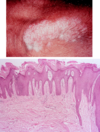
Jennifer is developing a scaly skin on her arms and legs. With respect to skin cell development, what is the normal vs disease state?

epidermal turnover rate
- 20-30 days = complete maturation
disease
- psoriasis
- increase # of proliferating cells
- decrease mitotic cell time
- develop
- increases epidermal thickness
- increase renewal rate (7 days)
- thick, scaly skin
photo on the bottom
- from left to right
- the epidermal ridges are normal in the last 1/4 on the right side, the ridge gets very deep.
- this is where the psoriasis starts

A patient spends 5 hours a day in the sun. They have done this for the past 45 years. what are they at risk for? name, location, stats

Malignant melanoma
- location
- melanocytes are found in the basal layer of epidermis
- disease
- when a melanocytes becomes malignant, the condition is called melanoma
- stats
- the most virulent of skin cancers, more tha 7k Americans die each yeart from melanoma
- closely linked with damaging exposure to solar radiation

byopsy of a skin graft found the following. What is this indicative of?
condtion, composition

squamous cell carcinoma
- condition
- dermal invasion by abnormal cells of the epidermis
- composition
- pleomorphism (structural changes in cell) of tumor cells
- keratinization within the cells results in abundantly pink cytoplasm, epithelial pearls

list the layers, physical structures and cellular structures

- dermis- CT deep to the epidermis with vessels, hair follicles, sweat glands, adn sensory receptors
- layers
- papillary layer
- superficial layer of loose irregular CT
- structures
- dermal ridge
- dermal papillae
- capillary loops
- meissner’s corpuscles
- free nerve endings
- associates with merkel cells
- reticular layer
- deeper layer of dense irregular ct
- structures-deep pressure
- pacinian corpuscle
- ruffini’s corpuscle
- papillary layer
- layers

Define the terms and give location, important structures and function.

besides supplying nourishment, the skin plays a major role in thermoregulation
- structures deep-superficial
- cutaneous plexus
- arteries and veins found at dermo-hypodermal junction
- AV shunts
- between cutaneous plexus and sub-papillary plexus.
- supplied by sympathetic nerves
- function
- restrict flow through superficial plexus to reduce heat loss as in extreme cold
- sub-papillary plexus
- in the papillary layer of dermis.
-
capillary loops extend into each dermal papilla and
- result in convective heat loss
- supply nutrients to epidermis
- cutaneous plexus

define the followin in the two layers.


Describe, locate and define the function of the 5 sensory receptors of the skin

- meisnener
- sensitive to touch
- NOT two point
- merkle cell
- responsible for two point discrimination
- free nerve ending
- pain
…rest is in the slide

Bill is in his third round of chemotherapy and has lost all of his hair.
Explain the normal anatomy/physiology of hair (unit), growth phases, and diseases associated.

Hair- a derivative of skin
- anatomy
-
pilosebaceous unit
- hair
- shaft of cornified cell and root within the hair follicle
- sebaceious gland
- hair
- muscle
-
arector pili
- smooth muscle attaches to the hair follicle and raises the muscle during
- innervated by sympathetic nervous system
-
arector pili
-
pilosebaceous unit
- growth phase
-
anagen-growth period, growing/always growing
- 80% of hair present in normal scalp are at this stage
-
catagen-breif period of ollicle regression or involution
- 3weeks
-
telogen-rest period or inactive phase
- 14% of hair
-
anagen-growth period, growing/always growing
- disease
- alopecia- baldness
- causes
- genetic/hormonal
- Cancer therapy
- therapeutic agents stop rapid cell growth. both WBC and cells associated with hair growth.
- how
- There is arrested mitotic activity in the hair matrix that dirupts both the function and the structural integrity of hair follicles and usually leads to rapid, reversible alpecia
- both of the above conditions are manageble
- causes
- alopecia- baldness
anatomy of sweat gland, what is it surrounded by and why?
eccrine sweat glands
- simple, coiled, tubular glands whose secretory units produce sweat which is delivered to the surface of the skin by long ducts
- secretory portions are surrounded by myoepithilial cells
-
function
- contractile cells (labeled My)
-
function


