Histo Pracs Sem 2 Flashcards
(36 cards)
For each feature does it relate to proximal or distal convoltued tuble of kidney?
- Smaller cells/more visible nuclei
- Brush Border
- Paler Cytoplasm
- larger well defined lumen
- More numberous cross section per field (longer)
- Distal
- Proximal
- Distal
- Distal
- Proximal
Proximal is long and brushy
Distal is larger lumen, visible nuclei and pale cytoplasm
5 functions of Mesagial cells and their matrix?
a: Participation in the tubuloglomerular feedback mechanism:
b: Structural support of glomerular capillary loops:
c: Secretion of vasoactive factors and cytokines:
d: Phagocytosis:
e: Contraction to control glomerular capillary blood flow:
In which glomerular region is the IgA deposited in a section of IgA nephropathy?
IgG deposits in membranous nephropathy?
IgA: within the mesangium
IgG: within the glomerulous basement membrane
What classes to the parafollicular or C cells of the thyroid belong?
epithelial? mesenchymal? neuroendocrine? glial?
Why?
Epithelial and Neuroendocrine.
C cells of the thyroid are secretory thus they are epithelial. They are endocrine as they secrete their hormone into the blood. They are particular type of endocrine cell, known as neuroendocrine (which have features of both secretory and neural cells), similar to the neuroendocrine (enteroendocrine) cells of the GIT.
What morphological type of epithelium normally lines the thyroid follicles?
simple cuboidal
What is the colour of the cut surface of the normal thyroid gland?
yellow
tan
white
deep red
pale brownish red
Pale brownish Red
(The red-brown colour of the gland is due to its iodine content and high vascularity. )
Clinical scenario: A 40-year-old woman presented to her general practitioner with several months of symptoms including intolerance to heat, increased appetite, weight loss, tremor, restlessness and feelings of anxiety with no apparent cause.
What general endocrine problem?
What is most common cause in Australia?
These symptoms suggest thyrotoxicosis, due to excessive amounts of circulating thyroid homone. Strictly speaking, hyperthyroidism refers to those cases caused by excess synthesis and secretion of hormones by the gland, however, there are other causes. The most common cause of both in western countries is Graves disease.
What type of epithelium lines the thyroid follicles in the patient’s thyroid?
Simple squamous
Simple cuboidal
Simple columnar
Stratified columnar
Simple Columnar
What features are demonstrated in the abnormal thyroid?
Chronic inflammation
Malignant change
Hyperplasia
Acute inflammation
Hypertrophy
Dysplasia
Atrophy
Metaplasia
Hypertrophy, Hyperplasia, Acute Inflam and Chronic Inflam
The follicular cells show hypertrophy (the cells are columnar rather than cuboidal, i.e. larger) and there are more of them than in the normal thyroid. There is much less colloid compared with a normal gland. Some scalloping of colloid at the periphery of follicles is noted. There are also patchy aggregates of lymphocytes and plasma cells i.e. chronic inflammation. Note also the vascularity of the gland.
Macrocopic Appearance of Thyroid in Graves Disease?
n Graves disease the gland will typically be diffusely enlarged (rather than nodular) and deep red/brown in appearance, the latter due to its vascularity.
Clinical scenario: A 40-year-old woman presented to her general practitioner having noticed a mass at the front of her neck and a reduction in the frequency of her periods. The GP took a history and found that she had also been suffering from increasing tiredness, intolerance to cold weather and weight gain. Her mother was on tablets for a thyroid problem. The neck mass was symmetrically enlarged and moved on swallowing.
What general endocrine problem do these clinical features suggest?
What is the most common underlying causative disease in Australia?
- Hypothyroidism
- Hashimotos
The clinical features suggest hypothyroidism. In Australia (where dietary iodine levels are generally sufficient) the most common cause is Hashimoto disease (autoimmune thyroiditis).
Describe the microscopic abnormalities seen in Hashimotos Thyroid Disease:
The thyroid reveals a marked reduction in follicles and colloid. There is a florid chronic inflammatory cell infiltrate with many l_ymphocytes and plasma cells._ Many germinal centres are present. Residual follicular epithelial cells exhibit granular eosinophilic cytoplasm (Hürthle cells).
Comment:
Note that the presence of germinal centres indicates the development of a humoral mediated immune response. Germinal centres are the regions where B lymphocytes commence their differentiation into plasma cells. The plasma cells produce antibodies that are involved in the disease process. Cell mediated immunity (via CD8+ T cells) may also be involved in damaging the thyroid epithelium.
The altered follicular epithelial cells (with eosinophilic granular cytoplasm) in Hashimoto disease are known as ‘Hürthle cells’. Their nuclei may appear quite enlarged and pleomorphic but they are not dysplastic.
How is the damage in Hashimoto’s Disease mediated?
Would the patient’s serum TSH level be expected to be high, low or ‘normal’ (within the reference interval)?
Cell and antibody mediated damage
High
Damage in Hashimoto disease is probably both cell and antibody mediated.
Serum TSH would be expected to be high.
Macroscopic features of early Hashimotos Thyroid Disease?
Diffusely enlarged
Atrophied
Paler than normal
Redder than normal
Nodular
Paler then Normal
and Diffusely Enlarged.
In Hashimoto’s disease the gland will typically be diffusely (rather than nodular) enlarged and paler than normal. Focal enlargement or nodularity may occur in a minority of cases.
Why does a Hashimoto’s Disease thyroid look paler then normal?
The lack of colloid and replacement of follicles by inflammatory cells will cause the gland to appear pale.
N.B.
The enlargement (due to presence of inflammatory cells) in Hashimoto disease is generally diffuse but it can be slightly nodular and even asymmetric. The typical case does not show much fibrosis, though ultimately if untreated, in many cases the gland will become fibrotic and atrophied.
Pathological Features of Multinodular Goitre on high power?
The main pathological features:
- On low power the gland looks nodular and there is large variation in the size of the follicles. The nodules are separated by bands of fibrous tissue
- Areas of haemosiderin-laden macrophages are present indicating previous haemorrhage

Which one of the following is believed to be the underlying cause of these pathologic changes in regions where iodine deficiency is not endemic e.g. Australia?
Stimulation of the TSH receptor by autoantbodies
Dietary iodine deficiency
Neoplastic dysregulaton of cell proliferation
Cell and antibody mediated destruction of thyroid
Dietary goitrogens and subclinical abnormalities in thyroid hormone production
Dietary goitrogens and subclinical abnormalities in thyroid hormone production
Macroscopic Features of multinodular goitre
Asymmetrical enlargement of the gland
Cystic or haemorrhagic
Nodular
Potential causes of a solitary thyroid nodule?
How do you diagnose?
Dominant nodule in a multinodular gland, a cyst, an adenoma, a carcinoma (various types) and focal thyroiditis.
Fine needle aspirate, ultrasound.
Patient diagnosed with diabetes mellitus.
What is the clinical term given to the patient’s symptom of pain in the calves on walking that settles on resting?
What is the likely pathologic cause (name the underlying disease process) of the patient’s pain on walking more than 50m and absent dorsalis pedis pulses?
Claudication
Atherosclerosis
Factors contributing to development and persistance of foot ulcer in diabetes
Atherosclerosis
Chronic ischaemia
Infection
Distal symmetric polyneuropathy
Impaired phagocyte activity
Recurrent trauma
Poor wound healing
What is the thickening of the basement membrane in small vessels causing the changes seen in the diabetic patient’s retina called?
Diabetic microangiopathy and hyaline arteriolosclerosis make the vessels leaky and also weakened, being susceptible to occlusion, exudates and aneurysmal change. The hyaline arteriolosclerosis can occur independently to hypertension in diabetes, and the mechanism of its formation in diabetes is different from that in hypertension.
In Diabetic Glomerulosclerosis what is this?

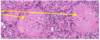
Proximal Convoluted Tubules
Have a lumen and are lined by simple cubiodal epithelium. Cells have abudnant eosinohphilc cytoplasm. These tubules are not atrophic, indicating that not all nephrons are affected to the same degree.
In Diabetic Glomerulosclerosis what is this?

Distal convoluted tubules.
These structures have a wide lumen and are lined by simple cuboidal epithelium. Cells are smaller then those linig the tubules indcated by black arrows and thats have more densely packed nuclei.