HISTO: Lung pathology Flashcards

1
Q

List 4 common airway diseases.

A

(1) Asthma
(2) COPD – Chronic Bronchitis
(3) Bronchiectasis
(4) Infections

2
Q

Define asthma. List some causes.

A

Widespread reversible narrowing of the airways that changes in severity over short periods of time.

Causes:

  • Allergens/atopy
  • Pollution
  • Drugs (NSAIDs)
  • Occupational (gases)
  • Diet
  • Physical exertion
  • Intrinsic
  • Genetics
3
Q

Describe the pathophysiology of asthma.

A
4
Q

Whata are the macroscopic features of asthma?

A
5
Q

What are the histological features of asthma?

A
  • Hyperaemia
  • Eosinophilic inflammation and goblet cell hyperplasia - mucus
  • Hypertrophic constricted muscle
  • Mucus plugging and inflammation
6
Q

Define COPD/chronic bronchitis. What are the causes?

A

COPD is chronic injury to airways elicits local inflammation and reactive changes

Definition = chronic cough productive of sputum; most days for ≥3 months over ≥2 consecutive years

Causes = smoking, air pollution, occupational exposures

7
Q

What are the histopathological features of CODP?

A
8
Q

What are the complications of CODP/chronic bronchitis?

A
9
Q

Define bronchiectasis. What are the most common causes?

A

Bronchiectasis – permanent abnormal dilatation of the terminal bronchi

Causes:

  1. Congenital (CF, ciliary dyskinesia [i.e. Kartagener’s syndrome])
  2. Inflammatory (post-infectious, obstruction, 2nd to bronchiolar disease and interstitial fibrosis, asthma)
10
Q

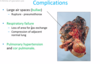
What are the complications of bronchiectasis?

A

RHF - cor pulmonale

11
Q

How common is CF?

A

1 in 2,500 but 1 in 20 are carriers (heterozygous carriers as it is recessive)

12
Q

What is the aetiology of CF?

A

Chr 7q3 = CFTR gene (ion transporter protein)–> defective Cl- ion transfer so less water transfer to secretions

Affects all organs (from abnormally thick secretions):

13
Q

What are the complications of CF? What are the most common infections?

A
  • Lung –> obstruction, respiratory failure, recurrent infection**, **bronchiectasis (90%), etc.
    • S. aureus H. influenzae
    • P. aeruginosa B. cepacia
  • GI tract - meconium ileus, malabsorption
  • Pancreas - pancreatitis, 2nd malabsorption
  • Liver - cirrhosis
  • Male infertility - blockage of sperm ducts
14
Q

What is the management of CF? What is the prognosis?

A

Imrpoved treatment with PT, antibiotics, enzyme supplements, parenteral nutrition

Survival to 4th decade

Increased with lung transplantation

15
Q

List 5 common parenchymal diseases.

A

(1) Pulmonary oedema and diffuse alveolar damage (include acute resp. distress syndrome and HMD)
(2) Infections
(3) COPD – emphysema
(4) Granulomatous diseases
(5) Fibrosing interstitial lung disease and occupational lung disease

16
Q

Define pulmonary oedema. What are the causes?

A

= fluid accumulation in alveolar spaces

  • Causes – LHF, alveolar injury, neurogenic, high altitude
  • Pathology:
    • Intra-alveolar fluid on histology (left)
    • “Heart failure cells” = iron-laden macrophages (right)
17
Q

What is seen on histopathology of pulmonary oedema?

A
  • Intra-alveolar fluid on histology (left)
  • “Heart failure cells” = iron-laden macrophages (right)
18
Q

Define alveolar damage. What is the pathogenesis?

A

Pattern of acute diffuse lung injury in which patient present with rapid onset respiratory failure, requiring ventilation on ITU. CXR shown “white out” of all lung fields.

Pathogenesis: Acute damage to endothelium ± alveolar epithelium leading to an exudative inflammatory reaction

19
Q

What are the causes of alveolar damage such as ARS and hyaline membrane disease?

A
  • Adults = acute respiratory distress syndrome / ARDS
    • Infection
    • Aspiration
    • Trauma
    • Inhaled irritant
    • Shock
    • Blood transfusion
    • DIC
    • Drug overdose
    • Pancreatitis
  • Neonates = hyaline membrane disease of the newborn / RDS (HMD of newborn = RDS)
    • Insufficient surfactant
    • Premature babies
20
Q

What is the prognosis of diffuse alveolar damage?

A
21
Q

What is the gross pathology of diffuse alveolar damage (DAD)?

A
  • Fluffy white infiltrates in all lung fields
  • Lungs expanded/firm
  • Plum-coloured lungs, airless
  • >1kg mass
22
Q

What is the micropathology of DAD?

A
    1. Capillary congestion
    1. Exudative phase
    1. Hyaline membranes
    1. Organising phase
23
Q

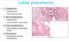
What are the patterns of baterial pneumonia?

A
  • Bronchopneumonia
  • Lobar pneumonia - aggressive phase, not seen as much
  • Abscess formation
  • Granulomatous inflammation
24
Q

Describe the difference between bronchopneumonia and lobar pneumonia.

A
25
Who is usually affected by **_broncho_**pneumonia? What organisms are implicated?
Elderly Low virulence organisms such as staph, H influenzae, strep and pneumococcus
26
What is the pathology of bronchopneumonia?
Patchy bronchial and peribronchial distribution, lower lobes, inflammation surrounding the airways themselves and is within the alveoli
27
What is the micro pathology of bronchopneumonia?
28
What is the pathology of lobar penumonia? What organism is the most likely culprit?
Affects entire lobe Infrequently due to antibiotics 90-95% due to pneumococci (i.e. strep)
29
What is the micropathology/staging of lobar pneumonia?
30
What are the complications of lung infection?
31
What are the atypical causes of pneumonia? What is the pathology?
* Mycoplasma, viruses (CMV if immunosuppressed, influenza), Coxiella, chlamydia, etc. * *Interstitial inflammation **without** accumulation of intra-alveolar inflammatory cells and chronic inflammatory cells within alveolar septa with oedema ± viral infections*
32
Define emphysema. What are the causes?
33
What is the pathogenesis of emphysema?
Alpha 1 antitrypsin usually protects but there is less in smokers or those with deficiencies.
34
What is the gross histopathology of emphysema in smoking/a1 deficiency?
35
What are the complications of emphysema?
36
Define granulomatous disease/granuloma. What are the causes?
Granuloma = collection of histiocytes, macrophages ± giant multinucleate cells Causes: * infection, * sarcoidosis, * foreign body aspiration / IVDU, * drugs, * occupational **TB must first be excluded before looking for other causes of granulomatous lung disease**
37
What parts of the body does sarcoid most commonly affect? What is the aetiology?
* **Affects**: lungs, skin, lymph nodes and eyes * **Pathogenesis**: abnormal host immunological response to variety of commonly encountered antigens, probably environmental in origin
38
What are the micro and macroscopic features of sarcoid? How is it diagnosed?
* **MICRO/MACRO -** discrete epithelioid and giant cell granulomas, usually in upper zones of lung with a tendency to peri-lymphatic and peri-bronchial; in advanced disease becomes **fibrocystic** * **DIAGNOSIS -** non-caseating granuloma, elevated serum ACE, _hypercalcaemia (1a-hydroxylase)_
39
Define idiopathic pulmonary fibrosis and give examples. What is it also known as? Describe the micro/macropathology.
40
List some causes of fibrosing interstitial/occupational lung disease.
1. Idiopathic pulmonary fibrosis/cryptogenic fibrosing alveolitis 2. Extrinsic allergic alveolitis/"Farmer's" lung (better over weekend away from work) 3. Industrial lung disease/"Pneumoconiosis" (asbestosis, silicosis, coal-miner's lung)
41
What is the prognosis of idiopathic pulmonary fibrosis? How is it diagnosed?
Over 50% die in 2-3 years High resolution CT and biopsy diagnosis
42
What are the macro/microscopit features of idiopathic pulmonary fibrosis?
* Macro = basal and peripheral fibrosis and cyst formation * Micro = interstitial fibrosis (varying stages)
43
What is seen on histology in asbestosis?
* Fine sub-pleural basal fibrosis with **_asbestos bodies in tissue_** * May also see pleural disease e.g. fibrosis, pleural plaques
44
List 3 common pulmonary vascular disease.
* (1) **Pulmonary hypertension** * (2) Pulmonary thromboembolism * (3) Pulmonary vasculitis
45
What are the causes of pulmonary hypertension? What is the definition in mm/Hg?
46
What are the morphological feaures of pulmonary HTN?
* Morphological changes: * Eccentric intimal fibrosis * Thickening muscle wall
47
What is the most common site for clot formation in PE?
DVT is the cause in 95%
48
What are the symptoms of PE in small emboli? What are the consequences?
49
What are the symptoms of PE in large emboli? What are the complications?
50
Give 5 examples of non-thrombotic emboli which can cause PE.
* Bone marrow * Amniotic fluid * Trophoblast * Tumour * Foreign body * Air
51
Give some examples of pulmonary vasculitis.
Small-medium vessel disease e.g. EGPA Leukocytoclastic vasculitis involving capillaries e.g. RhA
52
What is the epidemiology of lung cancer?
* Incidence in men dropping * Incidence in women rising (women took longer to quit smoking)
53
What sites can lung tumours arise in?
* Airways (_SCC_) * Peripheral alveolar spaces (_Adenocarcinoma_) * _SCLC_ can arise either centrally or peripherally * _Mesothelioma_ is a tumour of the pleura
54
Give examples of benign and malignant lung tumours. What are the most common types in smokers and non-smokers?
* **Benign Tumours –** *no metastasis, local complications (i.e. obstruction)* * Chondroma * **Malignant Tumours** – *metastasise, most commonly epithelial (90-95%):* * Squamous cell carcinoma (30%) = smokers, central, spread locally, late metastasis, *PTHrP* * Adenocarcinoma (30%) = non-smokers, peripheral, early metastasis * Large cell carcinoma (20%) * Small Cell Lung Carcinoma (20%)= smokers, central, SIADH, ACTH, Lambert-Eaton Myasthenic Syndrome (LEMS)
55
What % of lung cancer in smokers is due to passive smoking? What substances in smoke are carcinogenic?
25% of lung cancer in non-smokers is attributed to passive smoking Smoke contains: * Tumour initiators (polycyclic aromatic hydrocarbons) * Tumour promoters (nicotine) * Complete carcinogens (nickel, arsenic)
56
What % of lung cancer is in non-smokers? What are the risk factors?
10-20% * Asbestos * Radiation (radon exposure) * Air pollution * Heavy metals * Genetics (familial lung cancers are rare) * Susceptibility genes: * Chemical modification of carcinogens * Susceptibility to chromosomal damage * Nicotine addiction
57
What are the steps leading to development of carcinoma?
* Metaplasia --\> Dysplasia --\> Carcinoma in situ --\> Invasive carcinoma * Due to an accumulation of gene mutations
58
Describe the progression of the phenotype of SCC.
Squamous cells do not have cilia so mucus production can build up and carcinogens may be trapped within this and accumulate.
59
What % of lung cancer consists of SCC? What is its pattern of distribution? What hormone may be produced?
~30% of all lung cancers Central around bronchial epithelium, spreads locally with late metastasis. But there is increasing incidence of peripheral SCC PTHrP
60
Describe the macro/microscopic features of invasive adenocarcinoma of the lung.
* Gland formation * Papillae formation * Mucin * Histology shows evidence of glandular differentiation * Affect terminal airways with a multi-centric pattern (many tumours at different stages of differentiation)
61
Which molecular pathways are implicated in the development of adenocarcinoma?
In smokers- K-ras issues with DNA methylation and p53 In non-smokers - EGFR mutations (important to identify as they are a drug target)
62
Which populations is invasive adnocarcioma more common in?
* Incidence increasing * Far east * Females * Non-smokers
63
Is metastasis common in adenocarcinoma?
Yes, extrathoracic metastases common and early
64
Describe the progression of atypical adenomatous hyperplasia (AAH) of the lung.
Precursor lesion --\> proliferation of atypical cells lining the alveolar wall --\> increase in size and eventually becomes invasive 1. AAH 2. Non-mucinous adenocarcinoma in-situ 3. Mixed pattern invasive adenocarcinoma
65
Describe the macro/microscopic features of large cell carcinoma.
Peripheral or central No histological evidence of glandular/squamous cells BUT ON EM may show evidence of glandular, squamous or neuroendocrine differentiation i.e. poorly differentiated Composed of large cells
66
What % of lung cancer is made up by large cell carcinoma? What is the prognosis?
10% Poor prognosis
67
What % of lung cancer is made up by small cell carcinoma? What are risk factors for this?
20% Smoking is closely associated
68
Where are small cell lung carcinomas usually located? Describe their micro/macroscopic features.
Central near the bronchi Small poorly differentiated cells
69
Why is prognosis in small cell lung cancer poor? What are the complications?
80% present with advanced disease/ chemosensitive but come back very quickly Complications: * Can cause paraneoplastic syndromes * Necrotic core because they quickly outgrow their blood supply
70
Which mutations are commonly found in small cell carcinoma?
p53 and RB1
71
Compare the prognosis of small cell and non-small cell lung carcinomas.
Small cell: * 2-4 month survival untreated, 10-20 months on treatment usually by chemotherapy (surgery uncommon as usually spread by time of diagnosis) Non-small cell: * More chemosensitive and early stage 1 tumours have 60% 5-year survval BUT late stage 4 have 5% 5-year survival
72
What are the molecular targets for treating adenocarcinoma and SCC? What is the consequence of treating SCC as adenocarcinoma?
**Adenocarcinoma** molecular targets: * **EGFR mutation** (responder or resistance) - tyrosine kinase therapy * **ALK translocation** (responds to Crizotinib) * **Ros1 translocation** * **PD-L1 expression** **Squamous cell carcinoma targets:** * **PDL1 expression** ​If SCC is treated with biologics used for adenocarcinoma e.g. bebevacizumab, it can cause fatal haemorrhage.
73
What types of cytology and histology are used in lung cancer diagnosis?
* Cytology – looking at cells: * Sputum * Bronchial washings and brushings * Pleural fluid * Endoscopic fine needle aspiration of tumour/enlarged lymph nodes * Histology – looking at tissue: * Biopsy at bronchoscopy – central tumours * Percutaneous CT guided biopsy – peripheral tumours * Mediastinoscopy and lymph node biopsy – for staging * Open biopsy at time of surgery if lesion not accessible otherwise - frozen section * Resection specimen - confirm excision and staging
74
What are the complications of high levels of PD1/PDL1 expression in SCC?
High levels of PD1/PDL1 can inhibit the immune response
75
What is mesothelioma and what is the most common cause?
Malignant tumour of the PLEURA Associated with exposire to asbestos - there is a long lag between exposure and disease development
76
What % of cancer deaths are made up by mesothelioma? Who is it more common in? What is the prognosis?
\<1% of cancer deaths but peaked around 2010-2020 M:F ratio 3:1 (50-70years) Essentially fatal
77
What are the endocrine/non-endocrine features of paraneoplastic syndrome?
Endocrine: * (1) Antidiuretic hormone (ADH) --\> hyponatremia (SCC) * (2) Adrenocorticotropic hormone (ACTH) --\> Cushing’s syndrome (SCC) * (3) Parathyroid hormone-related peptide --\> hypercalcaemia (SCC) * (4) Other: * Calcitonin --\> hypocalcaemia * Gonadotropins --\> gynecomastia * Serotonin --\> “carcinoid syndrome” (especially carcinoid tumours; rarely SCC) Non-endocrine: * Haematological/coagulation defects, skin, muscular, miscellaenous