Health Assessment Review Flashcards
Physical Assessment Components
(Inspection, Palpation, Percussion, Auscultation)
-
Inspection:
- Use of sight to assess for size, color, share & symmetry.
-
Palpation:
- Use of touch to assess for temperature, turgor, texture, moisture, vibrations, shape, size. Use dorsal surface of hand to assess temperature. Assess most tender areas last.
-
Percussion:
- Tapping a person’s skin to assess for location, size, density of tissues
-
Auscultation:
- Listening with a stethoscope to assess pitch, loudness, quality & duration of body sounds.
Vital Signs: Pulse
What to assess?
How to take radial/apical pulse?
What is a pulse deficit?
Assess: Rate, rhythm (regular or irregular), equality (right vs. left side), and strength (0-4+)
Radial: Palpate using the index, middle & 4th finger on the wrist closest to the thumb.
Apical: Auscultate using the diaphragm of the stethoscope at the fifth ICS at left MCL
Pulse deficit = apical pulse - (minus) radial pulse; need two people to perform; one takes the apical pulse for a full minute; the other person takes the radial pulse for a full minute at the exact same time; the numbers should be the SAME; an actual pulse deficit is NOT an expected finding; it means that not every heart beat/ventricular contraction is not getting to the periphery.
Note: For an irregular pulse, count for a full minute.
Vital Signs: Blood Pressure
Key Points
- BP cuff sizing: The BP cuff width should be 40% of arm circumference. Bladder should surround 80% of arm circumference. If it is too large = false low reading. If it is too small = false high reading.
- Do not take BP on an arm with a patient who has a PICC line, a mastectomy or an AV fistula. Prefer not to take the BP on a side with an IV line if possible.
Pulse
Expected Rate, Rhythm & Strength
-
Rate:
- Adults - 60-100 bpm
- Bradycardia (<60)
- Tachycardia (>100)
- Adults - 60-100 bpm
-
Rhythm:
- Regular
-
Strength:
- 0 = absent
- 1+ = diminished
- 2+ = normal
- 3+ = strong
- 4+ = bounding/unable to obliterate w/ pressure
Orthostatic Hypotension
When & how to assess?
When to assess?
- When ordered by provider &/or when patient reports syncope, certain medications (ex., antihypertensive meds) or if hypovolemic.
How to assess:
- Have patient lie in supine 5-10 minutes; take BP & pulse in supine.
- Sit patient up, 2-3 minutes; take BP & pulse
- Have patient stand up, wait 2 minutes; take BP & pulse
- Monitor for dizziness, weakness, fatigue.
Pain Assessment
Components of Pain Assessment
- Location of pain?
- Quality of pain (patient’s word):
- Nociceptive pain (described as “aching” or “throbbing”)
- Neuropathic pain (described as “shooting” or “burning”)
- Intensity of pain (pain scale)
- Timing (onset, duration, frequency)
- Accompanying symptoms (e.g., nausea)
- What relieves pain?
- What makes pain worse?
Eye assessment: What to assess?
Conjunctive pink? Sclera white?
PERRLA (pupils are equal, round, reactive to light & accommodation)
- Check for accommodation by asking the patient to focus on a distant object (pupil dilates), then to a near object (pupil constricts).
How to check for consensual response?
- Shine a penlight into the patient’s right eye.
- Expected finding: Both the left “and” the right eye constrict.
S/S of Hypoxia are…
Apprehension, restlessness (often an early sign ), inability to concentrate, decreased level of
consciousness, dizziness, behavioral changes, fatigue, and agitation.
Cyanosis, blue discoloration of the skin and mucous membranes caused by the presence of desaturated hemoglobin in capillaries, is a late sign of hypoxia.
Respiratory:
Expected Rate
Pulse Oximetry
-
Rate (Adults):
- 12-20 breaths/minute
- Bradypnea (RR < 12)
- Tachypnea (RR > 20)
-
SpO2: 95-100%
- Low 90s is expected for COPD patients.
Abnormal Lung Sounds
-
Crackles (fine):
- High-pitched crackling/popping sound; take several strands of hair (if your hair is long enough :)) and rub them back & forth right at your ear. That is what fine crackles sound like. Indicates are moving through collapsed alveoli.
-
Crackles (coarse):
- Low-pitched bubbling/popping sound. Indicates air moving through collapsed alveoli.
-
Wheezes:
- High-pitched, muscial, squeaky; indicates air moving through narrowed passageways
-
Rhonchi:
- Low-pitched, continuous, snoring-like sounds; indicates air moving through fluid-field airways
-
Pleural friction rub:
- Loud, grating, scratching sounds; indicates inflamed pleura
- Absent breath sounds
Anatomy of Lungs
(Lobes & Apex vs Base)
-
Lobes:
- 3 on right lung
- 2 on left lung
-
Apex of lung:
- At top
-
Base of lung:
- At bottom (Note: the heart is opposite - apex at bottom & base at top of heart)

AP to Transverse Diameter
- Normal AP diameter is 1:2
- 2:2 diameter called a barrel chest; sometimes seen with patients who have chronic COPD or other obstructive pulmonary diseases

Posterior Chest Auscultation
Auscultate all lung sounds using the diaphragm (see picture; using the “S” pattern; are bilateral breath sounds equal & clear?)

Neck Vessels
Inspection, Palpation, Auscultation
-
Inspection:
- Assess for jugular venous distention (JVD):
- Position patient at 30-45 degree angle & inspect the jugular with penlight.
- Note visible pulsations or signs of JVD.
- Assess for jugular venous distention (JVD):
-
Palpation:
- Palpate each carotid artery (one at a time!).
- 4+ (0-4+ range) = “unable to obliterate with pressure; which is expected finding
-
Auscultation:
- Use bell to listen for bruits (swishing sound, not expected finding).
- Ask patient to inhale, exhale & hold breath while you listen.
- Use bell to listen for bruits (swishing sound, not expected finding).
Where are all the palpable pulses locations in the body?

Heart Auscultation
-
Auscultation:
- Auscultate with both bell & diaphragm
- Identify rate/rhythm, S1 & S2 sounds; listen for extra hear sounds, murmurs at 5 locations (APETM):
- Aortic valve: 2nd ICS, RSB
- Pulmonic valve: 2nd ICS, LSB
- Erb’s Point: 3rd ICS, LSB
- Tricuspid valve: 4th ICS, LSB
- Mitral valve (point of maximal impulse/PMI): 5th ICS, left MCL.
- Normal Heart Sounds:
S1 & S2 Heart Sounds
- S1 heard best at 5th ICS, LSB (PMI)
- S2 heard best at 2nd ICS, RSB

Heart: Auscultation Sites
“All People Enjoy Times Magazine”

- *When inspecting/palpating/auscultating the apical impulse, the nurse knows to assess at this
landmark: **
Apical pulse occupies the 5th intercostal space, at the left midclavicular line. (Varies slightly with individuals.)
Name the Structures on This Picture

A. Aortic area
B. Pulmonic area
C. Erb’s point
D. Tricuspid area
E. Mitral area
- 1st ICS
- 2nd ICS
- 3rd ICS
- 4th ICS
- 5th ICS
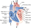
Where is the base/apex of the heart?
Where is the base/apex of the lungs?
The apex of the heart (see picture) is at the bottom of the heart. That is where the PMI (point of maximal impulse) is located. The base of the heart (see picture) is at the top of the heart.
The apex of the lungs (not denoted in picture) is actually at the top of the lungs; and the base of the lungs is located at the bottom of the lungs.

Anatomy & Blood Flow of the Heart
- Know blood flow (oxygenated & deoxygenated blood), chambers & valves
Musculoskeletal System
Inspection/Palpation, Spinal Curvatures, ROM, Muscle Strength
-
Spinal Curvatures:
- Scoliosis, kyphosis, lordosis
-
ROM of all joints:
- Specify
-
Muscle Strength:
- 0-5 range; strength should be equal bilaterally
Range of Motion of All Joints
- Flexion
- Extension
- Hyperextension (neck, waist, wrist)
- External rotation (hip, shoulder)
- Internal rotation (hip, shoulder)
- Abduction
- Adduction
- Circumduction (shoulder, hip, ankle)
- Supination
- Pronation
- Inversion
- Eversion
- Dorsiflexion & planter flexion (ankle)


