Head and neck Flashcards

1
Q

anatomy of the eye

A
2
Q

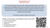
ways to differentiate causes of acute red eye

A
  • Vision- normal or reduced vision?
  • Painful or painless
  • Normal intraocular pressure or raised intraocular pressure
3
Q

painful acute red eye

(vission N= normal , R = reduced)

A
  • conjunctivitis (N)
  • sceleritis (N/R)
  • keratitis (R)
  • corneal foreign body (N/R)
  • epsicleritis (N)
4
Q

painless acute red eye

(vission N= normal , R = reduced)

A

subconjunctival haemorrhage (N)

5
Q

reduced vision and red eye with normal intraocular pressure

A

corneal abrasion

keratitis

6
Q

reduced vision and red eye with increased intraocular pressure

A

acute angle closure glaucoma

anterior uveitis

7
Q

what is the likely diagnosis

A

orbital cellulitis

8
Q

investigations required for orbital cellulitis

A
  • FBC
    • Raised WBC and CRP
  • Blood cultures
  • CT with contrast
    • Would show inflammation of orbital tissue deep to the septum
9
Q

management of orbital cellulitis

A
  • Abx
  • Admit for IV antibiotics
10
Q

how do we differentiate orbital cellulitis from periorbital cellulitis

A
  • Periorbital cellulitis- confined to tissues superficial to the orbital septum (and tarsal plates)
    • Secondary to infection e.g. bug bite
  • Orbital cellulitis- results of an infection affecting the fat and muscle posterior to the orbital septum, within the orbit but not involving the globe
    • Vision and movement affected
11
Q

what is the likely diagnosis

A

acute closed angle glaucome

12
Q

acute closed angle glaucome management

A
  • Drugs to reduce pressure
  • Surgical- use laser to make hole to allow flow of aqueous humour to the iris
  • opthalmological emergency
13
Q

outline how acute angle closure gluacoma occurs

A
  • Drugs to reduce pressure
  • Surgical- use laser to make hole to allow flow of aqueous humour to the iris
14
Q

likely diagnosis

A

Right oculomotor nerve palsy

  • Ptosis- LPS
  • Dilation of pupil- parasympathetic fibres lie on periphery
    • In diabetes usually not dilated (can receive blood supply from smaller blood vessels)
    • But with compression- pressure on parasympathetic fibres and the oculomotor nerve - dilation
  • Down and out
15
Q

likely diagnosis

A

Horner’s syndrome

  • Sympathetic fibres traveling up from the thoracic region
    • Superior tarsal muscle
    • Sweat glands
16
Q

likely diagnosis

A

right 4th nerve palsy (trochlear)

  • Superior oblique muscle affected
17
Q

label these eye muscles

A
18
Q

clinical testing of the extraocular eye muscle and their innervations

A

H in space for muscle isolation

19
Q

most of the extraocular eye muscles are innervated by

A

CN3- oculomotor

  • inferior oblique
  • superior rectus
  • inferior rectus
  • medial rectus
20
Q

which extraocular eye muscles are not innervated by the oculomotor nerve

A

Lateral rectus

Superior oblique

21
Q

how to remember Lateral rectus and Superior oblique

A

LR6 SO4

all other CN III

22
Q

lateral rectus innervated by the

A

CN6- abducens nerve

23
Q

supeiror oblique innervated by

A

CN4- trochlear

24
Q
A
25
muscle action of lateral rectus
abduction
26
muscle action of medial rectus
adduction
27
muscle action of superior rectus
elevation minor muscle actions : intorsion and adduction
28
muscle action of inferior rectus
depression minor muscle actions - extorsion - adduction
29
muscle action of inferior oblique
extorsion minor muscle action - elevation - abduction
30
muscle action of supeirior oblique
intorsion minor muscle movement - depression - abduction
31
triangles of the neck
32
midline neck structures
* thyroid * submental lymph nodes * hyoid bone
33
Conditions could lead to neck lump in the midline
* A cyst * Dermoid * Thyroglossal * Goitre * Hyper/hypothyroidisms * Thyroid malignancy
34
structures in the anterior triangle
* Anterior border of the SCM * Infrahyoid muscles * Lymph nodes * Anterior cervical chain * Submandibular glands * Carotid sheath * IJV * Carotid artery * Vagus nerve
35
**Conditions could lead to neck lump in the anterior triangle?**
* reactive lymphadenopathy * malignant lymphadenopathy * branchial cyst * carotid artery aneurysm * carotid body tumours * submandibular gland pathology
36
**structures in the posterior triangle**
* Trunk of the brachial plexus * Subclavian artery and veins * Pancoast tumour * Scalene , omohyoid inferior belly * Phrenic nerve * Lymph nodes
37
**Conditions could lead to neck lump in the posterior triangle?**
* Apical lung tumour * reactive lymphphadenopathy * malignant lymphphadenopathy
38
39
lumps which can occur anywhere in the neck
* lipoma * sebaceous cyst * haemangiomas * skin malignancy
40
**Assessment of the lump**
* Movement * Mobile/immobile * On swallow * On sticking out tongue * Texture * Hard or soft * Regular/irregular
41
lymph nodes feel
rubbery
42
hot lymph nodes
inflammatory
43
tender lymph node
not cancer
44
**Swallow and stick out tongue**
* Any lump related to thyroid will move on swallowing * If the lump moves when tongue being stuck out- thyroglossal
45
why does the patient have enlarged lymph nodes
infection lymphoma
46
questions to narrow down lymph nodes
**Onset** * Acute- days * Chronic- week- months **Associated symptoms** * Infection * Fever * Localising symptoms such as ear pain, cough or sore throat * Inflammation * Arthritis * Malignancy * Ref flag symptoms for all possible sites * May be a primary, local or distant metastasis
47
red flag symptoms
* age * weight loss * blood malignancy * symptoms such as night sweats and tiredness * local metastasis * persistant hoarse voice * difficulty swallowing * persisting sore throat * distant metastasis e.g. abdominal primary
48
investigations of lymph nodes
blood tests US refer
49
Describe what you see
* Hutchinson’s sign * **Hutchinson's sign** is the presence of vesicular lesions on the side or tip of the nose. It is classically associated with acute herpes zoster ophthalmicus, which may cause loss of vision, ocular inflammation, and debilitating pain. * **Nasociliary nevre (branch of V1)** * **Infection extends to the tip of the nose- sight threatening** * Unilateral * Va lesion- ophthalmic
50
what other questions do you want to ask?
* Onset/duration * Sensation * Pain * Immunocompromised * Contact with anyone with the same condition
51
likely diagnosis
* Varicella zoster infection causing ophthalmic shingles * Lies dormant in the trigeminal ganglion * Ophthalmic emergency- sight threatening
52
key symptoms
* Tinnitus- vestibulocochlear * Facial tingling * Facial nerve involvement * Progressive worsening
53
differentials
* Vestibulocochlear lesion * Schwannoma * Acoustic neuroma (vestibular schwannoma)
54
Acoustic neuroma (vestibular schwannoma)
* Compression of other structures * Facial nerve * Trigeminal nerve * Cerebellum * Very slow growing tumour
55
positive rinnes test
AC\>BC Normal response or sensineural hearing loss on tested side
56
negative rinnes test
BC \>AC conductive hearing loss on tested side
57
what does Rinnes test compare
compares BC to AC at each ear
58
what does webers test compare
compared BC bilaterally
59
normal webers test
sound heard in both ears equally
60
how will sound lateralise in sensinoural hearing loss in a Webers test
sounds lateralises to unaffected side
61
ow will sound laterlase with conductive hearing loss in webeers test
lateralised to affected side
62
* Vid shows tongue pointing to the right-likely diagnosis?
* Right hypoglossal nerve palsy - the tongue never lies
63
64
**C- MIDDLE EAR (grey part on diagram)** Explanation: * Right sided facial droop * Juice falling out of the mouth- orbicularis muscle weak * Crying- greater petrosal intact * Chorda tympani not functioning * Sensitivity to loud noises- nerve to stapedius not working **Think about this** ![]() * If you have a lesion before the internal acoustic meatus- all functions affected * Once we get to the geniculate ganglion- first branch of facial nerve leavesà (1)greater petrosal nerve- parasympathetics to a number of glands * 2 more intrapetrous branch * Nerve to stapedius (2) * Chordae tympani – special **sensory taste** and parasympathetic (3) * Once leaving stylomastoid foramen- motor to muscles of facial expression
65
A B C D
66
**Facial nerve lesion where the lesion is related to the Geniculate ganglion**
**- Ramsay hunt syndrome** * Look for vesicular rash on the ear
67
inflammation of the supraglottis (including epiglottis
epiglottitis - rare but airway threatening - 2-6 years - stridor, drooling, unwell, sniffling position - H . infuenzae but also strep pneumoniae
68
examples of conditions which would caused impaired actions of vocal cords
1. inflammation of cords (laryngitis), nodule cancer 2. paralysis of muscles moving vocal cords
69
paralysis of muscles moving vocal cords
* injury to external branch of superir laryngeal enrve * laryngospasm * laryngeal odema
70
inflammation of the larynx, trachea and bronchi
* croup * parainfluenza virus * 6 months - 3 years * seal like bark (sometimes stirdor, increased resp efffort)
71
innervation of the pharynnx
maxillary division of the trigeminal nerve (V)- sensory
72
**innervation of the Oropharynx** *
* Sit behind oral cavity * Includes posterior third of tongue * Gloss**o**pharyngeal (IX)- sensory and special sensory taste, some motor contribution (stylopharyngeus) * Glosso- tongue * O- oropharynx * Pharangeal-pharynx
73
innervation of the laryngopharynx and laryx
* Vagus (CN X) * Motor * Laryngopharynx * Larynx * Sensory * Laryngopharynx * Most important for swallowing
74
**Glossopharyngeal also innervates**
* **Middle ear and ET** * **Carotid sinus and bodies (carotid massage)** * **Parasympathetic to the parotid gland**
75
**Vagus also innervates**
* **So**ft palette * External ear * Parasympathetic
76
* B vagus (Q tip shouldn’t be able to get in to the middle ear)
77
both CNIX and CNX have some sensory function relating to the
ear, but different parts * IX- eustachian tube and middle ear * X- external ear (part) and external suface TM (part)
78
how do we clinically examine CN IX and X
* Speech * Swallow * Cough * Gag reflex * Uvula deviation
79