Hand Picture Cards Flashcards
Name the structure. Note: This is a palmar view

Palmar radiocarpal ligament

Dorsal radiocarpal ligament

Flexor retinaculum aka transverse carpal ligament

Flexor retinaculum aka transverse carpal ligament

Median nerve

Deep motor branch of the ulnar nerve

Ulnar nerve proximal to superficial and deep branches

Palmar aponeurosis
- Longitudinal fibers
- Transverse fibers

Flexor digitorum superficialis tendons


Flexor digitorum profundus tendons


Flexor digitorum superficialis

Flexor digitorum profundus

- Flexor digitorum profundus
- Flexor digitorum superficialis

Flexor pollicis longus

Common synovial sheath of the flexors

Extensor digiti minimi

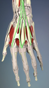
Extensor digitorum


- Extensor pollicis longus
- Extensor pollicis brevis

Name the artery.
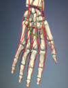
Superficial palmar arch
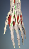
Extensor indices

Name the artery.

Deep palmar arch
Name the arteries

Common palmar digital arteries
On which joint do these lateral bands of extensor muscles act to extend?

The DISTAL interphalangeal joint. That way they can move ventrally within the finger during flexion so as to not stretch so much.

Name the nerves.
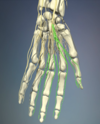
Proper palmar digital branches of the median nerve
Name the muscles, action, and innervation.

Dorsal interossei (1st, 2nd, 3rd, 4th)
Abduct the fingers (DAB). Ulnar innervation.
All are bipennate.
Name each muscle, which eminence they belong to, their function, and innervation.

- Abductor digiti minimi - acts to abduct the little finger.
- Flexor digiti minimi brevis - acts to flex the little finger at the MP joint.
These two are both innervated by the deep motor branch of the ulnar nerve and are part of the hypothenar eminence.
- Flexor pollicis brevis - acts to flex the thumb at both the MP joint and CMC joint.
- Abductor pollicis brevis - acts to abduct the thumb.
These are innervated by the recurrent branch of the median nerve and are part of the thenar eminence.
Name the muscle, which eminence it belongs to, innervation and action.

Opponens pollicis. Thenar eminence. Innervated by the recurrent branch of the median nerve. Acts to oppose the thumb (which includes flexion, abduction, and medial rotation).
Name the muscles, action, and innervation.

Palmar interossei (1st, 2nd, and 3rd)
Adduct the fingers (PAD). Ulnar innervation.
All are unipennate.
Name the muscle, innervation, action.

Adductor pollicis. Innervated by the deep motor branch of the ulnar nerve. Action is to adduct the thumb CMC joint. Note that this is NOT a thenar muscle.
Name the muscles, action, and innervation.
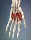
Lumbrical muscles.
Flex the fingers at the MP joint.
1 & 2 = median nerve, unipennate
3 & 4 = ulnar nerve, bipennate
Name the muscle, action, innervation, and which eminence it belongs to.

Opponens digiti minimi. Innervated by the deep branch of the ulnar nerve. Action is to flex and rotate the little finger CMC joint. Hypothenar eminence.


