Group 2, Column 2 Flashcards
(150 cards)
Name the clinical finding/diagnosis.
- What is the typical symptom?
- How does this heal?

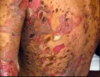
Atrophie blanche seen in livedoid vasculopathy.
- Burning pain along ankle prior to ulceration
- Painful ulcerations on lower legs +/- surrounding livedo reticularis
- Heal with atrophic hypopigmented scars
What systemic disorders are classically associated with livedoid vasculopathy?
- Hypercoagulable disorders (e.g., Factor V Leiden, protein C deficiency, prothrombin mutations, hyperhomocysteinemia)
- Autoimmune conditions (SLE, scleroderma, APLS)
- Atherosclerosis and stasis
What are the early and late histopathology findings of livedoid vasculopathy and atrophie blanche?
- What happens to the vessels?
- What happens to the epidermis?
- Segmental hyalinization (“pink-red crayon”) and thrombosis of small vessels in the upper and mid dermis
- Late stage with epidermal atophy and hyalinized vessels

What are the treatment options for livedoid vasculopathy?
- First line?
- In recurrent/recalcitrant cases?
- Aspirin
- Pentoxyfilline
- Dipyridamole
- In recurrent or recalcitrant cases:
- Anticoagulants (warfarin, heparin, etc.)
- Oral steroids
What labs should be obtained in suspected livedoid vasculopathy?
- What is part of the coagulopathy work up?
-
Coagulopathy workup
- Cryoglobulins
- Homocysteine
- Protein C and S
- Anti-thrombin III
- ANA
- Anti-cardiolipin antibody
- Factor V mutation
- Prothrombin mutation
Name the diagnosis.
- What do early lesions look like? Late lesions?
- How is hair and sweat affected?
- What indicates persistent disease activity with respect to lesion appearance?

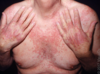
Plaque morphea (localized scleroderma)
- Begin as erythematous to violaceous patches on trunk and proximal extremities
- Evolve to indurated hyperpigmented or ivory plaques
- Often hairless and anhidrotic
- Violaceous border indicates persistent disease activity
What is the most common subtype of morphea (localized scleroderma) in adults and in children?
- Plaque in adults
- Linear in children
What is the suspected pathogenesis of morphea (localized scleroderma)?
- What cytokines are involved?
- What happens to vessels and the dermis?
- Genetic predisposition + environmental trigger
- Vascular injury leads to inflammation
- Triggers profibrotic Th2 cytokines (IL-4, IL-6 and TGF-beta)
- Fibroblast proliferation and collagen deposition
What are classic triggers of morphea (localized scleroderma)?
- What types of infections?
- What are general iatrogenic causes?
- Infections with Borrelia spp. (in Europe and Japan mainly; Borrelia afzelii and Borellia garinii)
- Radiation
- Medications
- Trauma
Name the diagnosis.
- What do early lesions look like? Late lesions?
- How is hair and sweat affected?
- What indicates persistent disease activity with respect to lesion appearance?

Plaque morphea (localized scleroderma)
- Begin as erythematous to violaceous patches on trunk and proximal extremities
- Evolve to indurated hyperpigmented or ivory plaques
- Often hairless and anhidrotic
- Violaceous border indicates persistent disease activity
Name the diagnosis.
- in which population is this the most common subtype?
- What autoantibodies is most common in this condition?
- What are possible complications of this condition?

Linear morphea (localized scleroderma)
- Associated with significant morbidity (especially in kids, in whom it is the most common subtype)
- Look for linear distribution often along Blaschko’s lines
- Legs (#1 site) > arms > head/trunk
- Anti-ssDNA autoantibodies most common
- Undergrowth of limbs, joint restriction/contractures
Name the diagnosis.

En coup de sabre
- Indentation of frontal, frontoparietal or parasagittal forehead or scalp
Name the diagnosis.
- What is the other term for this condition?
- What are other associations with this condition?

Parry-Romberg syndome (a.k.a. progressive hemifacial atrophy)
- Unilateral atrophy of face involving dermis, subcutaneous tissue, muscle and bone
- Can be associated with epilepsy, headache, exophthalmos, cerebral atrophy, alopecia
Name the diagnosis.
- What is the buzzword for this condition?
- What does the dermis look like on histology?

Atrophoderma of Pasini and Pierini
- Brownish-gray hyperpigmented oval, atrophic, well-demarcated plaques with sharp sloping borders (“cliff drop” edges)
- Begins as persistent single lesion with additional lesions over time
- Histology: significantly decreased dermal thickness compared to normal skin (biopsy at lesion edge)
Name the diagnosis.

Necrobiosis lipoidica
- Yellow to red-brown atrophic to indurated plaques typically over pretibial areas; prominent telangiectasis, +/- ulceration
Name the diagnosis.

Necrobiosis lipoidica
- Yellow to red-brown atrophic to indurated plaques typically over pretibial areas; prominent telangiectasis, +/- ulceration
How can you tell the difference between morphea and systemic sclerosis on rheumatologic serologies?
- All forms of morphea lack anti-Scl70 (Topo I) and anti-centromere antibodies (in contrast to systemic sclerosis)
What is the suspected pathogenesis of necrobiosis lipoidica?
- What happens to the vessels and dermis?
- What kind of inflammation occurs?
- Vascular compromise from immunodeposition in vessel walls or diabetes-related microangiopathic changes
- Subacute dermal ischemia leads to dermal collagen degeneration
- Secondary granulomatous inflammatory response
What are the key histopathology findings of necrobiosis lipoidica?
- What is the special “sign” on biopsy?
- What is the inflammatory pattern and how is it arranged in the dermis?
- How does its histology compare with granuloma annulare?
- What immune cells are abundant? What stromal component is absent?
- “Square biopsy” sign
- Horizontally arranged (“layered”) palisaded granulomatous inflammation with horizontal tiers of degenerated collagen fibers and dermal sclerosis
- Process diffusely involves entire dermis (vs. GA which is mainly superficial)
- Lacks mucin
- Plasma cells and multinucleated giant cells are abundant

What are the treatments for necrobiosis lipoidica?
- What are topical and systemic treatments?
- What does not affect disease course?
- Potent topical steroids and/or ILK (into inflammatory rim), TCIs
- Systemic treatments include PO steroids, colchicine, cyclosporine, TNF-alpha inhibitors and pentoxifylline for chronic/recalcitrant cases
- Note: none of these is borne out in the literature
- Glycemic control does not affect disease course
What is the inheritance pattern and genetic mutation of NF-1?
- Autosomal dominant inheritance (but can occur as a sporadic mutation)
- Mutation of neurofibromin (NF1), a tumor suppressor gene that downregulates Ras activation
What is another name for NF1?
von Recklinghausen’s disease
What is the diagnostic criteria for NF1?
Hint: think of the mnemonic!
“CA NN OT FAI L2 B 1ST”
Must have >2 of the following:
- CA: CAfe-au-lait (six lesions)
- NN: Neurofibromas
- OT: OpTic glioma
- FAI: Freckles (Axillary, Inguinal)
- L2: Lisch nodules x 2
- B: Bone (sphenoid or tibial wing dysplasia)
- 1st: (affected 1st degree relative)
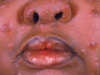
Name the clinical finding and disease association.

Lisch nodules found in NF1