Group 1, Column 2 Flashcards
(141 cards)
What are treatments for molluscum contagiosum?
- What are topical and oral treatments?

- Cryotherapy
- Cantharidin
- High-dose oral cimetidine
- Candida antigen immunotherapy
- Topical retinoids
- Imiquimod
What are the path findings of molluscum contagiosum?
Henderson-Patterson molluscum bodies (intracytoplasmic inclusion bodies)

What is the expected onset after drug initiation of a morbilliform drug eruption?
7-14 days
Name some of the most common culprit drugs of morbilliform drug eruptions.
- Beta-lactams (PNCs and CSNs)
- TMP/SMX
- Anticonvulsants
- Allopurinol
- Classically occurs within 7-14 days
Name the diagnosis.

Morbiliform drug eruption
What are some notable triggers/mutations that lead to the development of common acquired melanocytic nevi?
- UV exposure
- BRAF mutations (found in up to 80%; more common than NRAS mutations)
- Immunosuppression
How are congenital melanocytic nevi divided by size?
- < 1.5 cm = small
- 1.5 cm - 20 cm = medium
- > 20 cm = large
Note: > 40 cm by adulthood has recently been termed “giant”
What does FAMMM Syndrome stand for and what are the key features?
- Inheritance type
- Clinical features, family history
- Genetic mutation
Familial Atypical Multiple Mole Melanoma Syndrome
- AD inheritance
- Characterised by:
- 50+ melanocytic nevi
- Family history of melanoma
- CDKN2A gene (encodes p16 and p14)
How should large congenital nevi be treated?
- After what age?
- When should you screen the patient with an MRI - brain and for what potentially fatal condition?
- Surgical resection should be attempted if possible after 6 months of age
- If not possible, perform serial examinations with early biopsies of nodular areas
- If large posterior axial congenital nevi or multiple satellites, then obtain an MRI to screen for neurocutaneous melanosis, a potentially fatal condition
What are the classic histological features of dysplastic nevi?
- What is seen at the edges?
- How are the junctional nests arranged?
- What does the cytologic atypia look like?
- Asymmetry
- Junctional “shoulder” (extends > 3 rete ridges beyond dermal component)
- Irregular size and placement of junctional nests with bridging or lentiginous pattern
- Papillary dermal concentric and/or lamellar fibrosis
- Cytologic atypia: nuclei enlarged, “dirty grey” cytoplasm
What should you consider if an elderly patient has a new “atypical/dysplastic nevus” of a sun-damaged site?
It is most likely well-nested lentigo maligna!
Name the diagnosis.

Nummular dermatitis
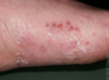
Name the diagnosis.
- What is the most common cause of this in adults and children, respectively?

White superficial onychomycosis, due to:
T. mentagrophytes (adults) or T. rubrum (children)
What is the classic histology of a dermatophyte infection, including tinea or onychomycosis?
- What does the corneum and dermis look like?

- Septate hyphae in stratum corneum or nail plate
- May have brisk dermal infiltrate (versus minimal in tinea versicolor)
- +/- neutrophilic microabscesses in epidermis or corneum/nail plate

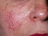
Name the diagnosis.
- What part of the face is usually spared?
- What is usually the associated symptom?

Perioral dermatitis
- Look for clusters of small, pink discrete scaly papules/pustules in perioral region with clear zone around vermilion border
- Can also involve nasolabial folds and cheeks
- Burning sensation, minimal itching
What is perioral/periorificial dermatitis most commonly attributed to?
- Topical fluorinated corticosteroids
- Facial cosmetics
What is the treatment of perioral/periorificial dermatitis?
- What are the oral medications used for adults and kids, respectively?
- What topical medications can be used?
- What should be avoided?
- Tetracyclines (or erythromycin in pediatrics) for 6-8 weeks with gradual tapering
- TCIs, topical metronidazole and other antibacterials
- Avoid cosmetics, steroids and other irritants
What parts of the face can perioral/periorificial dermatitis involve?
- Basically any part of face
- Variants include periorbital and periorificial (perioral + periorbital)
What are the notable causes of pityriasis rosea?
- What infections?
- What medications?
- HHV-6 and -7
- Drugs like ACE inhibitors (most common), gold, beta-blockers, NSAIDs and isotretinoin
- Note that ACE inhibitors, beta-blockers and gold salts are also triggers of drug-induced LP.
Name the diagnosis.

Pityriasis rosea
- Papulosquamous eruption
- Herald patch followed 1-2 weeks later with patches and plaques with trailing scale in “Christmas tree” pattern
What is the treatment for pityriasis rosea?
- What oral medication may hasten clearance?
- Not necessarily required
- Symptomatic treatment with topical steroids, antipruritics
- Oral erythromycin may hasten clearance
What is the notable histopathology of pityriasis rosea?
- What changes are seen in the corneum? In the epidermis?
- What may be present in the dermal papillae?
- Where is the infiltrate and what composes it?
- Non-adherent thin mounts of parakeratosis (wound be thicker in guttate psoriasis)
- Spongiosis
- RBC extravastion
- Perivascular lymphohistiocytic infiltrate

What are the treatments of prurigo nodularis?
- Oral and topical medications
- SSRIs/TCAs for underlying psychiatric conditions
- Doxepin
- Cryotherapy, ILK, TCS/TCI
- Methotrexate and cyclosporine have even been used!
Name notable triggers of psoriasis.
- Namely, what infection, electrolyte disturbance and drugs?
- Koebner phenomenon
- Infections (streptococcal pharyngitis #1)
- Hypocalcemia (pustular psoriasis)
- Pregnancy (impetigo herpetiformis)
- Drugs (lithium, beta-blockers, TNF-alpha inhibitors, steroid tapers, excess imiquimod use)
- TNF-alpha inhibitors may cause plaque or palmopustular psoriasis