GI Histology Flashcards
What are the features of connective tissue?
Few Cells
Lots of space
Lots of fibers

What are the 4 basic types of tissue?
Epithelial
Connective
Muscle
Nervous
What is the meaning of a simple epithelium?
One layer
What does epithelium always sit on?
Conective tissue
How do you tell if a gland is submucosal?
Will lie below the muscularis mucosae
Where are peyers patches found?
In the mucosa - they are often stained a purple colour
What is the epithelium in the oesophagus?
•Stratified squamous non-keratinised epithelium
What are theglands in the oesophagus that stain very poorly?
Mucus glands
These are located in the submucosa
What is the muscle in the oesophagus?
•Muscular layer – upper 1/3 skeletal, middle 1/3 – smooth+ skeletal, lower 1/3 - smooth
What is the epithelium in the stomach?
Simple columnar epithelium
What is the innermost layer of the muscularis externa in the stomach?
Oblique muscle layer
What colour do parietal cells stain?
Pink
What is the structure of the crypts of Lieberkuhn?
Lined with epithelium containing many types of cells:
Enterocytes (absorb water and electrolytes)
Goblet cells (secrete mucus)
Enteroendocrine cells (secrete hormones)
Stem cells
Paneth cells (secrete antimicrobial peptides)
Cup cells
Tuft cells
Where are Brunner’s glands located?
Submucosa of the duodenum
Where are peyers patches most prevalent?
In the Ileum
What is the abundance of villi and crypts in the large intestine?
No villi and lots of crypts
What is the structure of an acinar gland in the GI?
Roundish structure with columnar cells and nuclei at the base
Ducts
What is not found in the Islets of langerhans in the pancreas?
Acini

What are numbers 2 and 3 in the picture the pancreas?

2 - Exocrine part
3 - Pancreatic duct for exocrine part

What is the tissue seen here
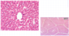
What is the tissue?
What are the characterising features?

Duodenum
Villi with a leaflike shape (wide)
Mucus secreting brunner’s glands int he submucosa
Crypts of lieberkuhn
What is the tissue type?
What are the characteristic features?

Villi have finger like shape (Longer)
Well developed plicae circulares
Crypts of Lieberkuhn
No glands in the submucosa
What is this tissue?
What are the characteristic features?

Villi are shorter when compared with the jejunum
Peyers patches extend through the lamina propria and the submucosa
Shorter villi
