Funtional anatomy and histology of the upper GI tract Flashcards
Identify the three types of lingual papilla

A: filiform papillae. elongated projections of connective tissue covered with highly keratinised stratified equamous epithelium. No taste buds
B: fungiform papillae. mushroom shaped projections. They project above the filiform papilla, visible as small spots to the naked eye. Tastebuds present in the stratified squamous epithelium. More near the tip of the tongue.
C: vallate papillae. large dome shaped structures in the mucosa anterior to the sulcus terminalis. 8-12 in the tongue. Surrounded by invagination lines with stratified squamous epithelium that contains numerous taste buds
Describe the structures highlighted.

Taste buds.
Consists of three principal cell types:
Neuroepithelial (sensory) cells contain taste receptors
Supporting cells
Basal cells (stem cells)
Blue arrow: taste pore, small opening onto the epithelial surface
How can you distinguish between mucus and serous salivary glands histologically?

A. Mucus acini: tubular cells, light staining
B. Serous acini: spherical cells, darker staining
Identify the salivary gland

Parotid gland
Has completely serous acini
Numerous narrow intercalated ducts
Nuclei dark staining and basally located
Identify the salivary gland

Sublingual galnd
Mainly mucus glands
large collecting duct
Low numbers of serous acini
Identify the salivary gland

Submandibular gland
Mixed gland, mostly serous
Less extensive intercalated ducts
Describe the basic structure of the alimentary canal

A: Mucosa, consisting of lining epithelium, underlying connective tissue (lamina propria), muscularis mucosa (smooth muscle)
B. Submucosa, consists of dense irregular connective tissue
C. Muscularis externa, consists of an inner layer of circular muscle and an outer layer of longitudinal muscle
D. Serosa, serous membrane layer of sinple squamous epithelium. Continuous with mesentery in intraperitoneal organs.
Label the floor of the oral cavity


State 3 functions of the oral cavity
- it is the inlet for digestive system involved with the initial processing of food, which is aided by secretions from salivary glands;
- it manipulates sounds produced by the larynx and one outcome of this is speech;
- it can be used for breathing because it opens into the pharynx, which is a common pathway for food and air.
Main sensory nerve that innervates the oral cavity
Trigeminal nerve (and branches)
 Cranial nerve V
Role of maxillary nerve [V2]
Innervtes upper parts of the cavity, including the palate, upper teeth, upper lip and cheek
Role of mandibular nerve [V3]
Sensory innervation: lower part or oral cavity - teeth and oral part of the tongue, lower lip and cheek
Motor innervation: muscles of mastication
Label the muscles of the tongue


Innervation of the tongue
Sensory:
Glossopharyngeal nerve [CN IX] - innervates posterior 1/3 of the tongue (pharyngeal part) and carries sensory information about taste and general sensation.
Lingual nerve - branch of mandibular nerve [CN V3] general sensation from the oral part of the tongue,mucosa on the floor of the oral cavity and gingiva associated with the lower teeth. Also carries parasympathetic and taste fibers from the oral part of the tongue that are part of the facial nerve
Facial nerve [CN VII] - carries sensory information about taste from the oral part of the tongue to the CNS
Motor:
Most muscles of the tongue are innervated by the hypoglossal nerve [CN XII] except for the palatoglossus which is innervated by the vagus nerve [CN X]
Sense of taste
Glossopharyngeal nerve [CN IX]
Lingual nerve [CN V3 branch]
Facial nerve [CN VII]
State the function of the highlighted structures

Buccinator: Forms wall of the oral cavity (cheek). Holds cheeks against the alveolar arches and keeps food between the teeth when chewing, involved in facial expression.
Motor innervation [CN VII], Sensory innervation [CN V3]
Orbicular oris: Lies within lips around oropharynx. Enables shape of lips to change, keeps mouth clsoed during swallowing.
Hyoid bone: connects floor of the oral canvity with pharynx and larynx. Involved in swallowing.
Hyoglossus: extrinsic muscle of the tongue. Depresses the tongue
Mylohyoid: provides structural support to the oral cavity, depresses mandible and opens mouth, elevates hyoid bone during swallowing,
Muscles of mastication (4)
Masseter: elevates mandible
Temporalis: Elevation and retraction of mandible
Medial pterygoid: Elevation and lateral movements of mandible
Lateral pterygoid: Protrusion and lateral movenents of mandible
Also…
Orbicularis oris: narrows mouth and closes lips
Buccinator: Contraction of the buccinator presses the cheek against the teeth. This keeps the cheek taut and aids in mastication by preventing food from accumulating between the teeth and the cheek.

Functions of saliva (4 points)
Solvent - taste and absorption
Lubrication - mastication, blous formation, articulation
Digestion - starch triglycerides
Protection - rinse, anti-abrasion, buffer, controls bacterial flora
What are the 8 components of saliva?
Ions - Na, K, Ca, F, Mg, PO4, HCO3
Mucin
Digestive enzymes
Antibacterial agents (lysozyme, lactoferrin)
Immunoglobulins
Phosphoproteins
Blood group factors
Identify the labelled structures


Function of the palate
Hard palate: bony plate covered by mucosa. Separates the nasal cavity from the oral cavity.
Soft palate: continues posteriorly from the hard palate and acts as a valve that can be depressed to help close the oropharyngeal isthmus or elevated to separate the nasopharynx from the oropharynx.
What are the roles of levator palati and tensor palati muscles?
Levator palati: elevates the soft palate above its neutral position
Tensor palati: tenses the soft palate, opens pharyngeotympanic tube (middle ear) during yawning and swallowing
Describe the skeletal structure of the palate

Palatine processes from the maxilla make up the anterior 2/3 of the hard palate
L-shape palatine bones also contribute to the roof of the oral cavity.
Sphenoid and temporal bones provide attachments for muscles of the hard and soft palate.
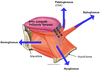
Role of pharyngeal constrictor muscles
Narrow or constrict the pharyngeal cavity. When the constrictor muscles contract sequentially during swallowing they move the bolus of food through the pharynx and into the oesophagus


