fetal monitoring/search methods Flashcards

1
Q

EFM

A

electronic fetal monitoring

2
Q

Why is continuous EFM controversial?

A
  • It makes the laboring mother more sedentary. Not optimal for giving birth.
  • It removes healthcare workers from the presence of mom, giving them a less complete picture of the overall condition of mom and baby.
  • Provides less intensive care for laboring mothers and babies.
3
Q

What is the best position for an electronic fetal monitor?

A

on the baby’s back

4
Q

tocodynamometer

A
  • device for monitoring and recording uterine contractions during labor
  • has button on back
5
Q

toco

A

contraction

6
Q

ctx

A

contraction

7
Q

tocolytic drug

A

drug that stops ctx

8
Q

fetal monitor

A
  • smooth on the back
  • measures FHR
9
Q

FHR

A

fetal heart rate

10
Q

vibroacoustic stimulator

A
  • device that uses sound waves and vibration to stimulate baby in utero
  • used to elicit FHR accel and ensure fetal well-being
11
Q

maternal-fetal unit

A

mom + uterus

placenta

cord + fetus

12
Q

uteroplacental junction

A
  • place where the placenta joins the uterus
  • forms chambers where maternal blood collects
  • nutrients and gases are exchanged through membrane of placental blood vessels lying in those chambers
  • uterus and placenta are interwoven, almost like a zipper
13
Q

umbilical cord

A

three blood vessels that carry fetal blood into and out of the uteroplacental junction for nutrient and gas exchange

14
Q

AVA

A
  • artery vein artery
  • the three vessels that should be found in the umbilical cord
15
Q

two-vessel umbilical cord

A
  • can be indicative of genetic anomaly
  • requires further investigation
16
Q

What happens to the placenta and fetus during a normal ctx?

A
  • placenta is squeezed, leaving less room for blood, and reducing gas and nutrient exchange
    ctx → placenta compressed → less blood to cord → less blood to fetus
  • baby’s head is squeezed, sometimes eliciting a vagal response and lowering FHR (early decel)
17
Q

What does a variable decel indicate?

A

cord compression

18
Q

What does a late decel indicate?

A

uteroplacental insufficiency

19
Q

What does an early decel mean?

A

baby’s head is squeezed during contraction, eliciting a vagal response and lowering FHR

20
Q

What factors affect fetal oxygenation?

A
  • normal maternal blood flow to placenta (volume)
  • normal maternal SpO2
  • functional placenta
  • functional umbilical cord
  • normal fetal circulation
21
Q

interruptions to fetal SpO2

A
  • changes in mom’s circulation
    • drop in BP
    • vena cava syndrome
    • drop in maternal O2
  • uterine activity
  • placental problems
  • umbilical cord problems
  • abnormal fetal conditions
22
Q

What causes drops in maternal O2?

A
  • epidural: drop in BP; IV fluid bolus prophylactic
  • vena cava syndrome: compression when supine; reposition mom
  • smokers, lung conditions, anaphylaxis
23
Q

What can cause placental insufficiency?

A
  • small placenta
  • blood clot in placenta
  • part of placenta that doesn’t function
24
Q

nuchal cord

A
  • umbilical cord wrapped around baby’s neck
  • doesn’t strangle baby
  • compresses cord, ↓ O2 delivery
25
Q

umbilical cord problems

A
  • nuchal: around neck
  • body: around body
  • true knot: actual knot in cord
26
Q

EFM pattern components

A
  • rate
  • variability
  • accels
  • decels
  • ctx pattern
27
Q

assessment frequency for laboring mother

A
  • < 4 cm: q30min
  • 4-10 cm: q15min
  • pushing until delivery: document q15min, review q5min
  • until delivery: q5min
28
Q

variable decel

A
  • caused by cord compression
  • uncorrected or severe = acidosis in baby
  • happens in 50% of labors, usually in 2nd stage (pushing)
  • duration and depth vary
  • shape: U, V, W
    • look for sharp drop and sharp return
    • may have shoulders
    • non-uniform: usually periodic, but can be episodic
29
Q

variability

A
  • measures fluctuations of FHR
  • 10-min block; subtract trough from peak in each one-minute block to find variability
  • documentation
    • absent: smooth line
    • decreased/minimal: 0-5 bpm
    • average/moderate: 6-25 bpm
    • marked/saltatory: > 25 bpm
30
Q

ctx monitoring factors

A
  • frequency
  • duration
  • peak IUP
  • resting tone
  • intensity
31
Q

ctx frequency

A

from beginning of one ctx to beginning of next

32
Q

ctx duration

A

first of ctx to end of same ctx

33
Q

peak IUP

A

highest pressure recorded by tocodynanometer

34
Q

resting tone

A

lowest IUP between ctx

35
Q

ctx intensity

A
  • external monitor: palpation required to measure intensity; mild, moderate, or strong
  • internal/spiral monitor: measures IUP accurately
36
Q

measuring baseline FHR

A
  • 10-min block of time
  • between ctx
  • normal range: 110-160 bpm
37
Q

causes of fetal tachycardia

A
  • maternal fever (infection)
  • maternal anxiety/fear/pain
  • dehydration
  • medications, street drugs
38
Q

causes of fetal bradycardia

A
  • maternal hypotension: epidural, vena cava compression, etc.
  • medications (maternal sedation)
  • cord compression
  • uteroplacental insufficiency
39
Q

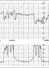
absent variability

A
40
Q

accel

A
  • brief, temporary increase in FHR
  • reassuring sign of responsive, non-acidotic fetus
  • usually occur with fetal movement
  • size, frequency, and occurrence increase with GA
41
Q

documenting accels

A
  • ++ = 15bpm x 15 sec
    • = < 15 bpm and/or < 15 sec
  • 0 = none
42
Q

decel patterns

A
  • type
    • early: slow and sloping with ctx
    • variable: U, V, or W shaped with sharp ↓ ↑
    • late: slow and sloping, starting after or lasting until after ctx
  • timing
    • episodic: spontaneously
    • periodic: with ctx
43
Q

VEAL CHOP

A

(FHR patterns)

Variable = Cord compression

Early = Head compression

Accel = OK

Late = Placental insufficiency

44
Q

early decel

A
  • caused by head compression, vagal stimulation
  • uniform, curved shape; mirror image of ctx
  • onset at beginning of ctx, FHR returns to baseline at end of ctx
  • during active phase of labor (4-7 cm)
  • commonly r/t cephalopelvic disproportion
  • nonpathalogic/benign
  • Tx: none, unless truly severe
  • document
45
Q

late decel

A

caused by placental insufficiency

46
Q

What does maternal pushing look like on a strip?

A

obvious sawtooth pattern during contractions

47
Q

prolapsed cord

A
  • vag exam to confirm
  • suspicious findings
    • baby high in pelvis when water breaks
    • variable decels
48
Q

variable decel Tx

A
  • stop Pitocin
  • vag exam to r/o cord prolapse
  • reposition mom
  • O2 40% (ATI: 8-10 L/min) via nonrebreather mask
  • possible amnioinfusion
49
Q

late decel

A
  • cause: uteroplacental insufficiency
  • consistent shape, usually slow and sloping
  • about 20-30 sec after ctx (not always obvious); check end of ctx against end of decel and look for gradual return to baseline
  • often seen with ↓ or no variability
  • leads to fetal hypoxia/acidosis
  • less common
50
Q

nadir

A

peak or trough of IUP or FHR on fetal monitor tracing

51
Q

shoulder

A

sharp, brief increase in FHR before or after variable decel

52
Q

placental abruption

A
  • characterized by late decels
  • placenta detaches from uterus
  • causes concealed or visible bleed
  • → major maternal AND fetal hemorrhage, possible sudden death for either
53
Q

late decel Tx

A
  • turn mom to left side
  • correct hypotension with IVF bolus
  • stop or ↓ Pitocin
  • give tocolytic
  • 40% O2 by Vinny or 8-10 L/min by nonrebreather