Eye Histology - Downing Flashcards
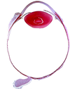
What are these 3 layers of the eye?

Cornealscleral layer (tunica fibrosa)
Uvea (tunica vasculosa)
Retinal layer (tunica interna or nervosa)

Label:
anterior chamber
posterior chamber
ciliary bodies
lens
vitreous body
optic papilla
OPtic nerve
fovea


Importance of corneal endothelium?
Important for brinign things back and forth between the cornea and anterior chamber
What is the vessel by which the aqueous humor can drain into the episceral veins?
The canal of schlemm
Three components of the uvea?
The choroid (bulk of it)
The ciliary body (ciliary processes and muscle)
The Iris
Aqueous humor is produced in what structures?
The ciliary processes
draw a simple picture of what happens to the lens upon contraction and relaxation of ciliary muscles

What type of cells form the inner and external limitting membranes?
Muller cells
What cells connect the inner and outer plexiform layers?
What layer is this?
Bipolar cells
Make up the inner nuclear layer
What cells make up the inner nuclear layer?
bipolar cells
amacrine cells
horizontal cells
muller cells
What cells of the retinal glial cells?
Muller cells!
have supportive function (probably…)
What is the macula lutea?
area of sharp visual ability
Light coming striaght through will hit the maculae lutea portion of the retina
Fovea centralis is area in center with greatest visual acuity (it only has cones)
What forms the blind spot of the retina?
What is the small central depression from which the central artery and vein of the retina emerge?
The optic disc
Physiological (or optic) cup
portion of disc that is slightly raised due to a heaping up of nerve fibers
optic papilla
Label This


Label This:


What retinal cells integrate information between rods and cones?
Horizontal cells


