Exocrine Pancreas Flashcards
What are the two majory pathways for developing sporadic colorectal cancer?
- Chromosomal instability PW (~80%)
- precursor lesion: conventional tubular or villous adenomas
- defect in APC genes
- beta-catenin/Wnt signaling K-flas
- Microsatelline instability PW
- precursor lesion: sessile serrated polyp (adenoma)
- defect in MLH1 gene
- hypermethylation of promoter region leading to a defect in DNA mismatch repain genes

What is the usual clinical phenotype of sporadic colorectal carcinoma that arise from the chromosome instability pathway?
Histopathology?
Provide the same information for the microsatellite instability pathway as well.
- Chromosme instability
- left-sided preominant
- tubular, tubulovillous, and villous adenomas
- moderately differentiated adenocarcinomas
- microsatelline instability (MSI)
- right-sided predminant cancers
- sessile serrated adenomas
- mucinous carcinomas
What are the two familial colorectal cancer syndromes?
- FAP
- familial adenomatous polyposis
- innumerable adenomatous colon polyps
- inherited germline APC mutation, followed by a “second hit” mutation
- 100% will get colon cancer prior to 30
- prophylactic colectomy
- still at ristk for adenomas/carcinomas elsewhere
- familial adenomatous polyposis
- HNPCC
- hereditary nonpoyposis colorectal cancer (“Lynch syndrome”)
- highly aggressive colon polyps
- most common syndromic form of CRC (2-5%)
- inherited germ-line mutations in DNA mismatch repair genes followed by 2nd somatic muation or epigastric silencing
- MSH2 or MHL1
What disease is shown in the provided image?

Look at the carpet of polyps
you can see the tubular adenoma in the histology slide

What are the 3 variant forms of FAP?
APC in germline mutation is present, but in a little different spot
- Attenuated FAP and Muthyl-associated polyposis
- lots of polyps, fewer than FAP
- develop cancers at young age, but older than FAP
- Gardner syndrome
- Same as FAP but also get
- osteomas, epidermal cysts, fibromatosis, abnormal dentition, duodenal and thyroid cancers
- Same as FAP but also get
- Turcot syndrome
- polyps/CRC and get brain tumors
Characteristics of Lynch syndrome?
- usually proximal colon cancer
- younger age (early to mid 40s)
- abundant mucin production & Tumor infiltrating lymphocytes (TIL)
- increased risk of malignancy at other sites
- esp endometrial cancer
- therapeutic difference between how sporadic cances vs. inherited cancers are treated
Variant forms of HNPCC? (Lynch syndrome)
- Turcot syndrome
- polyps/CRC
- brain tumors
- muir-torre
- polyps/crc
- sebaceous neoplasms
- constitutional mismatch repair deficiency syndrome
- baillelic mutations in MMR genes
- cancers often in first decade of life
Fill out the provided table

HNCRC tend to be right sided

What other junction does the recto-anal junction look like?

It looks like Barrett’s esophagus becasue you can see the transition from glandular mucosa with goblet cells to statified squamous
Which are the internal and which are the external hemrrhoids?

- internal
- glandular mucosa above it
- external
- squamous epithelium and a hair follicle above it
- lines of zahn = starting to thrombos

What types of tumors can develop in the anal canal?
- normal anal cell types
- columnar, transitional, suamous, melanocytes
- malignancy repcapitulates normal
- pure squamous cell carcinoma – HPV associated
- precursor condyloma
- primary melanoma
- often misinterpreted as hemorrhoids
- pure squamous cell carcinoma – HPV associated
The provied photo is an example of what?

- look like squamous cells anywhere else, get keratin curls, cellular bridges

What type of cancer is shown?

melanoma– notice the brown pigment which melanomas tend to do
If you get the response “extra-mammary Pagets Disease” form a pathologist, what should be your next step?
additonla imaging to see if malignancies anywhere else
- malignant glandular neoplasm confined to the epithelium

What histological features are related to the exocrine functions of the pancreas?
acini and ducts
grandular eosinophilic cytoplasm –multiple zymogen granules in cytoplasm with digestive enzymes

What are the congenital anomolies related to the pancreas?
- agenesis
- absence of pancreas – usually associated with other severe malformations incompatible with life
- pancreas divisum
- dorsal & ventral buds do not fuse properly so main pancreatic duct secretes it juices through minor papilla
- predisposed to pancreatitis
- annular pancreas
- pancrease has strangle hold around duodenum
- can lead to duodenal obstruction
- ectopic pancreas
What is the definition of acute pancreatitis?
Causes?
- reversible pancreatic parenchymal injury and inflammation
- 80% associated with biliary obstruction/alcohlism
- gallstones M:F = 1:3
- ETOH: M:F = 6:1
- Less common causes
- neoplasms (periampullary)
- cysts of bile duct
- biliary sludge
- parasites
- medications
- infections (mumps)
- metabolic derangements (hypercalcemia, hyper TGs)
- trauma
- inherited disorders (gene mutations resulting in early and prolonged activation of trypsin)
Clinical presentation of acute pancreatitis?
- abdominal pain
- anorexia, nausea, vomiting
- diagnosed by elevated lipase and amylase, and exclusion of oterh causes of abdominal pain
- full-blown acute pancreatitis is a medical emergency
- leukocytosis, hemolysis, DIC, vascular pooling, ARDS, diffuse fat necrosis –>> vascular collapse and shock

3 proposed pathogenesis of acute pancreatitis?
- duct obstruction
- cholelithiasis
- chronic alcohlism – secretions too thick
- dirct acinar injury
- alcohol, drugs, trauma, ischemia, mumps
- defective intracellular transport (activated inside cell before released)
- metabolic injury, alcohol, duct obstruction

When would you expect to see elevated amylase in a patient with acute pancreatitis?
What other conditions may cause elevated amylase?
- amylase increases w/in 6-12 hours
- window period where someone can have acute pancreatitis but do not have elvated amylase
- > 3x normal, prob the pancreas
- return to normal in 3-5 days
- can also be elevated in parotitis, intestinal obstruction or infarction
- decreased renal clearance may lead to falsely elevated levels
When would you expect to see elevated amylase in a patient with acute pancreatitis?
What other conditions may cause elevated amylase?
- lipase increases w/in 4-8 hours
- window period where someone can have acute pancreatitis but do not have elvated lipase, but shorter than amylae
- peaks 24 hrs
- return to normal in 8-14 days
- can also be elevated in salivary gland, intra-abdominal infarction
- decreased renal clearance may lead to falsely elevated levels
Once you have made the diagnosis of acute pancreatitis, what other tests would you run to figure out the cause or prognosticate the outcome?
- Liver enzymes (ALT, AST)
- >3x prob a gallstone
- Hematocrit
- if > 44% & rises over 24 hrs prop pancreatic necrosis
- C-reactive protein
- over 200 IU/L pancreatic necrosis
- useful after first 36-48 hrs
- Trypsinogen activation peptide
- > 30mmol/L in 6-12 hr prob severe disease
- Calcium
- persistently low = poor prognosis
- b/c have fat necrosis that is pulling in calcium, so if you have enough to be hypocalcemic you have a very injured pancreas
- persistently low = poor prognosis

What is shown in the provided slide of the pancreas?
- viable pancreatic acini
- sterile liquifactive necrosis
- enzymatic necrosis
- some of those enzymes kill off fat cells
- calcium is creating chalky deposits

What percent of people will die from acute pancreatitis?
Possible sequaelae?
- 5% will die form shock within first week
- ARDS, acute renal failure may develop
- sterile pancreatic abscess +/- pseudocyst
- infection, usualy by gram (-) organisms (gut flora), has high mortality rate
What is the difference between a pseudocyst and a true cyst?
- a true cyst has an epithelial lining
- a pseudocyst does not, usually an inflammatory lining or a fibrotic lining

What are the characteristics of chronic pancreatitis?
- Inflammation & irreversible destruction of exocrine pancreatic parenchyma w/ fibrosis
- may have destruction of endocrine pancreas in late stages
- most common causes is alcohol abuse
- other causes
- autoimmune
- obstruction pseudocysts, stones, tumors
- hereditary pancreatitis
- CFTR mutations (cystic fibrosis)
Why is it difficult to diagnose chronic pancreatitis?
Clinical features?
- If you are knocking out exocrine glands, you aren’t going to see elvated levels of amylase or lipase
- you need to have a high degree of suspicion
- not immediately difficult to diagnose
- develop severe exocrine pancreatic insufficiency and possibly diabetes mellitus (if knock out islets of langerhans)
- may have severe chronic pain in recurrent bouts
- pancreatic pseudocysts may result
- possibly increased risk of carcinoma

How doe cystic fibrosis impact the pancreas?
it makes the secretions very viscous which can cause concretions, which cause the enzymes to back up and self-digest the pancreas
proliferation of ductules trying to get the stuff out
with longstandign pancreatitis, the pancrease can be replaced by mature adipose tissue

What are the two general categories of cystic neoplasms of the pancreas?
- serous cystic neoplasms- always benign
- mucinous - have potential to becom malignant
- mucinous cystic neoplasmsm- premalignant
- do NOT communicate with a duct
- benign to having foci of invasive cancer
- intraductal papillary mucinous neoplasms (IPMNs)
- DO communicate with a duct
- communication with the main pancreatic duct = higher risk for malignant transformation
- mucinous cystic neoplasmsm- premalignant

What demographic is most commonly affected by cystic neoplasms?
Where are they commonly located?
Symptoms?
Treatment?
- Always benign
- 25% cystic neoplasms of pancreas
- elderly women
- tail of pancreas
- Symptoms
- nonspecific abdominal symptoms
- Treatment
- surgical resection is curative in most
- if asymptomatic, leave it alone
Characteristic stellate scar
lined with low cuboidal epithelial cells

What demographic is most commonly affected by mucinous cystic neoplasms?
Where are they commonly located?
Symptoms?
Treatment?
- do NOT communicate with pancreatic duct
- middle-aged women
- tail of pancreas
- 1/3 harbor one or more foci of invasive adenocarcinoma
- all have to come out
- w/ no invasion prognosis is excellent
- w/ invasive ~50% fatality
big cysts & lined with cuboidal epithelium – can get progressively dysplastic

What demographic is most commonly affected by intraductal papillary mucinous neoplasms?
Where are they commonly located?
Symptoms?
Treatment?
- within mucin cell lining & commnicates w/ pancreatic duct
- men
- head of pancreas
- 10-20% are multifocal
- may contain areas of invasive adenocarcinoma
- always removed
- Differe from mucinous cystic neoplasms
- absences of “ovarian” like stroma & involvement of duct

where do most cases of pancreatic ductal adenocarcinomas arise?
- not out of a duct that has been cystically dialated
- they arise in ductal epithelium that gets progressively dysplastic
- pancreatic intraepithelial neoplasia
- low grade to high grade
- not something you pick up on imaging
- no way to catch them early

Why are tumors in the head of the pancreas easier to diagnose early?
- they can cause blockage of the bile duct & cause you to be jaundiced
- if they are in the body & tail they are very hard to detect
What are risk factors fo pancreatic carcinoma?
- smoking !!
- alcohol (?)
- hereditary forms of pancreatitis
- CA-19-19 will be elevated but is not specific
- Migratory thrombophlebitis (trousseau sign)
- throw lots of clots
Adenocarcinoma just makes little gland lumina

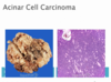
Characterisics of pancreatic acinar cell carcinoma?
- form zymogen granules like normal acinar cells
- 1-2% pancreatic neoplasms
- male
- 5-7th decade
- lipase hypersecretion paraneoplastic syndrome 10-15%
- aggressive tumors
- make little acinar type structure – make trypsin