Exam II - Canine Skull Flashcards
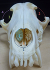
Identify the section labeled 1

Frontal Bone
Identify the section labeled 2

Maxillary Bone
Identify the section labeled 3

Nasal Bone
Identify the section labeled 4

Parietal Bone
Identify the section labeled 5

Incisive Bone
Identify the section labeled 6

Zygomatic Bone
Identify the section labeled 7

Occipital Bone
Identify the section labeled 8

Lacrimal Bone
Identify the section labeled 9

Temporal Bone
Identify the section labeled 10

Sphenoid Bone
Identify the section labeled 11

Palatine Bone
Identify the section labeled 1

Incisive Bone
Identify the section labeled 2

Maxillary Bone
Identify the section labeled 3

Palatine Bone
Identify the section labeled 4

Zygomatic Bone
Identify the section labeled 5

Sphenoid Bone
Identify the section labeled 6

Temporal Bone
Identify the section labeled 7

Occipital Bone
Identify the section labeled 8

Frontal Bone
Identify the section labeled 9

Pterygoid Bone
Identify the section labeled 10

Vomer
Identify the section labeled 1

External sagittal crest
Identify the section labeled 2

Temporal lines
Identify the section labeled 3

Nuchal crest
Identify the section labeled 4

External occipital protuberances
Identify the section labeled 5

Temporal fossa
Identify the section labeled 6

Zygomatic process of temporal bone
Identify the section labeled 7
Zygomatic process of frontal bone
Identify the section labeled 8

Zygomatic arch
Identify the section labeled 1

Pterygopalatine fossa (shaded)
Identify the section labeled 2

Temporal fossa (shaded)
Identify the section labeled 3

External sagittal crest
Identify the section labeled 4

Temporal line
Identify the section labeled 5

Nuchal crest
Identify the section labeled 6

External occipital protuberance
Identify the section labeled 7

Infrorbital foramen
Identify the section labeled 8

Fossa for the lacrimal sac
Identify the section labeled 9

Alveolar juga
Identify the section labeled 1

nasal aperture
Identify the section labeled 2

orbit

Identify the section labeled 3

orbital margin
Identify the section labeled 4

orbital ligament
Identify the section labeled 5
nasal conchae
Identify the section labeled 6

nasal septum
Identify the section labeled 1

Hard palate (shaded)
Identify the section labeled 2

paracondylar process
Identify the section labeled 3

tympanic bulla
Identify the section labeled 4

mastoid process
Identify the section labeled 5

retroarticular process
Identify the section labeled 6

mandibular fossa (shaded)
Identify the section labeled 7

palatine fissure
Identify the section labeled 8

major palatine foramen
Identify the section labeled 9

minor palatine foramen
Identify the section labeled 1
(canine skull, left side, caudal to rostral view)

Fossa for lacrimal sac (opening for nasolacrimal duct)
Identify the section labeled 2
(canine skull, left side, caudal to rostral view)

Maxillary foramen
Identify the section labeled 3
(canine skull, left side, caudal to rostral view)

Sphenopalatine foramen
Identify the section labeled 4
(canine skull, left side, caudal to rostral view)

Caudal palatine foramen
Identify the section labeled 5
(canine skull, left side, caudal to rostral view)

Zygomatic arch
Identify the section labeled 6
(canine skull, left side, caudal to rostral view)

Zygomatic process of the frontal bone
Identify the section labeled 7
(canine skull, left side, caudal to rostral view)

Left 2nd molar
Identify the section labeled 1
(canine skull, left side, ventro-rostral view of sphenoid area)

Optic canal
Identify the section labeled 2
(canine skull, left side, ventro-rostral view of sphenoid area)

Orbital fissure
Identify the section labeled 3
(canine skull, left side, ventro-rostral view of sphenoid area)

Rostral alar foramen
Identify the section labeled 4
(canine skull, left side, ventro-rostral view of sphenoid area)

Caudal alar foramen
Identify the section labeled 5
(canine skull, left side, ventro-rostral view of sphenoid area)

Alar canal
Identify the section labeled 6
(canine skull, left side, ventro-rostral view of sphenoid area)

oval foramen
Identify the section labeled 7
(canine skull, left side, ventro-rostral view of sphenoid area)

retroarticular process
Identify the section labeled 8
(canine skull, left side, ventro-rostral view of sphenoid area)

Foramen lacerum
Identify the section labeled 9
(canine skull, left side, ventro-rostral view of sphenoid area)

musculotubal canal
Identify the section labeled 1
(canine skull, left side, ventro-caudal view)

retroarticular foramen
Identify the section labeled 2
(canine skull, left side, ventro-caudal view)

External acoustic meatus
Identify the section labeled 3
(canine skull, left side, ventro-caudal view)

Stylomastoid foramen
Identify the section labeled 4
(canine skull, left side, ventro-caudal view)

hypoglossal canal
Identify the section labeled 5
(canine skull, left side, ventro-caudal view)

Tympanic bulla (left)
Identify the section labeled 6
(canine skull, left side, ventro-caudal view)

occipital condyle (left)
Identify the section labeled 7
(canine skull, left side, ventro-caudal view)

paracondylar process
Identify the section labeled 1
(canine skull, ventral view)

Foramen magnum
Identify the section labeled 2
(canine skull, ventral view)

Hypoglossal canal
Identify the section labeled 3
(canine skull, ventral view)

Tympano-occipital fissure
Identify the section labeled 4
(canine skull, ventral view)

External acoustic meatus
Identify the section labeled 5
(canine skull, ventral view)

musculotubal canal
Identify the section labeled 6
(canine skull, ventral view)

Foramen lacerum
Identify the section labeled 7
(canine skull, ventral view)

Oval foramen
Identify the section labeled 1
(Canine skull, ventral view, rostral-to-caudal)

foramen magnum
Identify the section labeled 2
(Canine skull, ventral view, rostral-to-caudal)

musculotubal canal
Identify the section labeled 3
(Canine skull, ventral view, rostral-to-caudal)

foramen lacerum
Identify the section labeled 4
(Canine skull, ventral view, rostral-to-caudal)

oval foramen
Identify the section labeled 5
(Canine skull, ventral view, rostral-to-caudal)

tympanic bulla
Identify the section labeled 6
(Canine skull, ventral view, rostral-to-caudal)

retroarticular process
Identify the section labeled 7
(Canine skull, ventral view, rostral-to-caudal)

paracondylar process
Identify the section labeled 1
(Canine skull, ventro-caudal view)

foramen magnum
Identify the section labeled 2
(Canine skull, ventro-caudal view)

hypoglossal canal
Identify the section labeled 3
(Canine skull, ventro-caudal view)

tympano-occipital fissure
Identify the section labeled 4
(Canine skull, ventro-caudal view)

foramen lacerum
Identify the section labeled 5
(Canine skull, ventro-caudal view)

musculotubal canal
Identify the section labeled 6
(Canine skull, ventro-caudal view)

oval foramen
Identify the section labeled 7
(Canine skull, ventro-caudal view)

caudal alar foramen
Identify the section labeled 8
(Canine skull, ventro-caudal view)

external acoustic meatus
What is the structure labeled 1? Which nerve(s) exit(s) the structure?

Optic canal
Optic nerve
What is the structure labeled 2? Which nerve(s) exit(s) the structure?

Orbital fissure
Oculomotor n., trochlear n., abducent n., opthalmic n.
What is the structure labeled 3? Which nerve(s) exit(s) the structure?

Rostral alar foramen
Maxillary a. and n.
What is the structure labeled 4? Which nerve(s) exit(s) the structure?

Oval foramen
mandibular n.
What is the structure labeled 5? Which nerve(s) exit(s) the structure?

Musculotubal canal
auditory tube
What is the structure labeled 6? Which vessel(s) exit(s) the structure?

Foramen lacerum
Loop of the internal carotid a.
What is the structure labeled 1? Which nerve(s) exit(s) the structure?

Tympano-occipital fissure
glossopharyngeal n., vagus n., accessory n.
What is the structure labeled 2? Which nerve(s) exit(s) the structure?

Hypoglossal canal
Hypoglossal n.
Identify the section labeled 1. (Left canine mandible, lateral view)

body
Identify the section labeled 2. (Left canine mandible, lateral view)

ramus
Identify the section labeled 4. (Left canine mandible, lateral view)

masseteric fossa (shaded)
Identify the section labeled 5. (Left canine mandible, lateral view)

coronoid process
Identify the section labeled 6. (Left canine mandible, lateral view)

mental foramen
Identify the section labeled 7. (Left canine mandible, lateral view)

condylar process
Identify the section labeled 8. (Left canine mandible, lateral view)

mandibular notch
Identify the section labeled 9. (Left canine mandible, lateral view)

angular process
