EXAM I Flashcards
Cause of pneumothorax
Lung collapse due to air infiltrating the pleural cavity breaking the surface tension between the parietal and visceral pleural cavities
Define hemothorax, hydrothorax, chylothorax
Hemothorax - blood in pleural cavity
Hydrothorax - serous fluid in pleural cavity
Chylothorax - lymph in pleural cavity
Cause lung collapse
Pleuritis
Inflammation of the pleura
Scraping sounds during ascultation, sharp stabbing pain
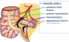
Pulmonary Embolism
Obstruction of pulmonary artery by blood clot, fat globule, or air bubble
DVT common cause
Bronchogenic Carcinoma
Lung Cancer
Cancer arising from bronchial epithelium
Smoking major cause
Highly metastatic due to association of lymphatics in bronchial tissue
Malingnant Mesothelioma
Lung cancer affecting pleura (mesothelium)
Asbestos exposure
Pulmonary Tuberculosis
TB
Bacterial lung infection; can spread to other organs
Airborne
Describe the location of the heart
Left of the body midline posterior to the sternum in the middle mediastinum
Rotated so that the right side faces anterior and left is more posterior
What forms the base of the heart?
Left atrium
Sits at the posterosuperior surface of the heart
The pericardium of the heart has three primary layers. If the pericardium of the heart is pierced by a needle, which of these primary layers would the needle first pass through?
Visceral pericardium
Pleural pericardium
Parietal pericardium
Fibrous pericardium
Epicardium
Fibrous pericardium
List the pericardium layers from superficial to deep
Outer portion - fibrous pericardium
Inner Portion - serous pericardium - parietal layer —> visceral pericardium layer; outermost layer of heart wall (epicardium) (pericardial cavity with serous fluid in between)
Pericardium is a membrane that encloses and protects the heart
Name the two layers of superficial fascia of the abdominal wall. What are they continuous with? Where is potential space within the abdominal wall?
Camper’s Fascia - fatty layer; continuous with superficial fatty layers of the thigh, thorax, and perineum (penis & scrotum)
Scarpa’s Fascia - membranous layer; deep, continuous with fascia lata of thigh and with deep layer of superficial perineal fascia
Potential Space can be found within the membranous layer (Scarpa’s fascia) of superficial fasica and deep fasica (Investing fascia superficial) of the external oblique muscle; where fluid can leak
List the anterior abdominal walls and state their functions collectively
External Oblique, Internal Oblique, Transverse abdominis, Rectus abdominis
Flex, stabilize, and laterally bend vertebral column
Describe the significance of the arcuate line and the abdominal musculature
Above the arcuate line, you have the internal oblique aponeurosis surrounding the rectus abdominis
Below the arcuate line, you have the rectus abdominis sitting above/directly on the transversalis fascia
Which abdominal muscle forms the inguinal ligament?
External oblique
List the posterior muscles of the abdominal wall and state their function collectively
Psoas major, Psoas minor, Iliacus, Quadratus lumborum
Flexors of the trunk and/or hip
Describe the location and pathway of the cutaneous branches of the anterior abdominal nerves
Via ventral rami or intercostal nerves, are between internal oblique and transverse abdominis and pierce the rectus sheath to supply the rectus abdominis, skin, muscles, and parietal peritoneum
List the 3 anterior abdominal wall nerves, which plexus are they apart of? What areas do they supply?
I.G.I. (Iggy) = via lumbar plexus (L1+L2)
Iliohypogastric - L1; lateral & anterior cutaneous branches, supplies suprapubic region
Genitofemoral - L1,L2; Genital branch exits inguinal canal thru superficial inguinal ring, supplies cremaster muscle or cutaneous to labium majus, Femoral branch is cutaneous to femoral triangle area
Ilioinguinal - L1; enters inguinal canal and emerges thru superficial inguinal ring, suppies groin & scrotum/labium majus
Describe characteristics of femoral hernias; location and more common in men or women?
Upper thigh, inferior to inguinal ligament, originating in femoral triangle
Medial portion of femoral triangle = weak
Mainly in females due to wider femoral triangle bc of wider hips
Whereas males more likely develop inguinal hernias
Distinguish between direct and indirect hernias
Direct - travels directly through the ab wall, passes medial to inferior epigastric vessels punching through peritoneum and transversalis fascia. Bulge in the lower anterior abdominal wall. Loop travels thru superficial inguinal ring but not the entire length
Indirect - travels ENTIRELY through inguinal canal, passes lateral to inferior epigastric vessels to enter deep inguinal ring; follows the path of spermatic cord
List the components of the foregut, midgut, and hindgut
PEGGY on LSD with a JAILED CAT who couldn’t Descend w/ RATS
F - pancreas, esophagus, gallbladder, liver, stomach, parts 1,2 duodenum
M - jejunum, appendix, ileum, 2-4 duodenum, cecum, ascending colon, proximal 2/3 transverse colon
H - descending colon, rectum, anus, transverse colon distal 2/3, sigmoid colon
Which layer of the peritoneum lacks pain fibers?
Visceral peritoneum
List the primary and secondary retroperitoneal organs
SAD* P*UC*KER.G (dpc)
S - suprarenal glands
A - aorta and IVC
D - 2-4 parts of duodenum (secondary)
P - pancreas (secondary)
U - ureter
C - ascending & descending colon (secondary)
K - kidneys
E - esophagus
R - rectum
G - gonads
What structures does THE Mesentery attach to? List the 3 other mesenteries that attach organs to the posterior body wall
Jejunum
Ileum
- Transverse mesocolon - fuses w/ posterior layer of greater omentum
- Sigmoid mesocolon
- Mesoappendix
What two peritoneal ligaments are found in this image?
Gastrosplenic ligament
Splenorenal ligament
What 2 ligaments form the lesser omentum? What is unique about this area?
Hepatogastric ligament
Hepatoduodenal ligament
Foramen of Winslow - opening to the inside of the lesser omentum behind the stomach but in front of the pancreas
What 4 ligaments are associated with the greater omentum?
- Greater omentum (via dorsal mesogastrium)
- Gastrocolic ligament
- Gastrophrenic ligament
- Gastrosplenic ligament
List the peritoneal ligaments of the liver (4)
CTFL
- Coronary ligament
- Left and Right Triangular ligament
- Falciform ligament (supraumbilical structure)
- Ligamentum teres (supraumbilical structure)
List the 3 umbilical folds and what they’re contained with, what is a fold?
Structures coursing thru extraperitoneal tissue forming elevations on interior ab wall
- Median umbilical fold - urachus
- Medial umbilical fold - medial umbilical ligaments, obliterated umbilical artery
- Lateral umbilical fold - inferior epigastric vessels, functional veins and arteries (Hasselbalch’s triangle)
What are the 3 fossae (between folds) that lie within the umbilical folds? What can form from them?
- Supravesical fossa (b/w median & medial folds) - site for supravesicle hernias
- Medial Inguinal fossa (b/w medial & lateral folds); inguinal triangle - site for direct inguinal hernias
- Lateral Inguinal fossa - (lateral to lateral umbilical folds) - site for indirect inguinal hernias
What 2 regions can the greater sac be divided into?
“closed” potential space b/w parietal and visceral layers of peritoneum
- Supracolic/supramesocolic - superior & anterior to liver and stomach (includes hepatorenal and subphrenic spaces and fossae of anterior wall)
- Infracolic/inframesocolic - inferior & posterior parts, upper & lower parts are divided by THE mesentery into right and left infracolic spaces
What structure connects the greater and lesser sacs?
Foremen of Winslow/Epiploic Foramen
At what levels do structures cross the diaphragm? (Hiatus)
T8 - IVC
T10 - esophagus
T12 - aorta
“Sliding” hiatal hernia
Esophageal hiatus of diaphragm enlarges or weakens and abdominal esophagus and parts of stomach herniate into the thorax
Paraesophageal hiatal hernia
Defect in the diaphragm next to esophageal hiatus; fundus of stomach herniates, not esophagus
What is the function of the heptaduodenal ligament?
Holds the portal triad
Portal hepatis “doorway” into the liver
What ducts enter the 2nd part (descending) of the duodenum? What structure does it enter?
Bile and pancreatic ducts at the major duodenal papilla
List features of the first part of the duodenum? What is it held in place by?
Begins at the pyloric sphinctor
Held in place by the hepatoduodenal ligament
Parts of the porta triad are posterior and superior
What are the features of the 3rd part of the duodenum?
Anterior: SMA and vein, root of mesentery
Superior: head of pancreas
Anterior and inferior: jejunum
Features of the 4th part of the duodenum (ascending)? What is anterior? Posterior? What holds it in place?
Not covered by peritoneum
Anterior = root of mesentery, jejunum
Posterior = left margin of aorta, medial border of psoas muscle
Held in place by the suspensory ligament of the duodenum (of Treitz) = anchors it to the right crus of the diaphragm
Where does the jejunum begin and where does the ileum end?
Duodenal-jejunal junction
Ileum = ileo-cecal valve
Vasa recta and plicae circularis are characteristic to what abdominal organ?
Jejunum and ileum
Jejunum = long vasa recta, more plicae circularis
Ileum = shorter vasa recta, fewer plicae circularis
Meckel’s diverticulum
A diverticulum about 2ft from the ileo-cecal junction
Remnant of the vitelline duct
What are the major structures of the large intestine? T.H.A.
Taenia coli - 3 bands of longitudinal muscle on the outside of the colon that converge at the root of the appendix and produce haustrae that slow the movement of feces
Appendices epiploicae are tags of fat, NOT found on the cecum
List 4 unique characteristics of the cecum
No epiploic appendages
Iliocolic valve - 2 folds that aren’t a true sphincter
Ileal papilla - projection of the ileum that connects it to the cecum
Opening for appendix - inferior to ileal papilla
What 2 structures does the spleen sit in between? What are its peritoneal relationships (2 ligaments)? What is posterior, anterior, and inferior to the spleen?
Stomach and Diaphragm
Left ribs 9-11; midaxillary line
Gastrosplenic ligament; Splenorenal ligament
Posterior - diaphragm
Anterior - tail of pancreas, stomach, left colic flexure
Inferior - left kidney, phrenicocolic ligament
What ducts form the major duodenal papilla? (entering the 2nd part of the duodenum)
Major pancreatic duct + bile duct = common hepatopancreatic ampulla = major duodenal papilla
Accessory pancreatic duct enters the Minor duodenal papilla
List the lobes of the liver, what does the falciform ligament do?
Left, Right, Caudate, Quadrate (gall bladder)
Falciform ligament = connects liver to anterior ab wall
Significance of the bare area of the liver
IVC is posterior to it and hepatic veins drain into it
List the muscles that elevate the ribs (4) ELIS
ELIS
Everyone Insane Loves Sex
External Intercostal
Innermost Intercostal
Levator Costarum
Serratus Posterior Superior
What muscles depress the ribs? (4)
SITS (sits for sex)
Subcostal
Internal Intercostal
Transversus Thoracis
Serratus Posterior Inferior
Where does the abdominal aorta begin and end along the vertebral column? What does it pass through to enter the abdomen? What structure crosses anterior to the aorta? What forms the aortic hiatus?
Begins at T12
Ends at L4
Enters abdomen via aortic hiatus
Left renal vein crosses anterior (therefore it’s longer than right renal vein)
Left & Right Crus of diaphragm forms aortic hiatus
What three structures lie anterior to the abdominal aorta? (3)
- Root of THE mesentery
- Pancreas
- Duodenum
Label the paired branches off the abdomindal aorta
Label the abdominal aorta paired branches
What are the 3 branches that come off the celiac trunk?
Left gastric artery (superior) (esophagus, lesser curvature)
Left splenic artery (stomach & esophagus)
Right common hepatic artery (lesser curvature of stomach, posterior duodenum, superior pancreas, liver)
What branches arise from the left gastric artery of the celiac trunk? (2) What does one anastomose with?
Esophageal branches
Branches to lesser curvature = anastomose with branches of the right gastric along the lesser curvature
What two arteries does the common hepatic artery branch into? (superior and inferior) What do they supply, what are their terminal branches?
Superior - Proper Hepatic artery (2 branches; left and right hepatic arteries)
Right gastric artery (anastomoses w/ left gastric artery along lesser curvature); Ends as right and left hepatic arteries
Inferior - Gastroduodenal artery
Supraduodenal artery, supplies posterior duodenum
Terminal branches: right gastro-omental artery and superior pancreaticduodenal branches
What are the branches/terminal branches of the splenic artery? What organ(s) does it supply?
Run posterior to stomach, superior to pancreas
Short gastric artery - upper part of greater curvature
Left gastro-omental artery - anastomoses with right gasto-omental on the greater curvature via gastroduodenal artery via common hepatic artery via celiac trunk
What are the contents of the hepatoduodenal ligament? (3; portal triad)
Bile duct (right)
Portal vein (posterior)
Hepatic artery (left)
From which organ does the SMA arise from posteriorly?
Neck of the pancreas
From what arterial branches does the pancreas receive blood?
Superior and Inferior Pancreaticduodenal artery
Superior (anterior & posterior) - Celiac trunk via gastroduodenal
Inferior (anterior & posterior) - SMA these branches anastomose w/ anterior & posterior surfaces of head of pancreas
What are the branches of the SMA? What do they supply?
- Inferior pancreaticduodenal artery
- Small intestine - jejunal (long vasa recta, simple arcades) & ileal arteries (short vasa recta, complex arcades)
- Large intestine - ileocolic artery, right colic artery, middle colic artery
What are the 3 branches of the large intestine via SMA? How about SMV? What’s different?
Superior - middle colic artery
Middle - Right colic artery
Inferior - Ileocolic artery (posterior & anterior cecal branch, appendicular branch)
SMV drains ilial and jejunal veins; everything else same
What 3 branches arise from the ileocolic artery via SMA?
Anterior & Posterior Cecal branches
Appendicular branch
Branches of the Inferior Mesenteric Artery
Left colic artery
Sigmoid arteries
Superior rectal artery
What are the 3 branches of the inferior mesenteric veins? (superior, middle, inferior) What do they drain?
Superior rectal veins - drain IMV
Middle rectal veins - drain internal iliac veins
Inferior rectal veins - drain external pudental veins
What veins make up the hepatic portal system? (3) Where does this structure form in relation to the pancreas?
Valveless; end up as capillaries in liver
Splenic vein
SMV (small intestine), IMV (via rectal veins)
Forms Posterior to neck of pancreas
Ends as right and left branches in liver
Runs through hepatoduodenal ligament to enter the liver
What two veins form the IVC?
What type of structures drain into the IVC?
What are the 4 main tributaries?
Left and right common iliac veins at L5
Posterior; kidneys, gonads, lumbar veins, median sacral vein, right and left common iliac veins, suprarenal veins, hepatic vein, inferior phrenic vein
Left suprarenal & gonadal veins enter left renal vein whereas right side is directly into IVC (hence why left renal vein is longer and runs anterior to aorta)
Tributaries: Common iliacs (lower limbs), lumbar veins, renal & hepatic veins
List the 4 portal-caval anastomoses
- Esophageal
- Paraumbilical
- Colic veins to retroperitoneal veins; veins draining ascending colon (via SMV) & descending colon (via IMV) drain posterior body wall (retroperi. organs)
- B/w superior rectal veins and middle and inferior rectal veins; empty into internal iliac veins —> common iliac veins = rectal varicosities/hemorrhoids
In terms of the portal-caval anastomoses, what can result in the esophagus? Where does the esophageal branches drain into?
Esophageal branches of left gastric veins connect w/ veins on lower thoracic esophagus = esophageal varicosities = via cirrhosis (portal hypertension)
Explain the drainage of the paraumbilical veins in terms of porta-caval anastomoses, what can result of this?
Paraumbilical veins are in the falciform ligament with subcutaneous veins around umbilicus in anterior abdominal wall = caput medusae
Drains umbilical area
Inferior & superficial epigastric veins empty into external iliac and femoral veins
What two structures are involved in bypassing the fetal lung?
Ductus arteriosus
Foramen ovale
What is the function of the fibrous skeleton of the heart? Where does it sit?
Separates the atria and ventricles
Anchors heart valves
Provides electrical insulation
Provides a rigid framework for the attachment of cardiac muscle tissue
What is another term for the right AV valve?
Tricuspid valve
Allows the flow of venous blood from right atrium into right ventricle
Prevents backflow into right atrium
Conus arteriosus of the pulmonary trunk
A conical region at the superior end of the pulmonary trunk
What does the right coronary artery branch into? (2)
Marginal artery - supplies right border of heart
Posterior interventricular artery - supplies LV & RV
What vessels branch from the left coronary artery?
Anterior Interventricular artery (LAD-left anterior descending) - supplies anterior of LV and RV
Circumflex artery - supplies LA and LV
