Embryology EXAM I Flashcards
What occurs during the early and late phase of lung development?
Early - positioning of lung primordium and primary lung bud formation
Late - mechanism of bronchial branching and cytodifferentiation
What week does development form and what forms during this in lung development?
Week 4
Laryngeotracheal/respiratory diverticulum via Tbx4 gene (endoderm of foregut)
Outgrowth of foregut (future esophagus) into surrounding splanchnic mesoderm
What direction does the laryngeotracheal diverticulum grow and what does it separate from, and what structure separates them?
Distally from the esophagus by the tracheoesophageal septum
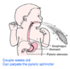
Tracheoesophageal Fistula, Esophageal Atresia; what fetal anomally is this related to?
Abnormal connection b/w trachea and esophagus
Improper formation of tracheoesophageal septum
Feeding tube cannot move beyond upper esophageal pouch
Related to: polyhydramnios (too much amniotic fluid)
Polyhydramnios
High volume of amniotic fluid
Baby is not swallowing enough amniotic fluid
Symptoms of esophageal atresia (occlusion) and tracheoesophageal fistula
Treatment?
Infant is drooling, with choking, coughing, sneezing
When fed infant swallows but begins to cough and struggle as fluid is regurgitated
Infant may become cyanotic (blue), stop breathing as overflow of fluid from blind pouch is aspirated in trachea and lungs
Treatment: Surgical fix
At what week do the bronchial buds form? What will be their final fate?
Week 5; left and right buds form
Will become main primary bronchi
After, a series of branchings will occur to become respiratory bronchioles (secondary)
What does the splanchnic mesoderm differentiate into?
Smooth muscle
Nerves
Blood vessels of lungs
What are the 5 stages of lung development? At which stage is the infant born viable?
EGCTP
Embryonic (4-7)
Pseudoglandular (8-16)
Canalicular (17-26) = viable
Terminal sac (27-birth)
Postnatal (Alveolar)
What occurs during the embryonic stage of lung development?
(4-7)
Initial formation of respiratory diverticulum —> formation of major bronchopulmonary segments
Lungs grow into pleural cavities
Pleural differentiation
What occurs during the pseudoglandular stage of lung development?
(8-16)
Formation and growth of duct systems within bronchopulmonary segments
No respiratory components or gas exchange
Resembles a gland
What occurs during the canalicular stage of lung development?
(17-26)
Formation of respiratory bronchioles & terminal sacs (primitive alveoli)
Increase in vascularization, capillaries = gas exchange = viable
What occurs during the terminal sac stage of lung development?
(27-birth)
Alveoli/terminal sacs develop from the respiratory bronchioles
Alveoli differentiates in Type I and Type II
Type I pneumocyte
Type II pneumocyte
Type I = blood-air barrier
Type II = produce surfactant (facilitates alveolar expansion)
For viability = capillaries, alveoli, surfactant
What occurs in postnatal/alveolar stage of lung development?
Alveoli differentiation
Infant respiratory distress syndrome
Deficiency/absence of surfactant
Immature/damaged Type II pneumocytes
60% born less than 28 weeks
5% born less than 37 weeks
Pulmonary agenesis
Complete absence of lungs, bronchi, and vasculature
Bilateral or Unilateral
Bronchial buds don’t develop
Pulmonary hypoplasia
Poorly developed bronchial tree
Partial or total (entire lung)
Congenital diaphragmatic hernia; which membranes is involved? Signs?
Abdominal contents herniated into pleural cavity; can cause pulmonary hypoplasia
Stomach, bowel can be in the thoracic cavity
Failure of pleuroperitoneal membranes to fuse with other components (i.e. septum transversum)
Signs: flat abdomen, breathlessness, cyanosis
What are the major trends of cardiovascular development? (4)
Converted into a 2 —> 4 chambered structure
Embryonic vascular system separates into systemic and pulmonary portions
Systemic arterial outflow —> Left
System venous retun —> Right
Describe the vascular circuit of the embryo
Series of aortic arches connect to dorsal aortae
= Cardinal, Vitelline, Umbilical
Dorsal aortae subdivide into smaller vessels to supply the embryo
Blood is drained by anterior and posterior cardinal veins
Common cardinal vein is formed by the left, right, anterior and posterior cardinal veins = embryonic circuit
What are the nutritional circuits for the embryo? What does the vitelline system do?
Umbilical and Vitelline
Nutritional circuits = Vitelline veins and arteries; supply and drain the yolk sac “nursery for blood cells”
Umbilical/placental arteries and veins
Name the adult structure from each of the embryonic structures
Truncus Arteriosus = Aorta, pulmonary trunk
Bulbus cordis = smooth part of right (conus cordis) and left ventricle (aortic vestibule)
Primitive ventricle = trabeculated part of right and left ventricles
Primitive atrium = trabeculated part of right and left atria (auricles)
Sinus venousus = smooth part of right atrium, coronary sinus, oblique vein of left atrium
Heart bends towards left
Heart loops ventrally around fulcrum
Dextrocardia
Positional abnormality
Heart bends to the left instead of the right
Usually not an issue
Function of endocardial cushions? When do they form? What does it initially sit in between?
Separates Atrium from Ventricles
Forms after ventral folding and dorsal and ventral blocks of tissue fold towards midline = endocardial tissue
Divides the single AV canal into separate right and left AV canals (which regulate blood blow from atria to ventricles)
Atrioventricular communis
Failure of the endocardial tissues to fuse you end up with a large communication between the left and right AV canals causing abnormal mixture of oxy- and deoxy- blood
What is the critical first step in the development of the 4-chambered heart?
Formation and fusion of endocardial cushions
What does growth of the bulbar region and muscular portion of the heart form?
Downward growth of bulbar region and upward growth of muscular portion towards the endocardial cushions forms the IV septum
Ventricular septal defects (VSDs)
Opening between the left and right ventricles
Associated with shunting of blood
Require surgical correction
Describe the formation of the atrial septum
The initial foramen between the atrium is the foramen primum; between the septum primum and dorsal endocardial cushions.
Septum primum grows downward and via apotosis forms the foramen secundum, developing septum primum from the endocardial cushion fuses with septum primum closing the foramen primum.
Septum secundum upper limb is developing from the top into the foramen ovale and eventually connects with the valve of foramen ovale (derived from septum primum)
At birth, high pressure on left atrium causes the septum primum to close valve = fossa ovalis
Atrial Septal Defects
Ostium/foramen primum - similar to endocardial cushion defects
Secundum type - involves foramen ovale and septum primum
Sinus venosus - usually near opening of SVC
Common atrium
Where do sinus venouses initially open into?
Dorsal wall of primitive atrium
Changes occur when right horn enlarges as blood is shunted from L –> R
Moves all systemic inflow to the right side
Shunts are created: vascular/nutritional circuit (vitelline & umbilical), embryonic circuits (cardinal)
What does the Vitelline Veins shunt turn into? Initially from the sinus venousus
Veins become incorporated into liver as hepatic sinusoids, hepatic veins, part of IVC and some GI veins
Loss at birth
What is the umbilical shunt that is derived from the sinus venosus?
Umbilical shunt bypasses liver and diverts oxygenated blood to heart
Loses direct connection with heart (ligamentum teres hepatis)
Joins the large venous shunt = ductus venosus = bypasses liver and diverts oxygenated blood to heart
Connects umbilical vein w/ IVC
What does the right anterior cardinal and common cardinal veins turn into after birth?
Anterior left and right cardinal veins connect and become the left brachiocephalic vein
Right anterior cardinal and common cardinal become the SVC
What do cardiac neural crest cells form?
Bulbus cordis
Truncus arteriosus
What problems are associated with tetralogy of fallot?
Pulmonary stenosis
VSD
Overriding aorta
Right Ventricular hypertophy
(large aorta, small pulmonary trunk)
Patent ductus arteriosus
No closure of the ligamentum arteriosum
Abnormal connection b/w aorta and pulmonary artery
What initiates specification of the gut tube formation?
RA gradient that causes TFs to be expressed in diff regions
SOX2 - esophagus
PDX1 - duodenum & pancreas
CDXC - small intestine
CDXA - large intestine
SHH initiates endoderm & mesenchyme/splanchnic mesoderm interaction
Where do post-ganglionics follow in hindgut innervation?
Arteries
in addition to IMA
Esophageal replacement options
Colon interposition
Gastric tube esophagoplasty
Gastric transposition
Describe how the esophagus and stomach develop, what do they begin as?
Straight tubes suspended by dorsal and ventral mesentery
Dorsal grows rapidly (towards left) with a simultaneous 90 degree rotation
Left side of stomach = anterior
Right side = posterior
Left vagus = anterior vagal trunk
Right vagus = posterior vagal trunk
Hypertrophic pyloric stenosis
Food never enters duodenum
Projectile vomiting/no bile
Duodenal atresia
Occluded lumen (2-3 part)
Double bubble sign = Gas distended stomach, no gas in proximal duodenum
Duodenal atresia, duodenal duplication cysts
Extrahepatic biliary atresia; symptoms, treatment
Incomplete canalization of the bile duct
Jaundice, Dark urine, pale stool surgical correction or transplant
What are the signals involved in pancreatic development?
PDX1 - pancreas & duodenum
PAX4 - insulin, somatostatin, pancreatic polypeptide
PAX6 - glucagon
3rd month = islets of langerhan
5th month = insulin
Annular pancreas
Ventral and dorsal pancreatic buds form a ring around the duodenum
Duodenal obstruction
Accessory ectopic pancreatic tissue
Most common in stomach or ileum (Meckle’s diverticulum)
Found in the distal esophagus thru the primary intestinal loop
Describe the rotation and fixation of the midgut
Rapid growth about 6 weeks
Normal physiologic herniation
Gut loops into umbilical cord and rotates 90 degrees counterclockwise around the SMA
10th week - returns to abdominal cavity and rotates an additional 180 degrees (270)
Malrotation of the midgut
What is it, symptoms
Volvulus
Partial rotation; abnormally positioned viscera
Increased risk of entrapment of portions of the intestine
Usually present within first week as duodenal obstruction with bilious vomiting
Recurrent abdominal pain, intestinal obstruction, malabsorption/diarrhea
Volvulus = abnormal twisting of intestine causing obstruction
Bilious Emesis
Omphalocele
Herniation of abdominal contents thru enlarged umbilical ring
Midgut loop fails to return to abdominal cavity
Normal if temporary
Covered by a membranous sac
Gastroschisis
Failure of anterior abdominal wall musculature to close during folding
Guts NOT surrounded by membrane
Meckel’s diverticulum
Remnant of vitelline duct
Asymptomatic
Gastric or pancreatic tissue
Ileal diverticulum
Fecal discharge through umbilicus
Hirschsprung’s Disease; HD
Congenital aganglionic megacolon
Failure of migration of neural crest cells
Functional intestinal obstruction, no parasympathetic innervation (colon cannot relax or do peristalsis)
Vagal neural crest cells invade anterior foregult and migrate to foregut, midgut, hindgut = enteric system
Surgical treatment
