Exam 3 - MS Flashcards
What are the four key features of the MSK exam?
Is the joint problem:
- Articular or extra-articular
- Acute (usually <6 weeks) or chronic (usually >12 weeks)
- Inflammatory or noninflammatory
- Localized (monoarticular) or diffuse (polyarticular)
How is acute joint pain classified? Chronic?
Acute: pain lasts up to 6 weeks
Chronic: pain lasts >12 weeks
Which are some examples of monoarticular disease processes?
Pain in a single joint suggests injury, monoarticular arthritis, or extra-articular causes like tendinitis, bursitis
Which are some examples of polyarticular disease processes?
Pain involving several joints (4+)
- Ex: rheumatic fever, gonococcal arthritis, RA (symmetric involvement), SLE, psoriasis
What is crepitus? What does it indicate?
Audible or palpable crunching during movement of tendons or ligaments over bone or areas of cartilage loss
D/t air in subcutaneous tissue
What are the four cardinal signs of inflammation?
Swelling, warmth, redness, pain
What history and exam findings are consistent with rheumatoid arthritis?
Chronic inflammation of synovial membranes
Tender, painful, stiff joints w/ symmetric involvement on both sides of body, limited ROM

What history and exam findings are consistent with osteoarthritis?
Degeneration and progressive loss of joint cartilage from mechanical stress
Hard and painless, Heberden nodes on dorsolateral aspects of DIP joints, flexion and deviation deformities may develop

What is the general approach to the newborn MSK exam?
Focuses on detection of congenital abnormalities (i.e. hands, spine, hips, legs, feet)
Combine MS exam with neurologic and developmental exam
How would the FNP perform a preparticipation sports physical? What would be abnormal findings and what would these findings indicate?
Focused, thorough MS examination looking for weakness, limited ROM, evidence of previous injury, asymmetry, swelling of joints
Common abnormalities from prior injury
- Asymmetry, swelling of joints
- Loss of ROM
- Weakness of shoulder, neck, or trapezius muscles
- Loss of strength of deltoid muscle
- Loss of external rotation and injury of glenohumeral joint
- Reduced ROM of elbow
- Reduced ROM from injury to forearm, elbow, or wrist
- Protruding knuckle, reduced ROM of fingers from prior sprain or fracture
- Inability to fully flex knees and difficulty standing up from prior knee or ankle injury
- Asymmetry from scoliosis, leg length discrepancy, weakness
- Asymmetry from scoliosis and twisting of back from low back pain
- Wasting of calf muscles from ankle or Achilles tendon injury
How would the FNP assess for gait, strength and coordination in the pediatric patient?
Note any asymmetry, weakness, undue tripping, clumsiness
Heel-to-toe walking, hopping, jumping
If concerned about strength, have child lie on floor and stand up –> kids w/ muscular dystrophy will roll over and push off floor w/ arms while legs are extended
How would the FNP perform the following tests and what do positive results indicate - crossover/cross body adduction test?
Maneuver - adduct patient’s arm across chest
Structure: acroclavicular joint
Positive test = pain w/ adduction
- Acromioclavicular joint tenderness and compression tenderness

How would the FNP perform the following tests and what do positive results indicate - Apley scratch test?
Maneuver - ask patient to touch opposite scapula
Structure: overall shoulder rotation
- Pain suggests rotator cuff disorder or adhesive capsulitis

How would the FNP perform the following tests and what do positive results indicate - painful arc test?
Maneuver - fully adduct patient’s arm from 0 to 180 degrees
Structure: rotator cuff
Positive = shoulder pain from 60 to 120 degrees
- Subacromial impingement/rotator cuff tendinitis disorder

How would the FNP perform the following tests and what do positive results indicate - Neer impingement sign?
Maneuver - Press on scapula to prevent scapular motion with one hand, raise patient’s arm with other, compress greater tuberosity of humerus against acromion
Structure: rotator cuff
Positive = pain
- Subacromial impingment/rotator cuff tendinitis disorder

How would the FNP perform the following tests and what do positive results indicate - Hawkins impingement sign?
Maneuver - flex patient’s shoulder and elbow to 90 degrees with palm facing down, then with one hand on forearm and one on arm rotate arm internally
Structure: rotator cuff
Positive = pain
- Supraspinatus impingement/rotator cuff tendinitis

How would the FNP perform the following tests and what do positive results indicate - external rotation lag test (strength test)?
Maneuver - with patient’s arm flexed 90 degrees with palm up, rotate arm into full external rotation
Positive = inability to maintain external rotation
- Supraspinatus and infraspinatus disorders

How would the FNP perform the following tests and what do positive results indicate - internal rotation lag test (strength test)?
Maneuver - ask patient to place dorsum of hand on lower back with elbow flexed 90 degrees, lift hand off back, ask patient to keep hand in this position
Positive = inability to hold hand in elevated position
- Subscapularis disoder

How would the FNP perform the following tests and what do positive results indicate - drop arm test (strength test)?
Maneuver - ask patient to fully abduct arm to shoulder level, up to 90 degrees, and lower it slowly
Positive = weakness
- Supraspinatus rotator cuff tear
- Bicipital tendinitis

How would the FNP perform the following tests and what do positive results indicate - external rotation resistance test (composite test)?
Maneuver - ask patient to adduct and flex arm to 90 degrees with thumbs up, stabilize eblow with one hand and apply pressure proximal to patient’s wrist as patient presses wrist outward in external rotation
Positive = pain or weakness
- Infraspinatus disorder
- Limited external rotation –> glenohumeral disease or adhesive capsulitis

How would the FNP perform the following tests and what do positive results indicate - empty can test (composite test)?
Maneuver - elevate arms to 90 degrees and internally rotate arms with thumbs pointing down, ask patient to resist w/ downward pressure on arms
Positive = inability to hold arm fully abducted at shoulder level of control lowering arm
- Supraspinatus rotator cuff tear

Which muscle groups make up the rotator cuff?
SITS muscles
- Supraspinatus
- Infraspinatus
- Teres minor
- Subscapularis
What exam findings would be consistent with a clavicle fracture in the newborn?
Asymmetric movement of arms and legs, feel break in contour of bone, tenderness, crepitus, lumps
In considering the elbow, what history and exam findings are consistent with lateral epicondylitis (tennis elbow)?
D/t repetitive extension of wrist or pronation/supination of forearm
Pain and tenderness 1cm distal to the epicondyle, possibly in extensor muscles
Pain increases as patient tries to extend wrist against resistance, decreased grip strength

In considering the elbow, what history and exam findings are consistent with medial epicondylitis?
Aka pitcher’s, golfer’s, Little League elbow - follows repetitive wrist flexion such as throwing
Tenderness is maximal just lateral and distal to medial epicondyle
Wrist flexion against resistance increases the pain, decreased grip strength

In considering the elbow, what history and exam findings are consistent with olecranon bursitis?
Swelling and inflammation of olecranon bursa from trauma, gout, or RA
Swelling superficial to olecranon process and can reach 6cm in diameter

What symptoms are consistent with carpal tunnel syndrome?
Nocturnal hand/arm numbness, dropping objects, inability to twist lid off jars, aching at wrist or forearm, numbness of first three digits, decreased grip strength
What tests can be used to assess carpal tunnel syndrome - thumb abduction test?
Thumb abduction - ask patient to raise thumb straight up while applying downward resistance
Positive = weakness on thumb abduction

What tests can be used to assess carpal tunnel syndrome - tinel test?
Tapping lightly over course of median nerve in carpal tunnel
Positive = aching and numbness in median nerve distribution

What tests can be used to assess carpal tunnel syndrome - phalen sign?
Ask patient to hold wrists in flexion for 60 seconds with elbows fully extended OR ask patient to press backs of both hands together to form right angles (compresses the median nerve)
Positive = numbness and tingling in median nerve distribution within 60 secs

What is snuffbox tenderness? What does this indicate?
Hollowed depression distal to radial styloid process formed by the abductor and extensor muscles of the thumb
More visible w/ lateral extension of thumb away from hand (abduction)
Indications: occult scaphoid fracture (w/ pain and tenderness), poor blood supply increases risk of scaphoid bone avascular necrosis

What are Dupuytren flexion contractures?
Thickened band overlying flexor tendon of 4th finger and little finger near distal palmar crease –> thickened fibrotic cord develops between palm and finger
Finger extension limited, flexion normal
Flexion contracture of fingers may gradually develop

What is stenosing tenosynovitis?
Inflammation of flexor tendon sheaths d/t local injury, overuse, infection
Tenderness and swelling develop along tendon sheath (NOT joint); from distal phalanx to metacarpophalangeal joint
Finger held in slight flexion (trigger finger); finger extension painful

What are Colles fractures?
Tenderness over distal radius after fall
May have bony step-offs

What are the muscle groups of the hips?
- Flexor group (i.e. iliopsoas)
- Extensor group (i.e. gluteus maximus)
- Adductor group
- Abductor group (i.e. gluteus medius and minimus)

How would the FNP perform the Barlow test? What are positive findings and what do they indicate?
Tests the ability to sublux or dislocate an intact but unstable hip
- Flex legs at hips and knees
- Place index finger over greater trochanter of each femur and thumbs over lesser trochanter
- Pull leg inward (toward midline) in adduction and apply posterior pressure to knee
Positive Barlow NOT diagnostic of dysplastic hip - indicates laxity and potentially dislocatable hip

How would the FNP perform the Ortolani tests? What are positive findings and what do they indicate?
Tests for the prescence of a posteriorly dislocated hip
- Flex legs to form right angle at hips and knees
- Place index fingers over greater trochanter of femur and thumbs over lesser trochanters
- Abduct both hips simultaneously until lateral aspect of each knee touches the table
Positive = palpable movement of femoral head back into place
Indicates possibility of developmental dysplasia of the hip

How would the FNP assess for leg shortening?
Compare distance from anterior superior spine of ilium to medial malleolus on each side
Have child stand straight and place hands horizontally over iliac crests from behind; note discrepancies
Observe from behind as child shifts weight from one leg to the other
- Negative Trendelenberg - pelvis remains level when weight is shifted
- Positive Trendelenberg - pelvis tilts toward unaffected hip during weight bearing on affected side
How would the FNP assess for tibial torsion?
Usually corrects itself during 2nd year of life after months of weight bearing
Have toddler lie prone on table w/ knees flexed 90 degrees, check position of malleoli (should be symmetric

What is a SCFE? In whom might this occur?
Disorder of adolescents - growth plate is damaged and the femoral head “slips” with respect to the rest of the femur (common in obese children)
Head of femur stays in the cup of the hip joint while the rest of the femur is shifted

What exam techniques can you use to assess for an effusion - bulge sign?
What is a positive result?
For minor effusions
- Extended knee, place L hand above knee, apply pressure on suprapatellar pouch (displacing or “milking” fluid downward)
- Stroke downward on medial aspect of knee, apply pressure to force fluid into lateral area
- Tap knee behind lateral margin of patella with right hand
Positive = fluid wave or bulge on medial side between patella and femur

What exam techniques can you use to assess for an effusion - balloon sign?
What is a positive result?
For major effusions
- Place thumb and index finger of R hand on each side of patella
- L hand compresses suprapatellar pouch against femur
- Palpate for fluid ejected or “ballooning” into spaces next to patella under R thumb and index finger
Positive = palpable fluid wave
- Palpable returning fluid wave into suprapatellar pouch confirms major effusion

What exam techniques can you use to assess for an effusion - balloting the patella?
What is a positive result?
For major effusions
- Compress suprapatellar pouch and “ballotte” or push patella sharply against the femur
- Watch for fluid returning to suprapatellar pouch
Positive = palpable fluid wave returning into pouch

How would the FNP perform the following tests and what do positive results indicate?
- McMurray test
- Patient supine, grasp heel and flex the knee
- Cup your hand over the knee joint w/ fingers and thumb along medial joint line
- From heel, externally rotate lower leg, push on lateral side to apply valgus stress on medial side of joint
- At same time, slowly extend lower leg in external rotation
- Same maneuver with internal rotation can be done
Positive = palpable click or pop along medial/lateral joint line
Structures involved: medial meniscus, lateral meniscus

How would the FNP perform the following tests and what do positive results indicate?
- Abduction (or Valgus) stress test
- Patient supine and knee slightly flexed, move thigh 30 degrees laterally to side of tale
- Place one hand against lateral knee to stabilze femur and other hand on medial ankle
- Push medially against knee and pull laterally at ankle to open knee joint on medial side (valgus stress)
Positive = pain or gap in medial joint line
Structures involved: MCL

How would the FNP perform the following tests and what do positive results indicate?
- Adduction (Varus) stress test
- W/ thigh and knee in same position, change position so you can place one hand against medial surface of knee and other around lateral ankle
- Push laterally against knee and pull medially at ankle to open knee joint on lateral side (varus stress)
Positive = pain or gap in lateral joint line
Structures involved: LCL

How would the FNP perform the following tests and what do positive results indicate?
- Anterior drawer sign
- Patient supine, hips/knees flexed to 90 degrees, feet flat on table, cup hands around the knee with thumbs on the medial and lateral joint line; fingers on medial and lateral insertions of hamstrings
- Draw tibia forward and observe if it slides forward (like a drawer) from under the femur
- Compare degree of forward movement with other knee
Positive = forward jerk showing contours of upper tibia
Structures involved: ACL

How would the FNP perform the following tests and what do positive results indicate?
- Lachman test
- Place knee in 15 degree of flexion and external rotation
- Grasp distal femur on lateral side with one hand and proximal tibia on medial side with other
- With thumb of tibial hand on joint line, simultaneously pull tibia forward and femur back
Positive = significant forward excursion
Structures involved: ACL

How would the FNP perform the following tests and what do positive results indicate?
- Posterior drawer sign
- Position patient supine, hips and knees flexed to 90 degrees, and feet flat on table
- Cup hands around knee with thumbs on medial and lateral joint line and fingers on medial and lateral insertions of hamstrings
- Push tibia posteriorly and observe the degree of backward movement in femur
Positive = proximal tibia falls back
Structures involved: PCL

What are symptoms and exam findings consistent with prepatellar bursitis (housemaids knee)?
Lies between patella and overlying skin
S/s: swelling over patella, pain with activity, tenderness, warm to touch
Triggered by excessive kneeling

What are some gait abnormalities and what do they indicate?
Most hip problems appear during weight bearing phase of walking
- Wide base - cerebellar disease or foot problems
- Pain during weight bearing or examiner strike on heel - femoral neck stress fractures
- Dopped pelvis on opposite side (waddling gait) - hip dislocation, arthritis, unequal leg lengths, abductor weakness
- Excess lordosis - flexion deformity of hip
- Loss of lordosis - paravertebral spasm
- Disparities in leg length - scoliosis
- Leg shortening and external rotation - hip fracture
What are some abnormalities of the toes and soles?
- Ingrown toenail
Sharp edge of toenail may dig into and injury lateral nail fold –> inflammation and infection
Tender, reddened, overhanging nail fold, sometimes granulation tissue and purulent discharge

What are some abnormalities of the toes and soles?
- Callus
Area of thickened skin that develops in region of recurrent pressure
Painless; if painful, suspect plantar wart
Tender to direct pressure

What are some abnormalities of the toes and soles?
- Hammer toe
Usually involves second toe
Hyperextension at metatarsophalengeal joint w/ flexion at proximal interphalangeal (PIP) joint

What are some abnormalities of the toes and soles?
- Plantar wart
Hyperkeratotic lesion caused by HPV, may look like a callus
Charasteristic small dark spot that give a stippled appearance
Normal skin lines stop at wart’s edge, tender if pinched side to side

What are some abnormalities of the toes and soles?
- Corn
Painful conical thickening of skin that results from recurrent pressure on normally thin skin, apex of cone points inward
Occur over bony prominances
when located in moist areas, such as pressure points between 4th and 5th toes, called soft corns

What are some abnormalities of the toes and soles?
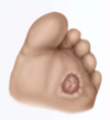
- Neuropathic ulcer
Develop at pressure points on feet; often deep, infected, indolent; painless
Diabetic neuropathy - diminished pain sensation or absent (detected using a nylon filament)
What are some common foot abnormalities of the young child?

What are some spine abnormalities the FNP should assess for during infancy?
- Midline hair tufts over lumbosacral spine - spinal cord defect (spinal bifida, meningomyelocele)
How would the FNP assess for scoliosis? What findings would be indicative of scoliosis?
- First assess standing assessing symmetry of shoulders, scapula, hips
- Have child bend forward w/ knees straight and head hanging straight down between extended arms
Suspect scoliosis w/ asymmetrical rise in thoracic or lumbar region
- Use scoliometer to test degree; angle >7 degrees is indicative
- Plumb line - string w/ weight attached; place at C7 and have child stand straight
