Electrical properties of the heart Flashcards
excitation-contraction coupling.
actin myosin fillaments overlap - cross bridges oull closer together. sarcolemma is membrane surrounding whole muscle.
t - tubule deviations rul down tubule (transferse)
sarcoplasmic reticulum acts as calcium store.
Why does cardiac muscle have a longer depolarisation time than skeletal muscle?
To allow for regulation of the amount of calcium getting into the cell, the calcium released inside the cell and so the strength of contraction
What does cardiac muscle form to allow it to function as a unit?
A functional syncytium
*A syncytium is a mass of cells that have merged together. The muscle cells in the cardiac syncytium are derived from the mesoderm. *
What allows the functional syncytium to work?
Gap junctions - electrical connections
Desmosomes - physical connections
What is the cardiac output of the heart?
5l / minute
CO = SV x HR
sarcoplasmic reticulum acts as__
calcium store
in skeletal muscle how would the sarcoplasmic reticulum get activated
sat at -90mv; activated by motor neuron releasing acetylcholine and binds to nicotinic receptors at end plate;
depolarises cell = evoke an action potential in muscle membrane
action potential will travel along being self-propagated by voltage-gated sodium channels
all along membrane then down t tubules and through interaction with sarcoplasmic reticulum which release calcium inside cells = calcium binds to troponin
Why does cardiac muscle have a longer action potential period than skeletal muscle regarding a long refractory period?
Stops the cardiac muscle from exhibiting tetanus
(twitch contraction which creates sustained contraction tetanus if you wish to keep a muscle contracted) skeletal good for holding things and avoiding dropping
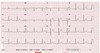
QRS in chest leads v1 and v2 are usually
negative.
When venous return decreases there is a corresponding decrease in .
cardiac output
is the type of penicillin used for prophylaxis against rheumatic fever and rheumatic heart disease.
Benzathine penicillin G
Infants with (dextro-transposition of the great vessels/levo-transposition of the great vessels) might not show symptoms at birth, but will eventually develop heart failure later in life.
(dextro-transposition of the great vessels/levo-transposition of the great vessels) levo-transposition of the great vessels
Infantile coarctation of the aorta is associated with both patent ductus arteriosus and (chromosomal anomaly) syndrome.
Turner syndrome.
There is a ____ to ____ shunt between the atria in hypoplastic left heart syndrome.
left to right shunt
The normal axis of the heart points downward and to the patient’s
left
__is the imaging test of choice to diagnose deep venous thrombosis.
Compression ultrasound with doppler
What are capillaries made up like
Exchange vessels
Very narrow lumen and thin wall
BP very low
What are veins made up like
Low resistance so blood can get back to the heart
Wide lumen
2/3 of blood stored in veins = capacitance vessels
What is MAP determined by
Cardiac output and total peripheral resistance
end-diastolic volume -
the left ventricle is filled with the maximum volume of blood,
Preload -
amount blood left in left ventricle before contraction (determined by end diastolic pressure) = volume work of heart
Stroke volume -
blood vol(l) pumped by heart per contraction - determined by blood filling ventricle, compliance of ventricular myocardium.
Cardiac output-
blood pumped by heart per minute (co=svxhr)
MAP -
is the average arterial pressure throughout one cardiac cycle, systole, and diastole. influenced by co and systemic vascular resistance
can be used for reversing witnessed ventricular fibrillation in the absence of defibrillators.
Precordial thump
What is a functional syncytium?
A group of cells that are synchronised electrically in an action potential
How is arteriolar resistance controlled
Extrinsic neural control
Extrinsic hormonal control
Intrinsic control - individual tissue
how long is the action potential of cardiac muscle
how long action potential for skeletal muscle
cardiac 250msec
skeletal 2 msec
what involvement does calcium have with contraction strength?
More calcium from ECF = more forceful contraction
calcium release does not saturate troponin, so regulation of calcium release can be used to vary the strength of contraction
What cells have unstable resting membrane potentials?
pace maker cells - spontaneously depolarise to threshold firing action potential
basic electrophysiology cell heart non-pacemaker
K+ higher in cell - leaky potassium channels
Ca+ and cl higher outside. - cannot get in without pumps
-90mv reached resting state.
basic electrophysiology cell heart non-pacemaker what makes it depolarise?
the neighbouring cells depolarising (syncytium)
0 - rapid depolarisation - rapid sodium influx
1 - early repolarisation - efflux potassium
2 - plateau - the slow influx of calcium
3 - final repolarisation - efflux of potassium
4 restoration of ionic concentration ions - resting potential restored by na+/k+atpase. slow entry of na+ into the cell until a threshold for new action potential

What else affects contractility
Inotropes - epinephrine,
Hypercalcaemia
Thyroid hormones
Glucagon
action potential pacemaker cells
Action potential Increase in Permeability to calcium (L type) channels (voltage-gated) open slower but stay open longer
Pre potential (pacemaker potential) - gradual decrease permeability to potassium (k+) (leaky channel shutting)
(early phase) Sodium (Na+) moved into the cell through “funny channels” (open in response to hyperpolarisation/repolarisation of previous action potential). = slowly depolarises until threshold met (responsible for instable resting membrane potential)
(later) increase permeability to Calcium (T-type) (voltage gated) transient small amount ca+ open more hyperpolarised potentials.
Strong inward calcium current responsible for rapid polarisation,
AV node conduction
0.05 m/sec
What separates atria from ventricles and what is significant about its conduction
annulus fibrosus - non conducting
depolatisation evoked in atria here cant get straight across to ventricles
Hyaline arteriolosclerosis of the ______ the glomerulus in diabetes mellitus leads to a high glomerular filtration pressure and resultant microalbuminuria
efferent arteriole
what wave caused the p wave?
the depolarization of the atria
what wave causes the QRS complex
depolarisation of the ventricles
what wave causes the T wave?
repolarisation of the ventricles
What decreases contractility
Ach by vagus nerve on m2
Hypocalcaemia
Ischaemia - hypoxia
Hyperkalaemia
Barbituarates
What does hyperkalemia do to the heart?
Fibrillation and heart block
ECG explain 1st-degree block
delayed p - q interval
the atrioventricular node is a delay box slowing the conduction of action potential to
ventricles this can slow/ stop so no depolarisation from the atrium to the ventricle. delay
normal interval p wave to start QRS complex should be less than 0.18 seconds. one large square is 0.2s

2nd degree block ECG
some depolarisations don’t get through at all. the increasing delay between Q to QRS interval and sometimes no QRS

3rd-degree heart block ECG
no transmission between atria and ventricles
depolarising from another place (not coordinating with atrium) hence inverted t wave

atrial flutter (conduction disorder) ECG
normal P QRS t sequence but each depolarising occurring quicker than should so superventricular tachycardia.

atrial flutter
atria and pacemaker isn’t depolarising spread of a wave of depolarisation across the atria. individual cells depolarising causing AF

ventricular fibrillation ECG
Lethan uncoordinated depolarisation in ventricles.
defibrillator - to depolarise all cells same time = enters a refractory period
pacemaker cells able to try to start rhythm again

standard ecg box
augmented limb leads aVR aVL aVF
are derived from the same three electrodes as leads I, II, and III, but they use Goldberger’s central terminal as their negative pole.

precordial/ chest leads
labelled v1-v6

standard ECG limb leads
SLL 1 - left arm wrt right arm
SLL 2 = left leg wrt right arm
SLL 3 - left leg wrt left arm
pr interval
time from atrial depolarisation to ventricle depolarisation (normally 0.12-0.2 seconds)
duration of QRS complex
time for whole ventricle to depolarise
0.08
time for wt interval
time spend while ventricle depolarised/repolarises (0.42) varies
what rhythm should the paper run at/be callibrated for?
25mm/sec
calibrating pulse should be __
0.2 sec = 1 large square
how to measure heart rate on the ECG
measure r-r interval and work out how many in 60 seconds.
- count 30 squares (6 seconds) and multiply by 10
what are the ranges in ecg: normal bradycardia tachycardia
normal 60-100
bradycardia < 60 bpm
tackycardia >100 bpm
what is more severe? st elevation or non st elevation
stemi is more severe
what direction does the x axir relate to
area of repolarisation
which of these is not a coronary artery
left marginal artery
diagonal artery
infra-cardiac artery
left anterior descenting artery
infra-cardiac artery
how long does 5 large squares relate to if the machine is callibrated correctly?
1 second. each square = 0.2 seconds
what anatomical plane do ecg measure in?
frontal
chest leads are unipolar what point do they measure voltage charge from?
mcburnys point
wilsons central terminal
sternum
midclavicular point
wilsons central terminal
what speed should ecg machine run as standard in mm/s
25mm/s
the qt interval encompasses which electrical events?
ventricular depolarisation
ventricular repolarisation+ atrial repolarisation
atrial depolarisation+atrial repolarisation + ventricular depolarisation
ventricular depolarisation + atrial repolarisation + ventricular repolarisation
what is standard paper setting for ecg machine
1mv oer 10mm 25mm/s
how many seconds does 5 small squared on ECG printout correspond to if the machine is running standard speed?
- 5s
- 2s
1s
10s
0.2s
which lead is not considered a limb lead?
v1
green
yellow
avf
v1
what is a normal pr interval length
150ms
- 9s
- 4s
- 5ms
150ms
which of these gives greatest detail of the size of the heart?
ap chest xray
12 lead ecg
cardiac MRI
cardiac ECHO
cardial MRI