DI Midterm Exam Material Flashcards
T/F: Pneumomediastinum may result in dyspnea
False
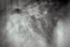
If you see an enlargement in this region, what should your top two differentials be?

Tracheobronchial lymphadenopathy and Left atrial enlargement

T/F: Ring Shadows (donuts) are often associated with a bronchial pattern
True
Bronchial pattern: donuts and tram lines
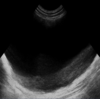
T/F: Hepatic veins have a hyperechoic wall on ultrasound
False
Hepatic veins have an isoechoic wall
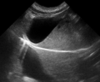
This is a sagittal image of the left kidney. Which side is cranial?

That one.

What view would be best for evaluating lesions in the right lung lobes?
Left Lateral
- In small animals, lung lesions generally are detected best in the non-dependent lung because the “up” lung is better aerated and therefore provides better contrast of lesions*
- Keep in mind, other lesions (not in the lung) generally are best seen on the “down” side because they are not distorted by magnification*
What echocardiographic modality would you use to measure wall thickness during systole and diastole?
M Mode
Name the MR sequence that nulls signal from free fluid (i.e. CSF):
FLAIR
_FL_uid _A_ttenuated _I_nversion _R_ecovery
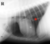
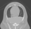
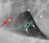
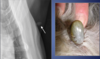
Is sternal lymphadenopathy present in this patient?

Yes.
Note the enlargement of the sternal lymph node

T/F: Ascites is commonly associated with mitral valve insufficiency
False
That is false
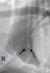
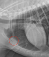
Is the circled lesion more likely in the lung or the mediastinum?

Lung
Note the acute angle to the body wall. If the lesion was in the lung you would not have such an acute angle
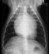
With which radiographic view is mediastinal shift best visualized?
VD/DV
Doppler measurements should be taken with the patient in ______ lateral recumbency
LEFT
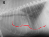
If you see a “bow-legged cowboy sign” on a DV view, what is your DDx?

left atrial enlargement

Identify the lymphatic structure indicated by the number 3:

Tracheobronchial

T/F: Diaphragmatic hernias cause caudal displacement of the gastric axis
False
Diaphragmatic hernias cause cranial displacement of the gastric axis
Long-axis left ventricular outflow view.
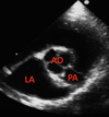
Identify the structure indicated by the number 2

right atrium

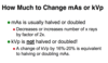
T/F: An overexposed radiograph is too light
False
An overexposed radiograph is too dark. Either kVp or mAs is too high.
T/F: If you suspect a lesion in the right lung of a dog, a left lateral thoracic radiograph should be made
True
What diagnostic imaging modalities might you use if you suspect a diaphragmatic hernia?
Radiographs, Ultrasound, Barium Study
RUB the hernia…
…creep
T/F: Atelectasis is associated with normal to increased size of the lung lobe
False
- Atelectasis is associated with decreased size of the lung lobe*
- Consolidation is associated with normal to increased size of the lung lobe*
This presentation is most often associated with _______ insufficiency

mitral insufficiency
Turbulent flow (regurgitation); often bright and a mixture of colors
T/F: Mammary adenocarcinomas are typically associated with mediastinal lymphadenopathy
False

T/F: Tracheobronchial lymphadenopathy is an example of a cranioventral disease
False
Tracheobronchial lymphadenopathy is an example of a dorsal disease
T/F: Pneumothorax may progress to pneumomediastinum
No!
Pneumomediastinum may progress to pneumothorax, but not the other way around
When using grids for radiographs, how should you adjust the mAs?
Increase mAs
The grid ‘intercepts’ scatter from patient before it reaches film. You need 2x-3x more photons when grid is used (higher mAs) due to absorption of primary beam by lead
If you suspect a right lung lesion, what radiographic views would you take for the dog? What about a horse?
Dog: R → L
Horse: L → R
Which has better contrast resolution: flat panel or film?
Flat panel

Identify the cardiac abnormality:

Pericardial Effusion
T/F: The cranial vena cava is normally visible radiographically in the mediastinum
False

T/F: Decreased mAs would contribute to increased film blackness
False
Increased mAs would increase film blackness
T/F: A diagnosis of cardiac failure can not be based on echocardiology alone
True
Are mediastinal masses typically more evident in a lateral view or a ventrodorsal view?
ventrodorsal (VD) view
T/F: The esophagus is normally visible on survey radiographs
FALSE
The esophagus is normally not visible on survey radiographs
What is the most common vascular ring anomaly?
Persistent right fourth aortic arch
If the distance between the film and the x-ray source decreases from 40” to 30”, how much does radiation intensity at the film change?
By 402/302
Intensity of radiation (x-rays/unit area) decreases with the square of the distance from the source
Identify the structure indicated by the red star:

caudal vena cava

This “scalloping” appearance of the lung margins indicates:

pleural effusion
Identify the lymphatic structure indicated by the number 2:

Cranial Mediastinal

For thoracic radiographs, what should your mAs and kVp settings be?
high kVp, low mAs
_________ is the extent to which a film, image plate or flat panel can be over and underexposed and still acheive an acceptable result
Exposure latitude
Identify the structure indicated by the red star:

right middle lung lobe

Is pneumomediastinum present in this radiograph?

Yes
Normally all of the tubular structures arent so readily visible, but in the case of pneumomediastinum gas acts as a contrast agent and allows for visualization of structures that would normally be undetected
Is this radiograph under-exposed or over-exposed?

Over-exposed
To correct this radiograph, you could decrease the mAs or decrease the kVp
The number of x-rays produced in a radiograph is quantified as:
mAs
T/F: Doubling the mAs doubles the amount of x-rays produced
True
Of the many structures present in the mediastinum of the normal thorax, only a few structures are seen radiographically.
Name ‘em, biatch!
- Heart
- Aorta
- Trachea
- Thymus (young animals)
- Caudal vena cava
- Occasionally Esophagus (left lateral)
“_H_ere _A_re _T_he _T_hings _C_ommonly _O_bserved” in the mediastinum
T/F: Border effacement is often associated with a bronchial pattern
False
Border effacement is associated with alveolar patterns
T/F: The diaphragm attaches to the ventral aspect of L3-L4
True
Pectus excavatum: detected or not detected?

Detected
Pectus excavatum is dorsal displacement of the sternum. It often results in narrowing of the thorax and is often associated with respiratory and CV anomalies
A change of kVp by _______ is equivalent to halving or doubling mAs
16-20%

Identify the lymphatic structure indicated by the number 1:

Sternal

If you see an interstitial pattern, is that considered airway or non-airway disease?
Non-airway

T/F: Grids are often used in radiography when the patient is less than 10 cm thick
False
Grids are often used in radiography when the patient is more than 10 cm thick. (Thicker patients create more scatter)
Which one of these radiographs is normal?
What do you observe in the abnormal one?

B is normal
In A, you can observe air bronchogram (this is indicative of an alveolar pattern) - there is air in the bronchus and the alveoli are filled with fluid. You can also observe border effacement, sillhouetting of the cranial margin of the heart.

Long-axis left ventricular outflow view.
Identify the structure indicated by the number 1

right ventricle

For interlobar fissures to be visualized radiographically when pleural effusion is present, how must the x-ray beam strike the fissures?
Tangentially
Is a bronchial lung pattern detected in this image?

No
T/F: Tracheal Stripe Sign is indicative of a pathological process

Not always
General anesthesia may result in signs that mimic megaesophagus and aspiration pneumonia
Identify the structure indicated by the red star:

cranial mediastinum

Note the V-sign on this radiograph. What is this indicative of?

Megaesophagus
On the VD view, when the enlarged esophagus is gas-filled, the left and right walls of the esophagus are sometimes visible as two soft tissue stripes that converge at the esophageal hiatus of the diaphragm

T/F: Pneumomediastinum does not result in dyspnea
True
For abdominal radiographs, what should your mAs and kVp settings be?
low kVp, high mAs
Radiographically, what do you expect to see with a bronchial pattern?
ring shadows (donuts) and tram lines

You should also see increased conspicuity of the bronchial tree
atelectasis or consolidation?

Atelectasis
Note the shift of the heart toward the rib (mediastinal shift), as well as less volume of the lung on the left hand side
What three structures are visualized in this image?

aorta, pulmonary artery, and left atria
When you see the Mercedes Benz sign, that indicates the aorta, pulmonary artery, and left atria

Identify the cardiac abnormality in this ultrasound view:

Dilated Cardiomyopathy (DCM)
Are x-rays positively or negatively charged?
Negatively charged
Right parasternal short axis view. What valve is indicated by the arrow?

Aortic valve
Creation of an opacity in a radiograph by overlapping of structures in the patient is termed:
summation effect
Is air bronchogram present in this image?

Yeah.

Name the MR sequence that nulls signal from fat:
STIR
_S_hort _T_au _I_nversion _R_ecovery

T/F: On the dorsal ventral view of the thorax of a dog, the pulmonary arteries are positioned lateral relative to the pulmonary veins
False
Pulmonary veins are ventral and central
Identify the artifact:

mirror image artifact

There is moderate pleural effusion in this patient.
Is this a DV view or a VD view?

VD
- The fluid is falling away from the heart, so the heart is visible*
- In a DV view, the view of the heart would be blocked by fluid*

There are two types of hiatal hernias. Which of the two is most common?
Sliding hiatal hernia
The other type is paraesophageal hiatal hernia
What are ‘the 5 opacities’ in radiology?
Air, Fat, Water, Bone, Metal

_A_lways _F_ind _W_ater _B_efore _M_eandering
___________ is the range of light intensities a medium can capture
Contrast Optimization
Is pneumomediastinum detected in this image?

No
Identify the cardiac abnormality:

Hypertrophic Cardiomyopathy
Note the thickening of the left ventricular walls

T/F: The caudal vena cava is normally visible radiographically in the mediastinum
True
Alveolar pattern right middle lung lobe: detected or not detected?

Detected
Hemothorax: detected or not detected?

Detected
What is the most common secondary thoracic lesion resulting from megaesophagus?
Aspiration Pneumonia
T/F: Fluid often accumulates in the esophagus in the right lateral view
False
Fluid often accumulates in the esophagus in the left lateral view
If you see air bronchogram on a radiograph, you should know it is a(n) _________ pattern

alveolar pattern
T/F: X-rays are devoid of mass
True
Name four characteristics of alveolar patterns on radiographs:
- Air bronchograms
- Border effacement
- Lobar distribution
- Labile
Identify the structure indicated by the red star:

trachea

T/F: An underexposed radiograph is too light
True
Either kVp or mAs is too low
In which radiographic view are hiatal hernias best visualized?
Left Lateral
Note the kink in the trachea on this VD view. This presentation is fairly characteristic for:

Persistent right fourth aortic arch

T/F: Tracheoesophageal stripe sign indicates tracheal pathology
False
Tracheoesophageal stripe sign indicates gas in the esophagus
What lung pattern is observed in this radiograph?

Alveolar
- Note the soft tissue opacity. Alveolar pattern is the only one that has this.*
- (Top DDx in this case would be pneumonia)*

T/F: MRI is the modality of choice to visualize bone
Hell no.
MRI doesn’t show bone, mutha f***a
T/F: Increased soft tissue opacity is observed with both atelectasis and consolidation
True
In which view is gas or fluid accumulation in the esophagus most visible?
left lateral
From what species was this radiograph taken?

Cat
Note the oblique lines in the caudal third of the esophagus due to a change to smooth muscle fibers - “herring bone pattern”
What is the most common cause of diaphragmatic hernias?
Trauma!
What thoracic lymph node would most likely be increased due to infection of the peritoneal space - peritonitis?
Sternal
What layer of the duodenum is the thickest/most hyperechoic?
Mucosal layer
What artifact is shown here?

Slice Thickness Artifact
T/F: Enlargement of the tracheobronchial lymph nodes may cause elevation of the tracheal bifurcation, depression of the tracheal bifurcation, or no change in tracheal bifurcation position
True
To decrease error % when measuring flow velocity, it is important to keep the doppler at a ___o angle
22o

What alveolar pattern characteristic is indicated by the arrows in this radiograph?
Air bronchogram

Long-axis left ventricular outflow view.
Identify the structure indicated by the number 3

left ventricle

Regarding ultrasound, is image resolution better with higher frequency or lower frequency?
higher frequency
Image resolution is better with higher frequency and smaller wavelength
Which view is more useful for determining whether a lesion is in the lung or the mediastinum: DV/VD or lateral?
DV or VD
T/F: Pyothorax is a common cause of unilateral distributions of pleural fluid
True
T/F: Sternal lymphadenopathy is an example of a cranioventral disease
True
T/F: A diagnosis of cardiac failure can be based on echocardiology alone
False!
Is pneumothorax detected in this image?

No
Is this image a film radiograph, digital radiograph, CT, or MRI?

CT
T/F: The mediastinum is a closed space
False
What echocardiographic modality would you use to determine normal and abnormal blood flow in the heart?
Doppler
T/F: Portal veins have a hyperechoic wall on ultrasound
True
Is tracheoesophageal stripe sign present in this radiograph?

Yes.

This indicates gas in the esophagus. This does NOT indicate tracheal pathology
Identify the artifact. Where is this most likely to be observed?

Slice Thickness Artifact
Seen at curved surfaces, such as the urinary or gall bladder
Bow-legged cowboy sign: detected or not detected?

Detected
This characteristic ‘wagon wheel’ appearance is characteristic for what portion of the intestine?

Ileum

Which view is this?

Left lateral
Roentgen Signs are six standard terms used to describe changes in a structure in a radiograph.
What are the Roentgen Signs?
- Size
- Shape
- Number
- Location
- Margination
- Opacity
What’s going on in the lungs?!

pulmonary osseous metaplasia
(mineralization)
Which is more hyperechoic: spleen or liver?
Spleen
- Order of increasing echogenicity:*
- (hypoechoic) Medulla - Cortex - Liver - Spleen - Prostate (hyperechoic)*
- “My cat loves sunny places”*
Is aortic insufficiency detected in this image?

Yes
- Blue = away; red = toward*
- This image should show all blue as blood should be from from the LV into the aorta*
T/F: Regarding pleural effusion: the space between lungs, and the lung and thoracic wall is radiolucent
False
Red arrow: nodule or blood vessel?

Nodule
The ones with the blue shit around them are blood vessels. The green nonsense is a bronchus

There is moderate pleural effusion in this patient.
Is this a DV view or a VD view?

DV
The fluid is blocking the view of the heart because it is between the lung and the pleural wall. The heart would be visible in a VD view

Holy shit, is that a lung nodule?!

No, calm down.
It’s a tick.

What pulmonary structure is indicated by “3”?

right pulmonary artery
The renal cortex is ______ when compared with the renal medulla
(hyperechoic, hypoechoic, anechoic, isoechoic)
The renal cortex is hyperechoic when compared with the renal medulla
The relative increase in echogenicity in the far field adjacent to an anechoic round structure is called:
acoustic enhancement
The spleen is ______ to the liver in the normal dog
(hyperechoic, hypoechoic, anechoic, isoechoic)
The spleen is hyperechoic to the liver in the normal dog
The image shows ultrasound image of two intestinal loops. What artifact is indicated by the red star?

Dirty Acoustic Shadowing
What two layers of the intestines are hypoechoic?
Mucosa and Muscularis
Pictured below is a sagittal sonogram of the liver. Identify the normal anatomic structure indicated by the red arrow:

Portal vein
The ultrasound probe is positioned on the ventral aspect of the abdomen with the patient in dorsal recumbency. What artifact is present here?

Mirror Image Artifact
Aortic insufficiency?

Yes.
What is the primary purpose of this piece of equipment in animal radiography?

Protect against scattered x-rays
And to make a fashion statement
If you set the kVp at 100, the mA at 50, and the time at 0.20 seconds, what is the mAs?
10
Lateral thoracic radiograph of an adult dog. What is the identity of the linear opacity indicated by the arrow?

pulmonary artery
Is pneumothorax detected in this image?

NO!
Pneumomediastinum is though