Dec18 Flashcards

1. to pass FRCR 2a neuro 2. To master neuro

1
Q

List the differences bwn ependymoma and medulloblastoma (6)

A

Medulloblastoma:

  1. Roof of 4th vent (midline- vermis)
  2. Hypredense
  3. 10% Ca
  4. Cysts are rare
  5. CSF seeding
  6. diffusion restriction
2
Q

Syndromes asso with medulloblastoma

A
  1. Cowden syndrome
  2. Gardner syndrome
  3. Turcot syndrome
  4. Gorlin syndrome
  5. Li-Fraumeni syndrome
  6. Coffin-Siris syndrome
  7. Rubinstein-Taybi syndrome
3
Q

What regions are affected in Pick’s dementia?

A
  • frontotemporal lobar degeneration
  • Caudate head volumes are also often reduced
4
Q

What is the diadnosis?

A

This is an exaple of frontotemporal lobar degeneration seen in Pick disease.

Striking asymmetry both of involvement of frontal vs temporal lobes, and involvement of left and right hemispheres.

5
Q

What is the diagnosis?

Describe the findings?

Syndrome associated with this tumour

A

This is a mass arising from the vermis and roof of the 4th ventricle causing hydrocephalus. Rarely cystic. 10% ca++

CSF seeding- imaging of whole spine needed.

Restricts diffusion

  1. Cowden syndrome
  2. Gardner syndrome
  3. Turcot syndrome
  4. Gorlin syndrome
  5. Li-Fraumeni syndrome
  6. Coffin-Siris syndrome
  7. Rubinstein-Taybi syndrome
6
Q

List the atrophied regions affected in PSP (3)

A

atrophy of midbrain, GP and frontal lobes.

Midbrain : pons reduced

Hummingbird sign and mickey mouse sign

7
Q

Traids of Wernicke encephalopathy:

A
  1. acute confusion
  2. ataxia
  3. ophthalmoplegia
8
Q

MRI findings in Wernicke

A

T2/FLAIR: symmetrically increased signal intensity in the:

  1. mamillary bodies
  2. dorsomedial thalami
  3. tectal plate
  4. periaqueductal area
  5. around the third ventricle

CT is usually normal.

9
Q

Deterioration of conscious level and agitation. Patient had gastric surgery 2 months ago for peptic ulcer.

What is the diagnosis?

A

Abnormal signal bilateral and symmetrical involving the medial and posterior aspect of both thalami displaying bright signal in DWI, T2 and FLAIR.

Diagnosis: Wernicke Encephalopathy.

Case Discussion

Previous gastric sugery or Bariatric sugeries are predisposing factors to various deficiency syndromes. Thiamine deficiency causes acute encephalopathy usually affects the medial thalamus, tectal plate and periacqueductal grey matter.

10
Q

What is the diagnosis?

Well circumscribed cysts, with an imperceptible wall, displacing adjacent structures, and following the CSF pattern (hypodense on CT and hyperintense on T2 with low on FLAIR). They can also have a remodelling effect on the adjacent bone.

A

Arachnoid cyst

Flair axial

11
Q

Best differentiating method bwn arachnoid and epidermoid

A

DWI- Epidermoid restricts diffusion.

Be aware of the white epidermoid.

12
Q

What is the diagnosis?

Progressive unintentional choreoathetoid movements, subcortical type dementia, behavioural changes, and psychosis in a 40 year old man.

On imaging, it is classically characterised by atrophy of the caudate nucleus with concomitant enlargement of the frontal horns of the lateral ventricles (<strong>BOX LIKE configuration</strong>). High SI in caudat and putamen on T2.

PET scan shows reduced FDG uptake in basal ganglia and frontal cortex.

A

Huntington disease

13
Q

Sign? Which condition?

the red nucleus and substantia nigra are surrounded by high T2 signal.
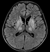
A

Panda sign

seen in Wilson disease

14
Q

Causes of cerebellar atrophy: (9)

A
  1. Phenytoin
  2. Alcohol
  3. Ataxic telangiectasia
  4. Paraneoplastc syndrome from bronchial ca
  5. Marijuanna
  6. Steroid
  7. Radiation
  8. Multisystem atrophy
  9. Gluten insensitivity
15
Q

Eye of tiger sign

A

Hallervorden spatz syndrome

NAA is also reduced

16
Q

Imaging protocols for mesial temporal sclerosis

How would you plan your scan to reduce volume averaging?

A

Coronal volume and coronal high resolution T2WI/FLAIR are best to diagnose MTS.

Thin section angled coronal sequences at right angles to the longitudinal axis of the hippocampus are required, to minimise volume averaging.

17
Q

Paediatric causes of ivory vertebrae (4)

A
  1. lymphoma: commonest cause, usually Hodgkin lymphoma
  2. osteosarcoma
  3. osteoblastoma
  4. blastic metastatic disease
    • neuroblastoma
    • medulloblastoma
    • Ewing sarcoma (rare)
18
Q

Adult causes of ivory vertebrae (7)

A
  1. osteoblastic metastases
    1. prostate cancer
    2. breast cancer
  2. lymphoma
  3. tuberculous spondylitis
  4. haemangioma
  5. chordoma
  6. Paget disease of bone
  7. SAPHO syndrome
19
Q

This line is used in evaluation of basilar invagination:

A

Mc Gregor line

From hard palat to the base of the occiput. The peg should lie 5mm below this level/

20
Q

Causes of basilar invagination: (4)

A
  1. Rickets
  2. Osteomalacia
  3. FD
  4. Developmental such as Klippel Feil
21
Q

Causes of drop mets in paeds (5)

A
  1. PNETs
  2. Medulloblastoma
  3. Ependymoma
  4. Germinoma
  5. Pineobastoma
22
Q

Causes of drop mets in adults:

ie irregularity and nodularity along the nerve root

A
  1. Melanoma
  2. Breast
23
Q

Most common locations for chordoma (2)

A
  1. sacrum
  2. clivus

Can mets to liver, lung, LN

24
Q

Name some features of Cleidocranial dysostosis

A

Delayed ossification of midline structures.

  • absent or hypoplastic lateral clavicles.
  • supernumeray ribs
  • Hemivertebrae
  • Widened pubic symphysis
  • absent/short radius
  • wormian bones
  • widened anterior fontanelle
  • large mandible
  • Small paranasal sinuses.
  • Bell shaped chest
25
Q

This suture can be affected in CD (cleidocranial dysostosis)

A

premature fusion of the coronal suture (brachycephaly)

26
Q

What area is affected in Lewy body dementia? (3)

A

parietal

occipital

and cerebellum

27
Q

Typical location for colloid cyst

A

Foramen of Monroe and 3rd ventricle

28
Q

Typical location for IV meningioma

A

trigone of Lateral ventricle

29
Q

Typical location for IV ependymoma and neurocytoma

A
  • Ependymoma: LV and 4th ventricle
  • Neurocytoma LV and septum pellucidum
30
Q

Described features of intracranial hypotension

A

The most common qualitative finding is

  1. pachymeningeal thickening and enhancement followed by
  2. dural venous engorgement,
  3. tonsillar herniation, and
  4. subdural collection;
31
Q

? Diagnosis

On brain imaging, it usually demonstrates vermian atrophy, compensatory enlargement of the fourth ventricle, cerebral infarcts and cerebral haemorrhage

A

Ataxia telangectasia

The main clinical characteristics include:

  1. cerebellar ataxia: progressive and present in all cases
  2. oculomucocutaneous telangiectasias
  3. greater susceptibility to types of infection (partial combined immunodeficiency) and neoplasms
32
Q

Valuable differentiator of ataxia telangectasia (AR)

A

MR spectroscopy:

increased choline signal in the cerebellum has been described as a valuable differentiator from other forms of ataxia

33
Q

What passes through the SOF?

A

3,4, 5(a) and 6

34
Q

Most common orbital mets in children

A
  1. Neuroblastoma
  2. Ewing’s
  3. Leukaemia
35
Q

Most common orbital mets in adults

A
  1. Breast
  2. Lung

Deposits are intra-ocular

36
Q

May be incidental but symptomatic masses produce pulsatile tinnitus, otalgia, or conductive hearing loss

A

Glomus tympanicum paragangliomas

37
Q

Diagnois?

CT: opacified frontal sinus with stranding and swelling of the overlying scalp. Bone algorithm will often demonstrate a defect in the anterior wall of the sinus.

Contrast may demonstrate a focal abscess, and may also allow intracranial complications to be better delineated.

A

Pott Puffy tumour

38
Q

What signal characteristics would be most characteristic on MRI for schwannoma?

  1. Iso to grey matter on T2 weighted images
  2. Hyperintense on T1 weighted images
  3. Hypointense on T2 weighted images
  4. Doesn’t enhance following contrast on T1 weighted images
  5. Isointense to the grey matter on T1 WI
A
  1. Isointense to the grey matter on T1 WI
39
Q

Cholesterol granuloma vs cholesteatoma (3)

A

Granuloma:

  1. T1 bright
  2. T2 bright
  3. No restriction

Cholesteatoma

  1. T1 dark
  2. T2 bright
  3. Restrict diffusion (- epidermoid) (DWI bright)

Both cause Expansile bony changes

40
Q

Abducens palsy (CN6) since last 1 week. Pain. Hearing loss. Tinnitus.

CT: A region of irregular lytic destruction in the left petrous apex

T1 weighted images it is of intermediate to low signal intensity with peripheral irregular enhancement.

A

Features are consistent with infection of the petrous apex (petrous apicitis) resulting in Gradenigo syndrome:

  1. otomastoiditis
  2. facial pain- trigeminal neuropathy.
  3. lateral rectus palsy (Dorello canal)
41
Q

Vertigo and tinnitus in a patient with previous history of VHL.

CT: Moth eaten, permeative lytic lesion in the right petrous temporal bone involving the vestibular aqueduct.

A

Endolymphatic sac tumor

  • almost always have internal amorphous calcification,
  • T2 bright
  • intense enhancement
  • very vascular with flow void and tumour blush on angio
42
Q

33 yrs old female patient with von Hippel-Lindau syndrome presents with an expansile lesion in jugular foramen and retro labyrinthine portion of the right temporal bone.

A

Endolymphatic sac tumour.

43
Q

This condition causes progressive sensorineural hearing loss that is associated with absence of bony modiolus in more than 90% of patients

A

Large vestibular aqueduct syndrome

The Aqueduct it Becomes enlarged > 1.5mm

Often bilateral

Associated with cochlear deformity- 100%

44
Q

75 Male, Flu 2 months previously. Since then, worsening sensorineural hearing loss in the left ear.

MRI: Diffusion restriction, enhancement, and T2 hypointense signal within the left cochlear aperture, basal turn of the cochlea, and vestibule and semicircular canals. Mastoid effusion with enhancement.

A

Abnormal signal and enhancement in the left membranous labyrinth. In the context of subacute hearing loss and with bilateral mastoiditis, these findings are most suggestive of infectious labyrinthitis

45
Q

? diagnosis

Ossification of the right lateral, posterior and superior semicircular canals as well as the apical turn of the cochlea.

A

Labyrinthitis ossificans

46
Q

Conductive hearing loss in adults female

A

Otosclerosis

47
Q

The Tullio phenomenon describes the precipitation of vertigo and nystagmus by a loud noise. What conditions this is seen in?

A
  1. superior semicircular canal dehiscence syndrome (SCDS)
  2. Ménière disease
  3. congenital syphilis with a semicircular canal fistula
  4. post-fenestration patient if the footplate is mobile
48
Q

35 M, enhanced hearing sensation, sometimes dizzy when exerting pressure, slight conduction loss at physical exam.

A

superior semicircular canal dehiscence

49
Q

euthyroid goitre associated with sensorineural hearing loss.

Inner ear malformation

modiolus deficiency

vestibular aqueduct enlargement

A

Pendred syndrome

50
Q

What causes abnormal enhancement of facial nerve?

What type of fracture damages your facial nerve?

A
  1. Bells palsy
  2. Lymes
  3. Ramsay Hunt
  4. Cancer

Transverse T bone fracture can damage your facial nerve

51
Q

Mc Cune Albright syndrome (3)

A
  1. Multi FD
  2. Cafe au Lait spots
  3. Precocious puberty
52
Q

16M, Right sided nasal obstruction and rhinorrhoea.

A

It tends to spread via the pterygopalatine fossa and osseosus destruction is common.

Highly vascular and biopsy is contraindicated due to the risk of haemmorhage.

53
Q

27M, nasal blockage; cold; headache

CT:

  • defined mass with mucin density arising from the maxillary sinus.
  • widening of the maxillary ostium and extending into the nasopharynx
  • no associated bony destruction but rather smooth enlargement of the sinus
A

Antrochoanal polyp

Widening of maxillary ostium

54
Q

40M patient with nasal stuffiness presented to ENT

A

Inverting papilloma

Classic location is the lateral wall of nasal cavity- related to middle turbinate.

Cerebriform pattern- alt bands on T1 and T2

10% squamous cell ca

55
Q

45 year old female with anosmia and nasal blockade since 2-3 months.

dumbell shaped with waist at the cribiform plate and avid enhancemet

A

Esthesioneuroblastoma