CORText Flashcards
Where do enchondromas occur?
Femur, humerus, tibia and small bones of hand/feet.
What is Reiter’s syndrome?
Triad of symptoms: urethritis, uveitis or conjunctivitis and arthritis.
Seen in reactive arthritis.
What treatment is best for mixed connective tissue disease?
Raynauds - calcium channel blockers.
If there is significant muscle or lung disease - immunosuppression.
What is an arthroplasty?
Removal of a diseased joint through excision or resection.
What is a Barlow test?
Dislocatable hip with flexion and posterior displacement.
How are undisplaced, minimally displaced and minimally angulated (stable) fractures managed?
Period of splintage or immobilisation.
Rehabilitation.
What is cauda equina syndrome?
Very large central disc prolapse compressing on all the nerve roots of the cauda equina - surgical emergency as the nerve roots involving defaecation and urination are affected.
Prolonged compression can cause permanent nerve damage requiring colostomy and urinary diversion.
What radiographic changes are seen in a late case of avascular necrosis?
Patchy sclerotic sclerosis of weight-bearing area of the femoral head.
Lytic zone underneath femoral head formed by granulation tissue from attempted repair.
What is the function of the lateral collateral ligament?
Resists varus force and abnormal external rotation of the tibia.
What are the red flags of back pain?
Neurological lower limb symptoms with bowel or bladder dysfunction.
Back pain in young patients (<20 years).
Sudden onset of NEW back pain in older patients (>60 years).
Nature of pain - constant, severe pain, worse at night.
Back pain with systemic symptoms of upset.
What is a large vessel vasculitis?
Primary vasculitis that causes granulomatous inflammation predominantly of the aorta and its major branches.
How is giant cell arteritis diagnosed?
Inflammatory markers (CRP, ESR or plasma viscosity) are almost always raised.
Temporal artery biopsy - mononuclear infiltration or granulomatous inflammation, usually with multinucleated giant cells.
What are the features suggestive of a benign soft tissue neoplasm?
Smaller size.
Fluctuation in size (malignant tumours don’t regress in size).
Cystic lesions.
Well-defined lesions.
Fluid-filled lesions.
Soft/fatty lesions.
What is carpal tunnel syndrome?
Swelling within the carpal tunnel resulting in median nerve compression.
What is the common organism that produces a late haematogenous prosthetic infection?
Staph. aureus.
Beta haemolytic strept.
Enterobacter.
What distribution of symptoms do cervical and lumbar nerve root compression give?
Dermatomal and myotomal distributions.
What are the upper limb compressive neuropathies?
Carpal tunnel syndrome.
Cubital tunnel syndrome.
What is the name of the radiographic sign associated with posterior dislocations of the humeral head?
The ‘lightbulb’ sign.
What loss of function are fractures of the distal radius that heal in a poor position (a malunion) may result in impaired grip strength associated with?
Loss of extension.
What is the action of infraspinatus?
External rotator.
What are the causes of cubital tunnel syndrome?
Tight band of Osborne’s fascia (fascia forming the roof of the tunnel).
Tightness at the intermuscular septum as the nerve passes through.
Tightness between the two heads at the origin of the flexor carpi ulnaris.
Where are the secondary lesions of osteosarcoma normally found?
Pulmonary mets.
Jaw, proximal humerus, proximal femur, mid-femur.
What is the management of proximal humeral fractures?
Minimally displace - conservative treatment with a sling and gradual return to mobilisation.
Displaced fractures - position often improves once muscle spasm settles.
Persistently displaced - internal fixation surgically, but stiffness, chronic pain and failure of fixation can occur particularly in the older patient.
Where does an acute intervertebral disc tear occur?
In the outer annulus fibrosis of an intervertebral disc.
What is an incomplete spinal cord injury?
Some neurological function (sensory and/or motor) is present distal to the level of injury.
What ligament can be ruptured in a varus stress injury?
Lateral collateral ligament.
What blood tests are raised in someone with large-vessel vasculitis?
ESR.
Plasma viscosity.
CRP.
What arthritis has this typical pattern?
Rheumatoid arthritis.
What is a gibbus?
Term used for an acute angular deformity in the spine in the sagittal plane.
What is Dupuytren’s contracture?
Palmar fascia undergoes hyperplasia with normal fascial bands forming nodules and cords progressing to contractures at the MCP and PIP joints.
What is the haematological presentation of SLE?
Leukopenia.
Lymphopenia.
Anaemia (may be haemolytic).
Thrombocytopenia.
What is an Ortolani test?
Reducing a dislocated hip with abduction and anterior displacement.
How is neurogenic shock treated?
IV fluid therapy.
What is the treatment for slipped upper femoral epiphysis?
Urgent surgery to pin the femoral head to prevent further slippage.
Greater the degree of slip the worse the prognosis, some cases may require hip replacement.
What investigations must you do in suspected cauda equina syndrome?
Rectal exam.
Urgent MRI.
What are osteoblasts?
Bone forming cells found on the surface of developing bone.
They have plentiful rough endoplasmic reticulum and prominent mitochondria.
What is the mechanism behind the pathology of Paget’s disease?
Increased osteoclastic activity (possibly due to an exaggerated response to vitamin D) causes increased bone turnover.
Osteoblasts become more active to compensate for this excessive bone resorption and form new bone.
This bone fails to remodel sufficiently -> despite increased thickness and bone density is brittle -> easily fractures.
What is the pathogenesis of polymyositis?
T-cell mediated cytotoxic process against unidentified muscle antigens -> CD8 T-cells and macrophages attack healthy non-necrotic muscle fibres.
Autoimmune response to nuclear and cytoplasmic autoantigens detected in 60-80% of patients.
What is pes cavus?
Abnormally high arch of the foot.
What are the risk factors for type 2 osteoporosis?
Reduce sunlight exposure (vitamin D).
Genetics.
Smoking.
Alcohol abuse.
Lack of exercise.
Poor diet.
What happens if developmental dysplasia of the hip is left untreated?
The acetabulum is very shallow.
In severe cases, a false acetabulum occurs proximal to the original one with a shortened lower limb.
Severe arthritis due to reduced contact area can occur at a young age and gait/mobility may be affected.
What investigations aid diagnosis of anti-phospholipid syndrome?
May be thrombocytopenic and prolongation of APTT.
Lupus anticoagulant, anti-cardiolipin and anti-beta 2 glycoprotein antibodies may be positive.
What is the function of the anterior cruciate ligament?
Prevent abnormal internal rotation of the tibia.
What shoulder problems tend to occur in the younger adult?
Instability.
Which bacteria is the most common cause of septic arthritis in adults?
Staphylococcus aureus.
What are the classical radiological findings associated with osteoarthritis?
Loss of joint space.
Subchondral cysts.
Osteophyte formation.
Subchondral sclerosis.
What arthritis has this typical pattern?
Inflammatory spondylitis.
What is a chrondrosarcoma?
Cartilage producing primary bone tumour.
What does a DAS 28 score of >5.1 indicate?
High disease activity.
What are the symptoms of cauda equina syndrome?
Bilateral leg pain.
Paraesthesiae or numbness (saddle anaesthesia).
Altered urinary function - retention/incontinence.
Faecal incontinence/constipation.
A patient has noticed gradually increasing tiredness of muscles on activity, particularly climbing stairs and drying her hair. She has also developed a purplish rash around her eyes and over her chest. What is the most likely diagnosis?
Dermatomyositis.
Where is osteomyelitis most likely to affect in children?
Metaphyses of long bones because they have many vessels that have sluggish flow resulting in an accumulation of bacteria allowing infection to spread towards the epiphysis.
What is gout and what usually causes it?
A crystal arthropathy caused by deposition of urate crystals within a joint, usually due to high serum uric acid levels (hyperuricaemia).
Which bacteria is a cause of septic arthritis in the elderly, the seriously ill and IV drug users?
Escherichia coli.
What is the clinical presentation of a mallet finger?
Pain.
Drooped DIP joint of the affected finger.
Inability to extend at the DIP joint.
What is a sequestrum?
Dead fragment of bone.
What can lead to joint instability?
Consequence of an injury.
Can be related to ligamentous laxity.
Can be predisposed by anatomic variation or underlying disease process (e.g. cervical spine instability in rheumatoid arthritis).
What types of presentation can occur in SLE?
Constitutional.
Musculoskeletal.
Mucocutaneous.
Renal.
Haematological.
Neuropsychiatric.
Cardiac.
Gastrointestinal.
What is Klumpke’s palsy?
Lower brachial plexus injury (C8 & T1 roots) caused by forceful adduction which results in paralysis of the intrinsic hand muscles +/- finger and wrist flexors and possible Horner’s syndrome.
What is a Galeazzi injury?
Fracture of the radius with dislocation of the distal radio-ulnar joint (DRUJ).
What is the clinical presentation of gout?
intensely painful, red, hot and swollen joint which may mimic septic arthritis.
Symptoms may last 7-10 days untreated.
What is a giant cell tumour of the tendon sheath?
Small firm swelling usually found on the flexor tendon sheath of a finger.
May or may not be painful but can erode bone if large enough.
What can chronic gout result in?
Destructive erosive arthritis.
What is the treatment for scoliosis?
Mild, non-progressive - no surgery.
Severe - surgery of vertebral fusions and long rods connecting the posterior elements of the spine.
What can cause pes cavus?
Often related to neuromuscular conditions e.g. hereditary sensory and motor neuropathy, cerebral palsy, polio (unilateral) and spinal cord tethering from spina bifida occulta.
What is the clinical presentation of an ACL rupture?
Pop felt or heard when it occurs.
Haemarthrosis (an effusion due to bleeding in the joint) occurs within an hour of injury.
Deep pain in the knee.
Chronically patient complains of rotatory instability.
You are a general practitioner and a patient presents to you with an acutely hot, painful and swollen knee. They made an urgent appointment this morning as the pain began suddenly overnight. There is no history of significant trauma. The patient is on long-term steroids for a respiratory condition. Examination reveals the patient is pyrexial and the knee range of movement is severely reduced due to pain.
What is the most appropriate management?
Urgent orthopaedic referral. This patient must be seen today as they may require washout of the joint.
A patient has had successful treatment of acute gout but has had 4 flares this year. What would be the most appropriate treatment to prevent further attacks?
Allopurinol.
When is a soft callus usually formed following a fracture?
2-3 weeks.
What is the management for Perthes disease?
No specific treatment.
Regular xrays and observation, avoidance of physical activity.
~50% cases do well.
Some cases occasionally require osteotomy of the femur or acetabulum.
What are the clinical features of ankylosing spondylitis?
Spinal pain.
Stiffness.
Knee/hip arthritis.
With times there is loss of lumbar lordosis and increased thoracic kyphosis.
What is meant by translation of a fracture?
Translation of a distal fragment can be described as anteriorly or posteriorly, and medially or laterally translated.
It is estimated with reference to the width of the bone (100% displacement is generally referred to as an ‘off-ended’ fracture.
What is the normal developmental milestone for loss of primitive reflexes (Moro reflex, stepping reflex, rooting, grasp reflex, fencing posture)?
1-6 months.
What is the usual treatment for primary bone tumours?
Surgery to remove a tumour with a wide margin of 3-4cm and a cuff of normal muscle all around; biopsy tract is also removed with the tumour.
Adjuvant chemotherapy and radiotherapy, and neo-adjuvant if appropriate.
What is the pathology of Dupuytren’s contracture?
Proliferation of myofibroblast cells and the production of abnormal collagen (type 3 rather than type 1).
What are predisposing factors of patellar dislocation?
Ligamental laxity.
Female gender.
Shallow trochlear groove.
Genu valgum.
Femoral neck anteversion.
High riding patella (patella alta).
What is fracture disease?
Term used to describe stiffness and weakness due to the fracture and subsequent splintage in cast.
Most cases resolve with time and may be helped with physio.
What part of the bone does localised osteomyelitis normally affect?
Affects cortex and medullary bone.
What are the functions of the tibialis posterior tendon?
Support the medial arch of the foot.
Plantarflexor and invertor of the foot.
What part of the bone does diffuse osteomyelitis affect?
A segment of bone is infected resulting in skeletal instability e.g. infected non-union.
What joints can be affected in rheumatoid arthritis?
Early: MCPs, PIPs in the hands and feet, wrists.
Late: atlantoaxial joint (C1-2), elbows, shoulders, TMJ, hips and ankles.
What can trigger uric acid crystals to precipitate in joints?
Dehydration.
Trauma.
Surgery.
What is the risk of recurrent shoulder dislocation following a shoulder dislocation?
Patients less than 20 have an 80% chance of re-dislocation.
Patients over 30 have a 20% risk of further dislocation.
What is the cutaneous presentation of systemic sclerosis?
Raynauds phenomenon.
Telangiectasia.
Calcinosis (subcutaneous deposits of calcium in the digits).
3 phases: (1) oedematous, (2) indurative, (3) atrophic - thickened and tight skin.
Major features: centrally located skin sclerosis that affects the arms, face and/or neck.
Minor features: Sclerodactyly and atrophy of the fingertips, bilateral lung fibrosis.
What is the management of Sjogren’s syndrome?
Largely symptomatic.
Lubricating eyedrops.
Saliva replacement products.
Regular dental care.
Pilocarpine can be used to stimulate saliva production - SE flushing.
Hydroxychloroquine - aids arthralgia/fatigue.
What is the clinical presentation for slipped upper femoral epiphysis?
Pain and a limp.
Pain felt in groin, but also pts can present with purely pain in the knee (due to obturator nerve supplying both the hip and knee).
Loss of internal rotation of the hip is predominant sign.
What are the clinical features of rheumatoid arthritis?
Symmetrical synovitis (doughy swelling).
Tenosynovitis.
Pain.
Morning stiffness.
What fracture is often associated with a Colles fracture?
Fracture of the ulnar styloid.
Where do osteosarcomas occur?
Seen in younger age groups (adolescence and early adulthood) with 60% involving the bones around the knee.
Other sites include proximal femur, proximal humerus and pelvis.
When would a patient be put on a biologic therapy?
When a patient’s DAS28 score is still >5.1 when on 2 DMARDs.
What is the gender ratio for polymyositis and dermatomyositis?
Females:males = 2:1.
Affects people over 20 years, especially those aged 45-60.
What are osteoprogenitor cells?
Located on bone surfaces, for example under the periosteum, these cells serve as a pool of reserve osteoblasts.
What is the treatment for a prolapsed intervertebral disc?
Analgesia and physiotherapy.
Symptoms usually resolve but can take 2-3 months to settle.
What is the gender ratio of SLE?
Female:male = 11:1.
What are osteocytes?
A bone cell trapped within the bone matrix.
What is fibromyalgia?
Disorder of central pain processing or a syndrome of central sensitivity.
Patients tend to have a lower threshold of pain and of other stimuli such as heat, noise and strong odours.
Commonest in young and middle-aged women.
What are the 3 main patterns of injury to the pelvis?
Lateral compression fracture.
Vertical shear fracture.
Anteroposterior compression injury.
What is an anteroposterior compression injury?
May result in wide disruption of the pubic symphysis.
Substantial bleeding from torn vessels occurs and the pelvis can hold several litres of blood (i.e. the entire circulating volume).
What are the flexor and extensor imbalances of claw toes?
Hyperextension at the MTPJ with hyperflexion at the PIPJ and DIP.
What is meant by angulation of a fracture?
It described the direction in which the distal fragment points towards and the degree of this deformity.
It can be described as medial or lateral and posterior or anterior.
Can be measured in degrees from the longitudinal axis of the diaphysis of a long bone.
What is the clinical presentation of Perthes disease?
Children present with pain and a limp.
Unilateral normally, bilateral cases represent an underlying skeletal dysplasia or thrombophilia.
Loss of internal rotation, followed by loss of abduction, followed by positive Trendelenburg test for gluteal weakness.
Why do neuromuscular disorders occur?
Due to abnormal or deficient motor neuron signals to skeletal muscle due to defect in either the brain, spinal cord, peripheral nerve, neuromuscular junction or muscle.
What is Erb’s palsy?
Injury to the upper (C5 & C6) nerve roots resulting in loss of motor innervation of the deltoid, supraspinatus, infraspinatus, biceps and brachialis muscles.
Internal rotation of the humerus (from unopposed subscapularis) -> classic waiter’s tip posture.
Where does chronic osteomyelitis tend to occur in adults?
In the axial skeleton (spine or pelvis) with haematogenous spread from pulmonary or urinary infections, or from infection of the intervertebral disc (discitis).
What is diffuse systemic sclerosis?
Skin changes develop more rapidly and can involve trunk.
Early significant organ involvement.
Anti-Scl-70 antibody association.
Which metacarpal fracture often occurs with a punching injury?
5th metacarpal fractures, particularly 5th MCP neck fractures (Boxer’s fracture).
What are the clinical features of Ehlers-Danlos?
Profound joint hypermobility.
Vascular fragility with ease of bruising.
Joint instability.
Scoliosis.
What fractures tend to occur in people with type 1 osteoporosis?
Colles fractures.
Vertebral insufficiency fractures.
What is the treatment for large-vessel vasculitis?
Corticosteroids (40-60mg prednisolone with gradual reduction).
Methotrexate and azathioprine may be added in.
What is scoliosis?
Lateral curvature of the spine which can be idiopathic or secondary to neuromuscular disease, tumour, skeletal dysplasia or infection.
What investigations can diagnose polymyositis?
Inflammatory markers raised.
Serum creatine kinase is usually raised - often 10x normal.
Autoantibodies = ANA, anti-Jo-1, anti-SRP.
MRI = localise extent of muscle involvement.
Electromyograph - short-duration, low-voltage, polyphasic motor unit action potentials with spontaneous fibrillation activity.
Muscle biopsy - muscle fibres with varying stages of inflammation, necrosis and regeneration.
What is trigger finger?
Tendonitis of a flexor tendon to a digit can result in nodular enlargement of the affected tendon (usually distal to a fascial pulley over the metacarpal neck (A1 pulley).
Movement of the finger produces a clicking sensation, as this nodule passes under the pulley - can be painful or finger may lock in a flexed position as the nodule passes under the pulley and can’t go back on extension.
What is mixed connective tissue disease?
A defined condition which features symptoms seen in other connective tissue diseases.
What is the clinical presentation of vasculitis?
Varies according to the histological type of inflammation, the size of the involved blood vessel segment and the distribution of the involved vessels.
What are the risk factors for type 1 osteoporosis?
Early menopause.
Genetics.
Smoking.
Alcohol abuse.
Lack of exercise.
Poor diet.
What does the primary survey of a major trauma include?
Airway management with cervical spine control.
Breathing & ventilation.
Circulation & bleeding control.
Disability (neurological examination).
Exposure & environmental control.
ABCDE evaluation.
What are the symptoms of upper motor neuron neuromuscular disorders?
Weakness.
Spasticity (rate dependent increased tone).
Hyperreflexia.
Extensor plantar response (Babinksi sign).
What are risk factors for clubfoot?
Boys twice as likely to get it than girls.
Potential genetic link (familial).
More common in breech presentation.
Oligohydramnios (low amniotic fluid).
What are the late local complications of a fracture?
Stiffness.
Loss of function.
Chronic regional pain syndrome.
Infection.
Non-union.
Mal-union.
Volkmann’s ischaemic contracture.
Post-traumatic osteoarthritis.
Deep vein thrombosis.
What type of local soft tissue swellings can present to orthopaedics?
Inflammatory swellings - bursitis, rheumatoid nosules.
Infection - abscess.
Cystic lesions - ganglion, meniscal cyst, Baker’s cyst.
Benign neoplasms.
Malignant neoplasms.
What is the treatment of bony nerve root entrapment?
Surgical decompression, with trimming od the impinging osteophytes can be performed in suitable candidates.
What is bursitis?
Inflammation of a bursa (small fluid-filled sac lined by synovium around a joint).
What is the treatment for developmental dysplasia of the hip?
Mild cases - observation.
Severe cases - unstable hips are reduced and held with a Pavlik harness to keep them in comfortable flexion and abduction -> maintaining reduction.
If persists over 18 months old, open reduction may be required.
What are causative factors of plantar fasciitis?
Diabetes.
Obesity.
Frequent walking on hard floors with poor cushioning in shoes.
What is neurogenic shock?
Occurs secondary to temporary shutdown of sympathetic outflow from the cord from T1 to L2, usually due to injury in the cervical or upper thoracic cord leading to hypotension and bradycardia.
Usually resolves within 24-48 hours.
Priapism from unopposed parasympathetic stimulation may be present.
What is the action of supraspinatus?
Initiation of abduction.
What is the name given to the space that chondrocytes occupy?
The extracellular matrix termed a lacuna.
How are unstable injuries managed?
Surgical stabilisation.
What are the clinical features of spinal claudication?
Claudication distance is inconsistent.
Pain is burning (rather than cramping).
Pain is less walking uphill - spine flexion creates more space for the cauda equina.
Pedal pulses are preserved.
What are the clinical features of inflammatory arthritis?
Joint pain with associated swelling.
Morning stiffness.
Improvement in symptoms with exercise.
Synovitis on examination.
Raised inflammatory markers (CRP and plasma viscosity).
Extra-articular symptoms.
What is a fibrosarcoma?
Malignant tumour of fibrous tissue.
What is the normal developmental milestone for eating with fingers or using a spoon?
14 months.
What causes avascular necrosis?
Idiopathic.
Alcoholism.
Steroid (ab)use.
Primary hyperlipidaemia.
Thrombophilia.
Sickle cells disease.
Antiphospholipid deficiency in SLE.
Caisson’s disease (or decompression sickness).
Condition X is an autosomal dominant or sporadic mutation of the fibrillin gene resulting in tall stature with disproportionately long limbs and ligamentous laxity. Associated features include a high arched palate, scoliosis, pectus excavatum, eye problems (lens dislocation, retinal detachment), aortic aneurysm and cardiac valve incompetence.
What is Condition X?
Marfan’s syndrome.
What score (or below) on the GCS implies a loss of airway control and should prompt the treating doctor to perform airway manoeuvres?
8 or less.
What features of ankylosing spondylitis can be seen on MRI?
Bone marrow oedema and enthesitis of the spinal ligaments.
What are the common sites affected by osteochondritis?
Second metatarsal head (Freiburg’s disease).
Navicular bone (Kohler’s disease).
Lunate of the carpus (Kienbock’s disease).
Capitellum of the elbow (Panner’s disease).
Vertebral bones (Scheuermann’s disease).
What is the treatment of calcium pyrophosphate deposition disease (CPPD)?
NSAIDs, corticosteroids (systemic and intra-articular) and occasionally colchicine.
What is an oblique fracture?
Occur with a shearing force (e.g. fall from height, deceleration).
Can be fixed with an interfragmentary screw.
Tend to shorten the bond and many also angulate.
What are the consequences of talipes equinovarus (clubfoot)?
Contractures of the soft tissues -> deformity consisting of ankle equinus (plantarflexion), supination of the forefoot and varus alignment fo the forefoot.
What forms the carpal tunnel of the wrist?
Carpal bones and the flexor retinaculum.
What are the main categories of arthritis?
Non-inflammatory arthritis (commonly osteoarthritis).
Inflammatory arthritis.
What are structures A and B?
A - perimysium.
B - endomysium.
What are the structures of the extensor mechanism of the knee from most proximal to most distal?
Quadriceps muscles.
Quadriceps tendon.
Patella.
Patellar tendon.
Tibial tuberosity.
A Colles fracture describes a dorsally angulated or displaced fracture at what site?
Distal radius.
What are the clinical signs of Achilles tendon rupture?
Weakness of plantar flexion and a palpable gap in the tendon are usually apparent.
No plantarflexion of the foot is seen when squeezing the calf (Simmonds test).
How can an ACL rupture occur?
High rotation force e.g. turning the upper body laterally on a planted foot -> internal rotation on the tibia.
What is the shoulder girdle formed of?
Scapular.
Clavicle.
Proximal humerus.
Supporting muscles including the deltoid and rotator cuff.
What is Henoch-Scholein purpura?
Acute IgA-mediated disorder characterised by a generalised vasculitis involving the small vessels of the skin, the GI tract, kidneys, joints, lungs (rarely) and the CNS.
Commonly affects children - common history of an upper respiratory tract infection predates the symptoms by a few weeks.
Which tissues cannot accept a skin graft?
Bare tendon.
Bone.
Any exposed metalwork.
What factors predispose to patellar dislocations and subluxations?
Generalised ligamentous laxity.
Valgus alignment of the knee.
Rotational malalignment (including femoral neck anteversion).
Shallow trochlear groove.
What is osteoporosis?
A quantitative defect of bone characterised by reduced bone mineral density and increased porosity.
Causes fragility of the bone and increased fracture risk.
How can osteomyelitis be caused?
Organisms can infect the bone directly by penetrating trauma or surgery.
Indirectly by haematogenous spread from an infection or bacteraemia at a distant site.
How can carpal tunnel syndrome be investigated?
Nerve conduction studies - slowing of conduction across the wrist.
What is joint instability?
Abnormal motion of a joint (rotation or translation) resulting in subluxation or dislocation with pain and/or giving way.
What is internal tibial torsion?
Tibia can be rotated inward about its vertical axis.
Ignore - normal variation.
What investigations can aid diagnosis of SLE?
FBC - anaemia, leukopenia, thrombocytopenia.
Immunology: anti-nuclear antibody, anti-dsDNA antibody (varies with disease activity), anti-Sm, anti-Ro, anti-La, anti-RNP, C3/4 (low when disease is active).
Urinalysis - glomerulonephritis.
CT chest - interstitial lung disease.
MRI brain - cerebral vasculitis.
Echocardiogram - pericardial effusion.
What is the most common neuromuscular disorder?
Upper motor neuron disorder.
Lower motor neuron disorder.
What is the function of the posterior cruciate ligament?
Prevent hyperextension and anterior translation of the femur.
What is the normal developmental milestone for sitting alone and crawling?
6-9 months.
What is an arthrodesis?
Surgical stiffening or fusion of a joint in a position of function.
Remaining hyaline cartilage of the joint and subchondral bone is removed and the joint is stabilised resulting in bony union.
What can predispose to slipped upper femoral epiphysis?
Hypothyroidism.
Renal disease.
What is tarsal coalition?
Abnormal bridge (bony, fibrous or cartilaginous) between the calcaneus and navicular or between the talus and calcaneus.
Can lead to painful fixed flat foot deformity.
What is a fight bite?
It is associated with punching injuries and describes a laceration sustained to the puncher’s hand from the punchee’s tooth.
What can systemic sclerosis do to the gastrointestinal tract?
Dysphagia.
Malabsorption.
Bacterial overgrowth of the small bowel.
What is a Salter-Harris I fracture?
Pure physeal separation.
Best prognosis and is least likely to result in growth arrest.
What is the management of greenstick fractures?
These fractures may be angulated and may require manipulation and casting if there is significant deformity, particularly in the older child.
What is spondylolisthesis?
Slippage of one vertebra over another and can be due to a developmental defect or a recurrent stress fracture of the posterior elements which fails to heal.
Usually L4/L5 or L5/S1.
What part of the bone does medullary osteomyelitis affect?
The medulla of the bone.
What arthritis has this typical pattern?
Psoriatic arthritis.
What is Ehlers-Danlos syndrome?
Heterogeneous condition often autosomal dominant.
Abnormal elastin and collagen formation.
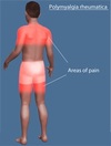
How is polymyalgia rheumatica diagnosed?
No specific diagnostic test but it is almost always associated with raised CRP and plasma viscosity/ESR.
What is the characteristic appearance of a giant cell tumour on an x-ray?
Soap bubble.
What are soft tissue procedures?
Ligament tightening/advancement.
Ligament reconstruction using a tendon graft.
Soft tissue reattachment.
What is a transverse fracture?
Occur with pure bending force where the cortex on one side fails in compression and the cortex on the other side n tension.
May not shorten the bone (unless completely displaced) but may angulate or result in rotational malalignment.
What are the cardinal signs of muscle ischaemia?
Increased pain on passive stretching of the involved muscle.
Severe pain outwith the anticipated severity in the clinical context.
Loss of pulses is a feature of end-stage muscle ischaemia.
What is a vertical shear fracture?
Occurs due to axial force on one hemipelvis (e.g. fall from height, rapid deceleration) where the affected hemipelvis is displaced superiorly.
Sacral nerve roots and lumbosacral plexus are at high risk of major injury and major haemorrhage may occur.
Leg on the affected side will appear shorter.
How is syndactyly treated?
Surgical separation of the digits/phalanges.
What is the treatment of tarsal coalition?
May improve with splintage/orthotics.
Pain resistant forms may require surgery to remove the abnormal connection.
What is the main treatment for inflammatory arthropathies?
Simple analgesia.
NSAIDs.
Steroids/steroid injections.
Disease-modifying anti-rheumatic drugs (DMARDs).
Biologic therapies.
What is the appearance of ganglion cysts?
Firm, smooth, rubbery and should transilluminate.
What risk of damage is increased in penetrating dorsal injuries to the hand?
Damage to the extensor tendons.
True or false: The striations present in skeletal muscle result from the orientation of the myofibrils, which run across the fibre.
False.
The myofibrils are orientated longitudinally in the muscle fibre. There is nothing that runs across the fibres, but the fact that components of adjacent myofibrils are held in registry with each other (i.e. dark band adjacent to dark band, etc.) gives the appearance of a ‘stripe’ running across the cell.
What should be included in the assessment of an injured limb?
Is the injury open or closed?
Assessment of the distal neurovascular status (pulses, capillary refill, temperature, colour, sensation, motor power).
Is there compartment syndrome?
Assessment of the status of the skin and soft tissue envelope.
What is the pathology of lateral epicondylitis?
Micro-tears in the common extensor origin.
What is the action of subscapularis?
Principal internal rotator.
What is a joint replacement?
Replacement of either part of the joint (hemiarthroplasty) or the whole joint.
Anti-neutrophil cytoplasmic antibody (ANCA) is associated with which condition(s)?
Small vessel vasculitis (GPA, EGPA, MPA).
What is pes planus?
Flat foot (loss of foot arch).
What is a radiograph of Paget’s disease likely to show?
The affected bone is enlarged with thickened cortices and coarse, thickened trabeculae with mixed areas of lysis and sclerosis.
Bone scans demonstrate marked increased uptake in the affected bone(s).
What is enteropathic arthritis?
Inflammatory arthritis involving the peripheral joints and sometimes spine - large joint asymmetrical oligoarthritis.
Occurs in patients with inflammatory bowel disease (Crohn’s disease and ulcerative colitis).
How can Duchenne muscular dystrophy be diagnosed?
Raised serum creatinine phosphokinase.
Abnormalities on muscle biopsy.
What are the late local complications of total hip arthroplasty?
Early loosening.
Late infection - due to haematogenous spread from a distant site.
Late dislocation - due to component wear.
What is myositis ossifications?
Condition where heterotopic ossification (bone forming outside the skeleton) occurs in muscles usually after injury.
What are the clinical consequences of Paget’s disease?
Arthritis (if close t the joint).
Pathological fractures.
Deformity.
Pain.
High output cardiac failure due to increased blood flow through pagetic bone.
A 60 year old lady presents with a complaint of right foot pain. On closer questioning, she tells you she first noticed it a month ago and it’s been getting worse since. She feels it on the instep of her foot (medial arch) and it’s worse if she does a lot of walking. On examination, she is acutely tender over the calcaneal tuberosity at the posterior end of the medial arch. What is the most likely diagnosis?
Plantar fasciitis.
How can spinal cord compression as a result of advanced spinal mets be treated?
Radiotherapy or surgical decompression (anterior or posterior).
How can the risk of osteoporosis be reduced?
Building up peak bone mineral density by way of exercise, good diet and healthy levels of sunlight exposure before bone density starts to decline.
What is the management of SLE if there is sever organ disease?
IV steroids and cyclophosphamide.
What is the treatment for transient synovitis of the hip?
Short course of NSAIDs and rest.
What is osteomyelitis?
Infection of the bone including compact and spongy bone as well as the bone marrow.
What is the treatment for mechanical back pain?
Analgesia.
Physiotherapy.
Bed rest not advised -> stiffness and spasm of the back.
What are the consequences of overproduction of PTH?
Hypercalcaemia -> fatigue, depression, bone pain, myalgia, nausea, thirst, polyuria, renal stones and osteoporosis.
Fragility fractures.
Lytic bone lesions (Brown tumours or osteitis fibrosa cystica).
What are the associated conditions of ankylosing spondylitis?
Anterior uveitis.
Aortitis.
Pulmonary fibrosis.
Amyloidosis.
How can gout be diagnosed?
Analyse synovial fluid with polarised microscopy and uric acid crystals will be seen.
Anti-cardiolipin antibody and lupus anti-coagulant is associated with which condition(s)?
Anti-phospholipid syndrome.
Does rheumatoid arthritis affect one gender more than another?
Yes.
Women:men = 3:1.
What are predisposing factors for extensor mechanism ruptures?
History of tendonitis.
Chronic steroid use or abuse (bodybuilders).
Diabetes.
Rheumatoid arthritis.
Chronic renal failure.
What are the two types of systemic sclerosis?
Limited.
Diffuse.
What imaging is useful to detect large-vessel vasculitis?
MR angiography - thickened vessel walls and stenosis.
PET CT - increased metabolic activity in the large vessels.
What is the treatment for hyperparathyroidism?
Removal of the adenomatous gland or treatment of the underlying cause (vitamin D supplements).
What is hallux valgus?
Deformity of the great toe due to medial deviation of the first metatarsal and lateral deviation of the toe itself.
What is acute calcific tendonitis?
Acute onset of severe shoulder pain.
Characterised by calcium deposition in the supraspinatus tendon just proximal to the greater tuberosity.
What extensor mechanisms can rupture after lifting a heavy weight, a fall or spontaneously in a severely degenerate tendon?
Patellar tendon or quadriceps tendon.
What is systemic sclerosis?
Systemic connective tissue disease.
What are the principal causes of osteomalacia and rickets?
Insufficient calcium absorption from the intestine because of lack of dietary calcium.
Deficiency of or resistance to vitamin D.
Phosphate deficiency caused by increased renal losses.
What sites are most commonly affected by avascular necrosis?
Femoral head.
Femoral condyles.
Heaf of the humerus.
Capitellum.
Proximal pole of the scaphoid.
Proximal part of the talus.
What is a mallet finger?
Avulsion of the extensor tendon from its insertion into the terminal phalanx.
Caused by forced flexion of the extended DIP joint.
What can cause dislocation of the acromioclavicular joint?
Usually occur after a fall onto the point of the shoulder.
What is osteopenia?
An intermediate stage where bone mineral density is between 1 and 2.5 standard deviations below mean peak value.
What are the characteristics of systemic sclerosis?
Vasomotor disturbances (Raynauds).
Fibrosis and subsequent atrophy of the skin and subcutaneous tissue.
Excessive collagen deposition -> skin and internal organ changes.
What is talipes equinovarus (clubfoot)?
Congenital deformity of the foot due to in utero abnormal alignment of the joints between the talus, calcaneus and navicular. (~50% cases are bilateral).
What is the presentation of giant cell arteritis?
Visual disturbances.
Headache.
Jaw claudication.
Scalp tenderness.
Fatigue.
Malaise.
Fever.
What will happen in a valgus stress injury?
Tear the medial collateral ligament.
Higher forces can potentially damage the ACL and risk lateral tibial plateau fracture.
What are the features of carpal tunnel syndrome?
Paraesthesiae of the thumb and radial 2.5 digits - usually worse at night.
Loss of sensation and sometimes weakness of the thumb or clumsiness in the areas of the hand supplied by the median nerve.
Tinel’s test positive (symptoms reproduced by percussing over the median nerve).
Phalen’s test positive (symptoms reproducible holding the wrists hyper-flexed (decreases the space in the carpal tunnel).
What is the constitutional presentation of SLE?
Fever.
Fatigue.
Weight loss.
What is the treatment for chronic osteomyelitis?
Antibiotics.
Debridement of bone.
A 60 year old lady presents with a painful bunion on the medial aspect of her left forefoot at the 1st MTPJ. There is lateral angulation/deviation of the great toe causing the second toe to over-ride the great toe. What is the underlying diagnosis?
Hallux valgus.
What is the treatment of impingement syndrome?
NSAIDs, analgesia, physiotherapy and/or steroid injection into the subacromial space.
If this doesn’t work -> subacromial decompression surgery to create more space for the tendon to pass through.
What diseases come under the umbrella of calcium pyrophosphate deposition disease (CPPD)?
Pseudogout.
Chondrocalcinosis.
What is a comminuted fracture?
Fractures with 3 or more fragments. This type of fracture is very unstable and tend to be stabilized surgically.
What will radiographs of ankylosing spondylitis show?
Sclerosis and fusion of the sacroiliac joints.
Bony spurs from the vertebral bodies (syndesmophytes) - these can bridge the intervertebral disc resulting in fusion, producing a ‘bamboo spine’.
What is polydactyly?
An extra digit is formed.
Anti-Scl-70 antibody is associated with which condition(s)?
Systemic sclerosis (diffuse).
What is the clinical presentation of a meniscal tear?
Patient localises pain to the medial (majority) or lateral joint line.
Effusion develops by the following day.
Catching sensation of ‘locking’ = difficulty straightening the knee with a 15 degree or so block to full extension.
What nerve is compressed in carpal tunnel syndrome?
Median nerve.
A 40 year old female patient consults you regarding the appearance of their big toe. You note slight widening of the forefoot with a mild to moderate hallux valgus. There is a small, non-tender bunion. The patient tells you the foot isn’t painful but they’re worried that the deformity is getting worse and it’s now affecting the type of shoe they can wear. What is the management for this patient?
Hallux valgus surgery should be performed for pain and restriction of function.
Advice for this patient may include wearing more accommodating footwear.
Why can’t xrays be used to diagnose developmental dysplasia of the hip?
The femoral head epiphysis is unossified until around 4-6 months.
What is cancellous bone?
A fine meshwork of bone that looks a bit like the inside of an Aero bar.
What is a liposarcoma?
Malignant tumour of fat.
A 20 year old girl presents with bilateral broad forefeet complaining of difficulty finding shoes that accommodate her wide feet. On examination, the toes look normal and there are no callosities but the forefeet do look wide. On xrays, the forefoot is broad with an increase in the intermetatarsal angle. What is the underlying diagnosis?
Primus varus.
What is a sarcoma?
Malignant soft tissue tumour of connective tissue.
Which condition can lead to a Popeye deformity and what is it?
Inflammation of the tendon of the long head of the biceps.
Tendon spontaneously ruptures and bunches up in the bicep.
What is the treatment for early (pre-collapse) diagnosis of avascular necrosis?
Drill holes can be made up the femoral neck and into the abnormal area in the head in an attempt to relieve pressure (decompression), promote healing and prevent femoral head collapse.
What does dermatomyositis increase a patients risk for?
Malignancy is found in 25% of patients and is greatest 5 years following diagnosis; should be screened for at time of diagnosis.
Common cancers = breast, ovarian, lung, colon, oesophagus and bladder.
What is Wolff’s law?
Bone laid down along areas of stress.
Anti-nuclear antibody(ANA) is associated with which condition(s)?
SLE, Sjogren’s syndrome, systemic sclerosis, mixed connective tissue disease (MCTD), autoimmune liver disease.
What is a slipped upper femoral epiphysis?
Mainly affects overweight pre-pubertal adolescent boys where the femoral head epiphysis slips inferiorly in relation to the femoral neck.
Growth plate (physis) not strong enough to support body weight -> femoral epiphysis slips due to the strain.
1/3 cases bilateral.
Where do aneurysmal bone cysts occur?
Metaphyses of long bones, flat bones (ribs, skull) and vertebral bodies.
What are the consequences of a femoral shaft fracture?
Substantial blood loss of up to 1.5L.
Fat from the medullary canal can enter the damaged venous system resulting in fat embolism with confusion, hypoxia risk of ARDS.
Why do children’s fractures heal more quickly than adults?
Children have thicker periosteum which is a rich source of osteoblasts.
They have a greater potential to remodel (change shape with bone laid down along areas of stress according to Wolff’s law) as they grow with bone being formed along lines of stress.
Children can correct angulation up to 10 degrees per year of growth remaining in that bone.
What are the clinical features of renal limited vasculitis?
Pauci-immune necrotising glomerulonephritis with no apparent extra-renal features at presentation.
What is the level of injury in a spinal cord injury?
Determined by the most distal spinal level with partial function (after spinal shock has resolved) as determined by the presence of dermatomal sensation and myotomal skeletal muscle voluntary action.
What area is affected if there is a L4/5 prolapse?
L5 root entrapment.
Pain down to the dorsum of the foot.
Reduced power to the extensor hallucis longus and tibialis anterior.
What is the treatment for giant cell arteritis?
Corticosteroids (usually prednisolone 40mg if no visual impairment, 60mg if there is).
Dose is gradually tapered over 2 years - by this time the condition will have resolved in most patients.
When would you use steroids in rheumatoid arthritis treatment?
Short term relief as a bridge awaiting the DMARDs to take affect; so the first 8 weeks of treatment.
For a flare-up of rheumatoid arthritis.
What factors associated with fracture can lead to increased risk of infection?
Higher energy injuries.
Increased amount of contamination.
Delays in appropriate treatment.
Problems in wound closure.
What are the symptoms and signs of fracture healing?
Resolution of pain and function.
Absence of point tenderness.
No local oedema.
Resolution of movement at fracture site.
What visual disturbances can occur in giant cell arteritis?
Unilateral visual blurring or vision loss (often painless).
Occasionally diplopia.
How is Sjogren’s syndrome diagnosed?
Confirmation of ocular dryness (Schirmers test).
Positive anti-Ro, anti-La antibodies.
Typical lip features on a lip gland biopsy.
What are the investigations and treatment for dermatomyositis?
Same as polymyositis.
In patients with a prolapsed intervertebral disc, what makes pain worse?
Coughing - increases disc pressure.
What conditions are termed seronegative inflammatory arthropathies?
Ankylosing spondylitis.
Psoriatic arthritis.
Enteropathic arthritis.
Reactive arthritis.
What is the neuropsychiatric presentation of SLE?
Seizures.
Psychosis.
Headache.
Aseptic meningitis.
Which tissues can accept a skin graft?
Muscle.
Fascia.
Granulation tissue.
Paratenon.
Periosteum.
What nerve can be damaged in a humeral shaft fracture?
Radial nerve.
What conditions can also present as a lytic bone lesion?
Brodie’s abscess (subacute osteomyelitis).
Hyperparathyroidism (Brown tumours).
What other diseases may occur with Sjogren’s syndrome?
Peripheral neuropathy.
Interstitial lung disease.
Increased risk of lymphoma.
Which patients are at risk of osteomyelitis of the spine?
Poorly controlled diabetics.
IV drug abusers.
Immunocompromised patients.
What is the treatment for giant cell tumours?
Intralesional excision with use of phenol, bone cement or liquid nitrogen to destroy remaining tumour material and reduce risk of recurrence.
Where are chondrosarcomas usually found?
Pelvis or proximal femur.
What are the different types of benign bone tumours?
Neoplastic.
Developmental.
Traumatic.
Infectious.
Inflammatory.
How are small and medium vessel vasculitis categorised?
What is catastrophic anti-phospholipid syndrome?
Often fatal manifestation characterised by multiorgan infarctions over a period of days to weeks.
At what age do children’s fractures tend to be treated as an adult’s fracture would and why?
When children have reached puberty (around 12-14 years).
Remodelling potential is less.
What are the symptoms of cervical spondylosis?
Slow onset stiffness and pain in the neck radiating locally to the shoulders and the occiput.
What investigations can be done to diagnoses rheumatoid arthritis?
Blood tests: rheumatoid factor, anti-CCP antibody, CRP, ESR, plasma viscosity.
Xrays (at onset of disease will show no abnormality) - can show peri-articular osteopenia (bone thinning), soft tissue swelling and peri-articular erosions.
Ultrasound - detects synovial inflammation.
How are fractures described?
Site of fracture.
Type of bone involved, if it extends into the joint.
Fracture displacement: depends on degree of translation, angulation and rotation.
Fracture pattern.
Where does pain from impingement syndrome tend to radiate to?
Deltoid and upper arm.
Anti-Sm antibody is associated with which condition(s)?
SLE.
Anti-centromere antibody is associated with which condition(s)?
Systemic sclerosis (limited).
What are the clinical signs and symptoms of rickets and osteomalacia?
Bone pain (pelvis, spine and femur).
Deformities from soft bones (particularly rickets).
Sustain pathological fractures easily.
Symptoms of hypocalcaemia (paraesthesiae, muscle cramps, irritability, fatigue, seizures, brittle nails).
Which is the commonest site for a prolapsed intervertebral disc to occur?
L4-S1.
Which bacteria was the most common cause of septic arthritis in children but now is uncommon in areas where Haemophilus vaccination is practised?
Haemophilus influenzae.
What is the likely knee alignment of a child around the age of 3?
Valgus alignment of around 10-15 degrees.
What is patellar tendonitis?
Inflammation of the patellar tendon.
Self-limiting needing rest and possibly physiotherapy.
How is rheumatoid arthritis diagnosed?
Clinical presentation.
Radiographic findings (normal x-ray can be found in patients in the early stages of the disease so this is not definitive).
DAS28 scoring.
What is the region of the sarcomere defined as?
The sarcomere is defined as the region from one z-line to the next.
A Smith’s fracture describes a volarly angulated or displaced extra-articular fracture at what site?
Distal radius.
What serum autoantibodies can be detected in polymyositis?
ANA, anti-RNP, anti-Jo-1, anti-SRP antibodies.
What cell type do the arrows point to?
Chondrocytes (immature chondroblasts).
What is dermatomyositis?
Clinically similar to polymyositis but also has typical cutaneous manifestations:
- V-shaped rash over chest.
- Gottron’s papules.
- Heliotrope rash.
What conditions is fibromyalgia seen as a secondary condition?
Rheumatoid arthritis (25% patients).
SLE (50% patients).
What is mechanical back pain?
Recurrent relapsing and remitting back pain with no neurological symptoms.
Pain is worse with movement (mechanical) and relieved by rest.