Congenital Heart Defects Flashcards
What are the more common congenital heart defects?
- Patent ductus arteriosis (PDA)
- Atrial septal defect (ASD)
- Ventricular septal defect (VSD)
- Tetrology of Fallot
- Coarctation of the aorta
- pulmonary stenosis with right to left shunt
Which defects cause Left to right shunting?
Is this cyanotic?
- ASD, VSD, PDA
- Blood flows from the L heart to the R heart
- May lead to Tardive cyanosis, but does not cause cyanosis from the outset
What is Tardive cyanosis?
What is the other name for it?
Common cause?
- Eisenmenger Syndrome
- When a long-standing L to R shunt causes pulmonary hypertension and the pressure builds on the R side enough to switch the shunt to R to left
- R to L shunt is cyanotic
- Common cause is an unrepaired VSD, half of which will ultimately result in Eisenmenger syndrome
ASD
When does it form?
Who is at greater risk?
- Defect in the septal wall between the L and righ Atrium of the heart
- due to foramen ovale not closing properly or completely
- Allows for blood to flow from L to R
- Can have mitral insufficiency
- Most common of the cardiac malformations that are diagnosed in adulthood
- Usually forms between weeks 4-6 of embryonic life
- higher risk in moms with diabetes or who drink during early pregnancy

VSD
When does it form?
What does it cause?
- VSD develobs between 4-8 weeks gestation
- Can cause severe L to R shunts with pulmonary HTN and CHF as well as infective endocarditis
- May close in early childhood, but surgery is needed for larger VSDs
- Most common heart defect at birth

PDA
What is it?
What should happen?
What does it cause?
- Arterial channel that goes between the PA and aorta, allowing for blood to skip the unoxygenated lungs in utero
- It should constrict and close at birth due to:
- increased O2 level
- decreased pulmonary resistance
- decrease in PGE2
- Causes a high pressure L to R shunt which can lead to:
- Pulm HTN
- cyanosis and CHF with bigger ones
- infective endocarditis

What defects cause R to L shunts?
What does this mean?
- Tetralogy of Fallot, Transposition of great vessels
- Cyanotic at birth
- poorly oxygenated blood from the R side of the heart is introduced directly into arterial circulation via the L heart
Tetralogy of Fallot
Cause?
What are the four components?
- Caused by abnormal division of the truncus arteriosus into a pulmonary trunk and ortic root
- Four components:
- VSD- this is needed to survive
- Dextraposed aortic root that overrides the VSD
- RV outflow obstruction- thought to be the beginning of the problem
- RV hypertrophy

What are the clinical signs of Tetrology of Fallot (TET)?
How does this manifest?
- R to L shunt
- decreased blood flow to the lungs as well as an increased blood flow to the aorta
- Extent of shunting depends on the degree of the outflow obstruction
- Manifestations- Chronic cyanosis, causing:
- Erythrocytosis
- increased blood viscosity
- digital clubbing
- infective endocarditis
- systemic emboli
- brain abscesses
- TET spells
What is transposition of great vessels?
What must these patients have?
Clinical features?
- Aorta rises from the R ventricle and PA rises from the L ventricle
- Must be associated with ASD, VSD or PDA for the pt to be able to survive extrauterine life
- Clinical features:
- cyanosis

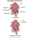
What is Coarctation of the Aorta?
More common in ____
- Abnormal narrowing of the aorta
- can be preductal or postductal, named based on location in relation to the ductus arteriosis
- postductal is more common
- more common in males than females

Preductal Coarctation of the aorta:
Who?
Clinical signs
treatment
- Infantile- more severe form of COA
- Weak femoral pulses, cold extremeties
- cyanosis esp in lower extremeties
- CHF
- Treatment- need surgery to survive
Postductal COA
Who?
Clinical manifestations?
- Older children and young adults
- collaterals have developed
- Manifestations:
- decreased perfusion to kidneys
- activation of RAAS
- high pressures in upper extremeties and low pressures in lower extremeties
- Intermittent claudication can occur
- decreased perfusion to kidneys

Anesthesia management for patients with congenical heart defects?
- Invasive monitoring in patients presenting with:
- severe pulm HTN or Eisenmenger syndrome
- unrestricted or long standing shunting
- heart failure
- severely depressed exercise tolerance
- Intra-op focus on optimization of O2 to all organs
- maintain cardiac contractility (esp of R ventricle)
- balancing pulmonary to systemic blood flow ratio
- treat dysrhythmias
- maintain adequate BP and O2 sat


