Classic Labs/Findings Flashcards
Onion skin periosteal reaction
Ewing sarcoma
Hair on end appearance on xray
Beta-thalessemia, Sickle cell anemia
Wire loop glomerular capillary appearance on light microscopy
Lupus nephropathy
Antidesmoglein antibodies
Pemphigus vulgaris
Decreased AFP in amniotic fluid or maternal serum
Down syndrome
Thyroid like appearance of kidney
Chronic bacterial pyelonephritis
Soap bubble in femur or tibia on xray
Giant cell tumor of bone
Generally benign
Podocyte fusion or effacement on EM
Minimal change disease
Intranuclear eosinophilic droplet-like bodies
Cowdry Type A bodies (HSV or CMV)
Rectangular crystal like cytoplasmic inclusions in Leydig cells
Reinke crystals (Leydig cell tumor)

Lumpy bumpy appearance of glomeruli on IF
PSGN (immune complex deposition of IgG and C3b)
Glomerulus-like structure surrounding vessel in germ cells
Schiller-Duval bodies (yolk sac tumor)
Circular grouping of dark tumor cells surrounding pale neurofibrils
Homer-Wright rosettes (neuroblastoma, medulloblastoma, retinoblastoma)
Optochin response
Sensitive: Strep pneumo
Resistant: Virdans strep
Anti GBM antibodies
Goodpastures
Antiplatelet antibodies
Idiopathic thrombocytopenic purpura
Cardiomegaly with apical atrophy
Chagas disease (Trypanosoma cruzi)
Giant B cells with bilobed nuclei with prominent inclusions
Reed-Sternberg cells (Hodgkin lymphoma)
Heterophile antibodies
EBV
Hilar lymphadenopathy
Peripheral granulomatous lesion in middle or lower lung lobes (Can calcify)
Ghon complex
Increased uric acid levels
Gout
Lesch-Nyhan syndrome
Tumor lysis
Loop & Thiazide diuretics
Linear appearance of IgG deposition on glomerular basement membrane
Goodpasture syndrome
Lytic hole punched bone lesions on xray
Multiple myeloma
Mucin filled cell with peripheral nucleus
Signet ring (gastric carcinoma)
RBC casts in urine
Acute glomerulonephritis
Apple core lesion on axr
Colorectal Cancer
Basophilic stippling of RBCs
Lead poisoning or sideroblastic anemia
Bacitracin response
Sensitive: Strep pyogenes (Group A)
Resistant: Streptococcus agalactiae (Group B)
Large lysosomal vesicles in phagocytes + immunodeficiency
Chediak-Higashi
Polished ivory-like appearance of bone at cartilage erosion
Eburnation (osteoarthritis resulting in bony sclerosis)
Heart nodules (Granulomatous)
Aschoff bodies (rheumatic fever)

Antineutrophil cytoplasmic antibodies
MPO-ANCA/p-ANCA: MPA and Churg Strauss
c-ANCA/PR-3 Anca: Wegener
Necrotizing vasculitis (lungs) and necrotizing glomerulonephritis
Granulomatosis with Polyangitis (wegener; cANCA)
Goodpasture syndrome (anti-GBM antibodies)
Eosinophilic globule in liver
Councilman body (toxic or viral hepatitis, often yellow fever)
Yellowish CSF
Xanthrochromia
Thrombi made of white/red layers
Lines of Zahn (arterial thrombus, layers of platelets/RBCs)

Renal epithelial casts in urine
Acute toxic/viral renal injury
Protein aggregates in neuron from hyperphosphorylation of tau protein
Neurofibrillary tangles (Alzheimer) and Pick bodies (Pick disease)
High levels of D-dimers
DVT, PE, DIC
Antinuclear antibodies (Anti-Smith, anti-dsDNA)
SLE (type III HS)
Disarrayed granulosa cells in eosinophilic fluid
Call-Exner bodies (Granulosa-Theca cell tumor of ovary)
Antihistone antibodies
Drug-induced SLE (Hydralazine, INH, phenytoin, procainamide)
Antimitochondrial antibodies
Primary Biliary Cirrhosis
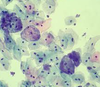
WBCs that look smudged
CLL (almost always B cell)
Anti-topoisomerase antibodies
Diffuse systemic scleroderma
Branching gram + rods w/sulfur granules
Actinomyces Israelii
Bloody tap on LP
SAH
Spikes on BM, dome like subepithelial deposits
Membranous glomerulonephritis

Silver staining spherical aggregation of tau proteins in neurons
Pick bodies

Azurophilic peroxidase positive granular inclusions in granulocytes and myeloblasts
Auer rods
(AML - especially PML type M3)
Bronchogenic apical lung tumor on imaging
Pancoast tumor
Increased AFP in amniotic fluid/maternal serum
Dating error, anencephaly, spina bifida
Novobiocin response
Sensitive: Staph epidermidis
Resistant: Staph saprophyticus
Waxy casts with low urine flow
Chronic ESRD
Elevated hCG
Choriocarcinoma
Hydatidiform mole (+/- embryo, multiple pregnancy)
Nodular hyaline deposits in glomeruli
Kimmelstiel-Wilson nodules (diabetic nephropathy)
Dysplastic squamous cervical cells w/nuclear enlargement and hyperchromasia
Koilocytes (HPV)
Thumb sign on lateral xray
Epiglottitis (H Flu)
Brown tumor of bone
Hyperparathyroidism or osteitis fibrosa cystica
Desquamated epithlium casts in sputum
Curschmann spirals
(Bronchial asthma - can result in whorled mucus plugs)
Psamomma bodies
- Meningioma
- Papillary thyroid carcinoma
- Mesothelioma
- Papillary serous carcinoma of endometrium and ovary
Eosinophilic cytoplasmic inclusion in liver cell
Mallory body (alcoholic liver disease)
Narrowing of bowel lumen on barium xray
String sign (Crohn disease)
Lead pipe colon on barium enema
Ulcerative colitis (loss of haustra)
Hexagonal, double-pointed, needle-like crystals in bronchial secretions
Bronchial asthma (Charcot-Leyden crystals: Eosinophilic granules)

Keratin pearls on skin biopsy
SCC
Iron-containing nodules in alveolar sputum
Ferruginous bodies (asbestosis: Increased chance of mesothelioma)
Enlarged thyroid cells with ground-glass nuceli
Orphan Annie eyes nuclei - papillary carcinoma of thyroid
t[8;14]
Burkitt lymphoma
C-myc activation
Depigmentation of neurons in substantia nigra
Parkinson disease
Basophilic nuclear remnants in RBCs
Howell-Jolly bodies (due to splenectomy or nonfunctional spleen)
Pseudopalisading tumor cells on brain biopsy
Glioblastoma multiforme

Migrating DVTs and vasculitis
Trousseau syndrome - adenocarcinoma of pancreas or lung
Stippled vaginal epithelial cells
Clue cells (Gardnerella vaginalis)
Periostum raised from bone, creating triangular area
Codman triangle on x-ray (osteosarcoma, Ewing sarcoma, pyogenic osteomyelitis)
Anti-IgG antibodies
Rheumatoid arthritis
Hypochromic microcytic anemia
Iron deficiency anemia
Lead poisoning
Thalessemia
Eosinophilic cytoplasmic inclusion in nerve cell
Lewy body (Parkinson disease)
Hypertension, HypoK, metabolic alkalosis
Conn syndrome
Mammary gland (blue domed) cyst
Fibrocystic change of breast
Rhomboid crystals, positively birefringent
Pseudogout (calcium pyrophosphate dihydrate crystals)
Trousseau syndrome
Adenocarcinoma of pancreas or lung that causes hypercoaguability
Anticentromere antibodies
CREST Scleroderma
Eosinophilic inclusion bodies in cytoplasm of hippocampal and cerebellar nerve cells
Negri bodies of rabies
Monoclonal antibody spike
- Multiple myeloma
- MGUS
- Waldenstrom (IgM)
- Primary amyloidosis
WBC casts in urine
Pyelonephritis
Tram-track appearance of capillary loops of glomerular basement membranes on light microscopy
Membranoproliferative glomerulonephritis

Nutmeg appearance of liver
Chronic passive congestion of liver due to RHF
