Chapter Ten - Surgical Interventions for Coronary Artery Disease Flashcards
Name four common cardiovascular surgical procedures and their acronyms.
Coronary Artery Bypass Graft (CABG)
Percutaneous Coronary Intervention (PCI)*
Aortic Valve Replacement (AVR)
Mitral Valve Replacement (MVR)
* Percutaneous Transluminal Coronary Angioplasty (PTCA) +/‐ Stent
Ed is 53 years old
He developed crushing substernal chest pain, which radiated to his left arm
He was short of breath
When the paramedics arrived they found him cool, clammy and bradycardic (low heart rate) and hypotensive
What does Ed have?
(Women don’t have the dame presentation of symptoms, so it is underdiagnosed. We will see that next semester)
Cardiovascular event
What is plaque? What does it result in?
Plaque: calcified narrowing of the arteries. They become so narrow that if you have a blood clot or vasoconstriction, blood flow to the heart is severely obstructed, and that results in ischemia or death

What is the balance needed to avoid putting the myocardium at risk? How can we tip that balance? Why would someone have a heart attack after eating? What is the classic sign of ischemia?
We can decrease oxygen supply or increase oxygen demand during activity. Tipping that balance one way or another is enough to put the myocardium at risk
Not uncommon to have a heart attack after they’ve eaten: blood flow directed to the gut for digestion, and if you have bad cardiac circulation, that could be enough to tip the balance
Classic sign of ischemia is angina: chest pain. Ischemia is reversible

What are the main consequences of ischemia (relative lack of O2 to the muscle)? Be specific.
Angina
Stunned myocardium
- Acute, transient ischemia (for months, usually)
- Prolonged systolic dysfunction
- Function recovers
Hibernating myocardium
• Persistent decrease blood supply
- Chronic contractile dysfunction
- Improved function after blood supply reestablished (Low metabolic state: anabolic metabolism)
Stunned and hibernating myocardium: sort of like a self-protection state, so that over time the myocardium can recover
Infarction
• Tissue necrosis
* When someone has a heart attack, they can recover from that more that we would expect
What are the common areas of referred pain linked to angina?
We need to knw these.

What is atherosclerosis? When does it start and why are we worried when symptoms start?
We believe this process starts in our twenties. Typically it doesn’t progress to this point if you maintain a healthy lifestyle or are not genetically predisposed. Symptoms start when you are about 90% obstructed. By the time people get symptomatic with angina, they are usually very obstructed.

Describe the Canadian Cardiovascular Society Functional Classification of Angina Pectoris. Give its classes and give a brief description of each.
We also need to know this. In this case, higher numbers are a worst situation

Give the four stages of angina and their respective characteristics. Which one can you predict, and which one can’t you?

What are the coronary artery syndromes that are linked to angina?
* Stemi: ST segment elevation MI

What are the objective diagnostic criteria?
- ECG
- Lab results - biomarkers
- Imaging
What are some common ECG abnormalities? What causes them?

Why do a blood test when you suspect a heart attack? What do we look for and what do high levels of this molecule indicate? Why is it a good biomarker compared to others?
Some of those ECG changes never go away, so we make blood tests. Some heart attacks are silent, so the changes will stay there so we need some blood tests (biomarkers) to confirm the current state. Troponin is part of your sarcomeres, so if the muscle dies, then it becomes necrotic and the troponins are released in the blood supply as part of the associated metabolic cleanup process. So troponin blood levels go high and they go high early. Acceptable level (normal range) is always in the chart, so don’t have to know that, it will always be there

What is the common diagnostic imaging technique for heart attacks that uses dye?
Contrast Angiography
Radiopaque contrast to visualize regions of the cardiovascular system (very large catheter in your femoral (sometimes brachial) artery and they inject a die, and they x-ray and they can then pick up the opacity of the vessels. You can see the narrowings, if present)
Uses: Coronary artery disease
Valve disease
Aortic stenosis
* Angiogram may lead automatically to a procedure (PCI)
Heart failure

Give the three main steps of a coronary angiography.

What is a diagnostic imaging technique that allows you to see the heart structures?
Echocardiography
Echos: ultrasound wave bounces back = echo
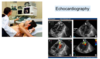
What technique does an echocardiography use? What are its uses?
Ultrasound
Uses:
Size of the ventricular chamber
Wall/septum thickness
Stroke volume
Ejection fraction
Wall motion
Valve function
Used to look at anatomy but also allows you to see movement: allows you to see things like valve movement. Also called a wall motion study sometimes
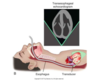
What is a diagnostic technique that is a less common procedure?
Transesophageal Echocardiogram
Less common procedure. Gives you more imaging options
Name all the diagnostic techniques that are mentioned in class.
ECG
Lab results: biomarkers
Imaging:
- Contrast Angiography
- Echocardiography
- Transesophageal Echocardiogram
- Computed Tomography (CT)
- Magnetic Resonance Imaging (MRI)
^ (CT and MRI) Not so common
- Nuclear Imaging (More common: allows you to look at the metabolic activity of the heart. That’s all she wants us to know)
- Single Photon Emission Computed Tomography (SPECT)
- Positron Emission Tomography (PET)
What does Single Photon Emission Computed Tomography (SPECT) allow us to see? What would change in stress vs rest?
MIBI (Radionucleotide)
(myocardial perfusion)

What is a Percutaneous Coronary Intervention (PCI)? Describe the technique. What are two potential problems linked to that?
Compresses the plaque. They inflate and deflate the balloon a number of times, which allows them to get a better widening of the artery. Prevents renarrowing. Two potential problems with this: clotting factors may detach, so stent can be covered in a drug coating that decreases clotting (some people say it slows the healing process). Second, sometimes plaque breaks off and could obstruct the vessel distal (you can use an umbrella filter to try to catch it).

What is bypass grafting? What are the two main options?
One option is the internal mammary artery, and the other option is the saphenous vein. One surgeon will be harvesting the graft from the leg, and the other one is in the heart. The amount of vein that they take depends on the amount of grafting they have to do. CABG times four sometimes.

What cut do you do to perform a CABG?
You perform a CABG via Sternal Split
They do this through the median sternotomy

What are two reasons why you would do a cardiopulmonary bypass? What is the most common way to go to do one? What is cardioplegia?
Bypass surgery for two reasons: bypasses around the obstruction, but during the surgery, we are bypassing the heart and lungs: blood out of the body, oxygenated, and passed back to the body, so the heart is not beating during the surgery (makes the surgical process easier). Sometimes you can have a beating heart, and you could even do it through scopes, BUT usually = median sternotomy and non-beating heart (faster). The longer you are on bypass, the bigger the consequences can be
Special solution that is cold: chills the heart and decreases its metabolic rate: allows you longer time on bypass

Do you ever take out sternal wires?
No, they stay in there forever

What are some cardiovascular post-CABG complications?
Cardiovascular
• VTE (venous thromboembolism)
- Myocardial infarct
- Hypotension (one of the two most common ones)
- Arrhythmia (one of the two most common ones)
- Pulmonary edema
- Cardiogenic shock
- Wound dehiscence (Wound, and sutures, come apart. Sternal wires didn’t hold. They dehiss if there is infection, so we need to reassure people that these sutures can hold)
- Graft failure
What are some non-cardiovascular post-CABG complications?
Infection
Respiratory
• Pleural effusion
• Atelectasis
• Pneumonia
• Respiratory failure
Neurological
- Stroke
- Neurocognitive impairment (‘’Pump head’’: bypass pump -> higher incidence or neurocognitive impairment. Much of it clears, but there is an ongoing investigation on this. Sometimes it is transient, sometimes it isn’t)
Hematologic
• Coagulopathy, hemolysis, mediastinal bleeding
What are the three main aspects of respiratory care?
Air entry
Secretions
Ability to Cough
Where are the inpatients in Day 1 post-op? Day 2? Days 3-5?
Day 1 -> CCU : Sitting on edge of bed
Up to chair
Day 2 -> IMCU : Walking in room
Days 3-5 -> Ward :
Walking 5‐10 min several times a day
Self care activities
Trial of stairs pre‐d/c
* Don’t forget U/E ROM and Posture throughout the whole process

What are some common at-home Sternal Precautions?
* Think walkers!

Give the four main Measures to Ensure Safe Mobility.
Chart review
Communication with ward staff
Physical Examination
Objective monitoring pre‐ and post‐activity
What are some aspects to consider during Chart Review?
PMH
HPI
Sx report
Meds
Labs (cardiac enzymes, Hb/HCT)
ECG
CXR
Home situation
What are some things to look out for during Physical Examination?
Physical Examination
chest wall inspection
auscultation of lung sounds
range of motion
examination of extremities (pulses, edema)
balance
What are some key things to look out for while doing Objective monitoring pre- and post-activity?
HR
BP
ECG
Oxygenation
What would make you think that you need to consider modifying the activity schedule of a patient?
HR > 100 bpm (tachycardia)
BP: no change or decrease with activity
ECG abnormalities
Angina with self-care activities
Undue fatigue
What are some contraindications for activity?
Acute heart failure:
- peripheral edema
- unusual shortness of breath
- falling BP
- evidence of failure on CXR
Tachy-arrhythmias, significant bradycardia
Angina at rest
Unstable angina
Heart block
What are the five aspects that are addressed in a Cardiac Rehabilitation program? Who gets a referral to such a clinic?

Read the summary.
Risk reduction: pre‐ & post‐op
Education: patient and their support network
Post‐op: prevent & manage complications
- Respiratory
- Mobility
Discharge Planning
- Home exercise program
- Referral to cardiac rehabilitation
