Chapter 8 Flashcards
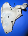
Left Scapula, 1-10

- Suscapular fossa
- Glenoid cavity
- Coracoid process
- Acromion
- Superior border
- Suprascapular notch
- Superior angle
- Medial border
- Inferior angle
- Lateral border
1-10, Ignore 4 and 7
Which view is this?
Which side is this?

Posterior aspect, Right scapula
- supraspinous fossa
- Spine
- Infraspinous fossa
- Ignore this one
- Superior angle
- Medial border
- Ignore this one too
- Lateral border
- Glenoid cavity at lateral angle
- Acromion
Which side? Which view?

Left scapula, lateral aspect
- Coracoid process
- Glenoid cavity
- Supraspinous fossa
- Acromion
- Infraspinous fossa
- Inferior angle
- Subscapular fossa

Humerus of the right arm
- Greater tubercle
- Lesser tubercle
- Radial fossa
- Head of the humerus
- Anatomical neck
- Surgical neck
- Deltoid tuberosity
- Capitulum
- Trochlea
- Medial epicondlye
- Coronoid fossa
- Lateral epicondyle
- Olecranon fossa
Which side, only Ulna and radius and only #
21, 22, 23, 28, 29, 30, 31, 32, 38 and 39

- Head of the radius
- Neck of the radius
- Radial tuberosity
- Styloid process of radius
- Ulnar notch of the radius
- Trochlear notch
- Coronoid process
- Head of ulna
- Styloid process of ulna
Right hand, posterior view

- Phalanges (Distal, middle, proximal)
- Metacarpals
- Capitate
- Hamate
- Pisiform
- Triquetrium
- Lunate
- Schapoid
- Trapezium
- Trapezoid
Anterior view of right hand

- Distal Phalanges
- Middle Phalanges
- Proximal Phalanges
- Hamate
- Pisiform
- Tirquetrum
- Lunate
- Capitate
- Scaphoid
- Trapezium
- Trapezoid
- Metacarpal 1
- Metacarpal 2
- Metacarpal 3
- Metacarpal 4
- Metacarpal 5
Ignore H, D and F are the same

A. Ischium
B. Pubis
C. Coccyx
D. Ilium
E. Sacrum
F. Iliac Crest
G. Iliac fossa
I. Pelvic brium
J. Acetabulum
K. Pubic tubercle
L. Pubic symphysis
Right

- Head
- Fovea capitis
- Intertronchanteric crest
- Greater trochanter
- Lesser trochanter
- Neck
- Linea aspera
- Adductor tubercle
- Medial epicondyle
- Medial condyle
- Intercondylar fossa
- Lateral condyle
- Lateral epicondyle
What is H and I?

Pattellar surface
The tibia and fibula of the right leg
3 (between the two bones)

- Interosseous membrane
- Tibia
- Proximal tibiofibular joint
- Medial condyle
- Articular surface of lateral condyle
- Medial mallelous (the flat part on the very end of the tibia is the articular surface)
- Head of fibula
- Lateral malleolus
Superior view of the right foot

- Trochlea of talus
- Calcaneus
- Navicular
- Cuboid
- Lateral cuneiform
- Intermediate cuneiform
21 Medial cuneiform
- Tarsals
- Metatarsals
- Phalanges

A. Calcaneus
B. Talus
C. Cuboid
D. Navicular
E. Lateral cuneiform
F. Inermediate cuneiform
G. Fifth metatarsal
H. Phelanges

A. Tibia
C. Talus
D. Calcaneus
E. Navicular
G. Medial cuneiform
H. Intermediate cuneiform
J. First metatarsal
K. Other metatarsals
O. Proximal Phalanges
This arch curves well above the ground. The talus, near the talonavicular joint, is the keystone of this arch, which originates at the calcaneous, rises to the talus, and then descends to the three medical metatarsals.
The medial longitudinal arch

This arch is very low. It elevates the lateral edge of the foot just enough to redistribute some of the body weight to the calcaneus and some to the head of the fifth metatarsal. The cuboid bone is the keystone of this lateral arch.
lateral longitudinal arch

The two longitudinal arches serve as illars for this arch, which runs obliquely from one side of the foot to the other, following the line of the joints between the tarsals and the metatarsals.
Transverse arch


