Ch 7-8 Carbs and Nucleotides Flashcards
carbohydrates: basic formula, functions, produced from?
any organic molecule that has the basic formula: Cn(H2O)n
produced from CO2 and H2O via photosynthesis in plants
isn’t just sugar; can be glyceraldehyde, pyruvate, amylopectin
functions include: energy source and energy storage; structural component of cell walls and exoskeletons, and informational molecules in cell-cell signaling (ABO blood types)
can be covalently linked with proteins to form glycoproteins and proteoglycans
aldoses vs ketoses
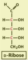
aldose: contains and aldehyde functional group (glucose, galactose, ribose)
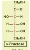
ketose: contains a ketone functional group (fructose)

epimers
2 sugars that differ only in the configuration around one carbon atom
memorize glucose and galactose structures!

fructose structure
a simple change in structure (from glucose) results in a huge difference in taste (sweeter)

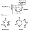
ribose/deoxyribose structure
deoxyribose does not have an –OH at C?

hemiacetals and hemiketals
aldehyde and ketone carbons are electrophilic
the alcohol oxygen atom is nucleophilic
hemiacetals: when aldehydes are attacked by alcohols
hemiketals: when ketones are attacked by alcohols
If the –OH and carbonyl groups re on the same molecules, a five- or six-membered ring results. The addition of the second molecule of alcohol produces the full acetal

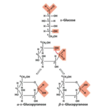
cyclization of monosaccharides
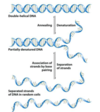
in aqueous solution, all monosaccharides with 5+ carbon atoms in the backbone occur predominantly as cyclic structures; cyclic sugars are opening and closing constantly (the ring form exits in equilibrium with the open-chain forms)
- pyranoses: six-membered oxygen-containing rings (glucose and galactose)
- furanoses: five-membered oxygen-containing rings (fructose)
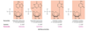
the former carbonyl carbon becomes a new chiral center, called the anomeric carbon
The former carbonyl oxygen becomes a hydroxyl group; the position of this group determines if the anomer is α or β
- more β-glucopyranose than α-glucopyranose (85:15 ratio)

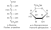
Fisher/Hanworth projections of glucose
to convert the Fischer projection formula to a Hanworth perspective formula
1) draw the six-membered ring (5 carbons, and 1 oxygen at the upper right)
2) number the carbons in a clockwise direction, beginning with the anomeric carbon
3) if a hydroxyl group is to the right in the Fischer projection, it is placed pointing down in the Hanworth perspective; if left, placed pointing up
4) the terminal –CH2OH group projects upward for the D-enantiomer, downward for the Lenantiomer

Draw the Haworth perspective formula for D-galactose
a) six-membered (oxygen atom at the top right)
b) number the carbon atoms clockwise
c) place the hydroxyls below, above, and above the ring for C-2, C-3, and C-4, respectively
d) the hydroxyl at C-1 can point either up or down
Draw the Haworth perspective formulas for ß-L-galactose
a) use the Fischer representation of D-galactose to draw the L-galactose (mirror image)
b) draw the Haworth perspective: –OH groups on C-2, C-3, and C-4 are oriented up, down, and down, respectively
c) the –OH on the anomeric carbon points down
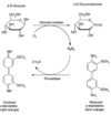
Reducing Sugars and 3 tests
the aldehyde can reduce Cu2+ to Cu+ (Fehling’s test)
the aldehyde can reduce Ag+ to Ag0 (Tollens’ test)
colorimetric glucose analysis: glucose oxidase catalyzes the conversion of glucose; hydrogen peroxide oxidizes organic molecules into highly colored compounds; concentrations of such compounds is measured colorimetrically; 1 for 1, measure how much glucose is present by the intensity of the orange color
allows the detection of reducing sugars, such as glucose
the oxidation of sugar by the cupric ion occurs only with the linear form, which exists in equilibrium with the cyclic form

disaccharide formation
Two monosaccharides join between an anomeric carbon and a hydroxyl carbon, the acetal or ketal formed is a disaccharide. The bond formed is a glycosidic linkage, which is less reactive than the hemiacetal at the second monomer (reducing)
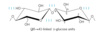
maltose: disaccharide formed upon condensation of two glucose molecules via 1 → 4 bond

nonreducing disaccharides
when the anomeric carbons are involved in a glycosidic bond, the interconversion of linear and cyclic forms is prevented, rendering the sugar nonreducing.
sucrose: glucose and fructose (two acetal groups and no hemiacetals)
- formed by plants but not by animals
- its stability (no reducing ends = resistance to oxidation) makes it suitable for the storage and transport of energy in plants (from leaves to other parts of the plant body)
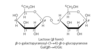
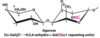
lactose
reducing disaccharide
Gal (B1 →4)Glc
disaccharides are not absorbed from the small intestine; without lactase, the undigested lactose passes into the large intestine
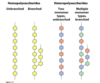
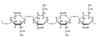
Polysaccharides
homopolysaccharides vs heteropolysaccharides
linear vs branched
do not have a defined molecular weight; unlike proteins, no template is used to make polysaccharides
size-exclusion columns are built out of a sugar polymer called Sephadex; sugar casted in a form of a bead

glycogen
main storage homopolysaccharide in animals (muscle and liver)
glucose monomers form (α1 →4) linked chains and (α1 →6))-linked branches every 8-12 residues (more branched and compact than starch)
molecular weight reaches several million
starch
main storage homopolysaccharide in plants
contains two types of glucose polymer:
- amylose: long, unbranched chains of glucose connected by (α1 →4) linkages
- amylopectin: highly branched (α1 →6; every 24-30 residues) and has a high molecular weight (up to 200 million)
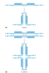
strands of amylopectin form double-helical structures with each other or with amylose strands

metabolism of glycogen and starch
glycogen and starch often form granules in cells that contain enzymes that synthesize and degrade these polymers
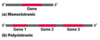
glycogen molecule with n branches has n + 1 nonreducing ends, but only one reducing end
glucose residues at the nonreducing ends of the outer branches are removed enzymatically for energy production; many degradative enzymes can work simultaneously on many branches
cellulose
a tough, fibrous, water-insoluble substance found in cell walls of plants; most abundant polysaccharide in nature; cotton is nearly pure fibrous cellulose
linear, unbranched homopolysaccharide
glucose residues are linked by (β1 →4) glycosidic bonds in contrast to the (α1 →4) bonds of amylose (gives them a different structure and physical properties)
humans cannot use cellulose as a fuel source because we lack an enzyme to hydrolyze the (β1 →4) linkages
hydrogen bonds between adjacent monomers and lots between chains

chitin
linear homopolysaccharide composed of N-acetylglucosamine residues in (β1 →4) linkage
forms extended fibers similar to cellulose; the only difference is the replacement of the hydroxyl group at C-2 with an acetylated amino group, which allows for more H-bonding and a stronger structure
tough, flexible, water-insoluble, cannot be digested by vertebrates
found in cell walls in mushrooms and in exoskeletons of arthropods; second-most abundant polysaccharide, next to cellulose

agar and agarose
agar: a complex mixture of heteropolysaccharides containing modified galactose units (sulfated)
- serves as a component of the cell wall in some seaweeds
- agar solutions are used to form a surface for the growth of bacterial colonies; bacteria cannot dissolve the (β1 →4) linkages in agar
agarose: one component of agar (highly purified)
- agarose solutions (heated and cooled), form gels that are commonly used in the lab for separation of DNA by electrophoresis
- galactose sugar with ether bridge and addition of sulfur group

What are the 3 classes of glycoconjugates?
glycoconjugates are proteins or lipids with sugar molecules attached to them
glycoproteins
proteoglycans
mucins
glycoproteins
mostly protein by mass
modified after protein synthesis
often found on secreted proteins and on cell surface proteins, in the ECM, and in the blood; forming highly specific sites for recognition (blood groups proteins, HIV virus coat, erythropoietin)
erythropoietin (EPO): hormone involved to signal for more RBC production; Ser and Asn involved in linking protein and sugar

proteoglycans
macromolecules of the cell surface or ECM in which one or more sulfated glycosaminoglycan chains (long unbranched polysaccharide chains of repeating disaccharide) are joined covalently to a membrane protein or a secreted protein
mostly carbohydrate by mass
a major component of cartilage and tendons, the adhesion of cells to the ECM, lubricants in synovial fluid of joints, and horny structures formed from dead cells in horn, hair, and hoofs (keratan sulfates)
mucins
heavily glycosylated mucin proteins
used for lubrication and protective barrier (mucus)
carbohydrates mostly linked through Ser, Thr or Asn
lectins
proteins that bind carbohydrates with high specificity and with moderate to high affinity; lectins have a subtle molecular complementarity that allows interaction only with their correct carbohydrate-binding partners
lectin multivalency: a single lectin molecule has multiple carbohydrate-binding domains; in a cluster of oligosaccharides found on a membrane surface, each oligosaccharide can engage one of the lectin’s CBDs, strengthening the interaction
serve in a wide variety of cell-cell recognition, signaling, and adhesion processes and in intracellular targeting of newly synthesized proteins
in vertebrates, oligosaccharide tags “read” by lectins govern the rate of degradation of certain peptide hormones, circulating proteins, and blood cells
Describe the common structural features and the differences for each of the following pairs:
a) cellulose and glycogen
b) D-glucose and D-fructose
c) maltose and sucrose
(a) Both are polymers of D-glucose, but they differ in the glycosidic linkage: (β1→4) for cellulose, (α1→4) for glycogen.
(b) Both are hexoses, but glucose is an aldohexose, fructose a ketohexose.
(c) Both are disaccharides, but maltose has two (α1→4)-linked D-glucose units, and sucrose has (α1↔2β)-linked D-glucose and D-fructose.
The fructose in honey is mainly in the β-D-pyranose form. This is one of the sweetest carbohydrates known, about twice as sweet as glucose; the β-D-furanose form of fructose is much less sweet.
The sweetness of honey gradually decreases at a high temperature. Also, high-fructose corn syrup (a commercial product in which much of the glucose in corn syrup is converted to fructose) is used for sweetening cold but not hot drinks. What chemical property of fructose could account for both these observations?
Fructose cyclizes to either the pyranose or the furanose structure. Increasing the temperature shifts the equilibrium in the direction of the furanose, the less sweet form.
Lactose exists in two anomeric forms, but no anomeric forms of sucrose have been reported. Why?
Sucrose has no free anomeric carbon to undergo mutarotation. The anomeric carbons of both monosaccharide units are involved in the glycosidic bond (nonreducing disaccharide)
The almost pure cellulose obtained from the seed threads of Gossypium (cotton) is tough, fibrous, and completely insoluble in water. In contrast, glycogen obtained from muscle or liver disperses readily in hot water to make a turbid solution.
Despite their markedly different physical properties, both substances are (1→4)-linked D-glucose polymers of comparable molecular weight. What structural features of these two polysaccharides underlie their different physical properties? Explain the biological advantages of their respective properties
Native cellulose consists of glucose units linked by (β1→4) glycosidic bonds, which force the polymer chain into an extended conformation. Parallel series of these extended chains form intermolecular hydrogen bonds, aggregating into long, tough, insoluble fibers.
Glycogen consists of glucose units linked by (α1→4) glycosidic bonds, which cause bends in the chain and prevent the formation of long fibers. In addition, glycogen is highly branched and, because many of its hydroxyl groups are exposed to water, is highly hydrated and disperses in water.
Cellulose is a structural material in plants, consistent with its side-by-side aggregation into insoluble fibers. Glycogen is a storage fuel in animals. Highly hydrated glycogen granules with their many nonreducing ends can be rapidly hydrolyzed for energy
functions of nucleotides?
energy for metabolism (ATP, GTP, CTP, UTP; any of the ribonucleotides)
enzyme cofactors (NAD+)
signal transduction (cAMP)
function of nucleic acids?
storage of genetic info (DNA because more stable than RNA)
transmission of genetic info (mRNA; short half-life compared to DNA)
processing of genetic information (ribozymes)
protein synthesis (tRNA and rRNA)
first molecules that were created in the deep sea hot vents were molecules that look like RNA (RNA can be used to transfer information and can be used as an enzyme)
nucleotides vs nucleosides vs nucleobase
ribo vs deoxyribo
nucleotide: nitrogenous base, pentose, phosphate
nucleoside: nitrogenous base and pentose
nucleobase: nitrogenous base
in DNA, 2’ will have hydrogen instead of –OH; lack of this –OH gives stability

Name the purines and draw their structures

Name the pyrimidines and draw their structures
never find thymine in RNA
but there are circumstances where uracil is in a DNA molecule
thymine = 5-methyluracil

ß-N-Glycosidic Bond
- the linkage between base and sugar
- N1 in pyrimidines
- N9 in purines
- the bond is formed to the anomeric carbon of the sugar in ß configuration
- cleavage is catalyzed by acid
- free rotation can occur around the bond in free nucleotides
- the torsion angle about the bond is denoted by the symbol c
- syn conformation: angle near 0°
- found in Z-DNA
- anti conformation; angle near 180°
- favored due to steric hindrance/electrostatic repulsion
- found in normal B-DNA (found in our cells)

Tautomers of Nucleobases
tautomers are structural isomers that differ in the location of protons
keto-enol tautomerism: common in ketones
lactam-lactim tautomerism: occurs in some heterocycles
lactam form predominant at neutral pH; other forms become more prominent as pH decreases; the other free pyrimidines and free purines also have tautomeric forms but they are more rarely encountered
In a 1000 to 1 ratio of lactam to lactim, DNA polymerase might misrecognize enol form and put in the wrong nucleotide (C instead of a U)

nucleotide bases and UV light
all bases absorb UV light and nucleic acids are characterized by a strong absorption at wavelengths near 260 nm
used when purifying DNA to estimate DNA/RNA concentration in solution
we can distinguish nucleotides because they have a slightly different maximums of absorption

Deoxyribonucleotides: names, structures, and symbols
the more common name is followed by the complete name in parentheses; the nucleoside portion of each molecule is shaded in light red
The nucleotide units of DNA are usually symbolized as A, G, T, and C, sometimes as dA, dG, dT, and dC
in their free form, the deoxyribonucleotides are commonly abbreviated dAMP, dGMP, dTMP, and dCMP.

ribonucleotides: names, structures, and symbols
the more common name is followed by the complete name in parentheses; the nucleoside portion of each molecule is shaded in light red
The nucleotide units of RNA are usually symbolized as A, G, U, and C.
in their free form, the ribonucleotides are commonly abbreviated AMP, GMP, UMP, and CMP.

minor nucleosides in DNA
- modification done after DNA synthesis (epigenetic markers)
- 5-methylcytosine: common in eukaryotes; a way to mark which genes should be active (finger vs liver expression); on genes that are turned off
- 5-hydroxymethylcytidine: found in bacteria to detect viruses in cell and destroy foreign DNA; sometimes found in eukaryotes (rare)
- N6-methyladenosine: common in bacteria, not found in eukaryotes
minor nucleosides in RNA
allows for non-watson-crick base pairs that help stabilize RNA structure
inosine
- sometimes found in the “wobble position” of the anticodon in tRNA
- instead of A, there’s inosine by deaminating adenosine
- provides richer genetic code
pseudouridine (Ψ)
- found widely in tRNA (stabilize structure) and rRNA (help folding)
- more common in eukaryotes but also found in bacteria
- made from uridine by enzymatic isomerization (base reattached to ribose ring at a different position) after RNA synthesis
polynucleotides: bonds and polarity
- covalent bonds formed via phosphodiester linkages
- linear; no branching or cross-links
- negatively charged backbone
- DNA: fairly stable; hydrolysis accelerated by DNAse
- RNA: unstable
- lasts for a few years in water;
- mRNA degraded in few hours in cells
- directionality
- 5’ (phosphate) → 3’ (unprotected alcohol group(s))
- DNA is complementary and antiparallel

hydrolysis of RNA
- alkaline conditions
- ribonucleases (RNase): nonspecific and specific
- S-RNase: in plants, prevents inbreeding
- RNase P: enzyme made of RNA (ribozyme) that processes tRNA precursors into mature tRNAs
- Dicer: cleaves double-stranded RNA into oligonucleotides (smaller functional RNAs that regulate gene expression)
B-DNA and its grooves
the more hydrated form of DNA
not symmetrical (major and minor grooves)
the distance between the two strands that make the major groove is the same distance as a diameter of an alpha-helix of proteins; a number of proteins that bind DNA bind through this major groove with an alpha-helical strand of the protein molecule
proteins that bind the minor groove by binding to the DNA and bending it to open up the strands and be able to interact with the bases

Nucleic acid structure: A, B, and Z
- A-form structure:
- slightly dehydrated; shortest
- wider right-handed helix;
- base pairs not perfectly perpendicular to the backbone; deepens the major groove and makes minor groove shallower
- forms when there’s base pairing between DNA and RNA molecules
- B-form structure:
- more elongated, than A-DNA
- right-handed helix
- the natural form of DNA found in our chromosomes;
- 10.5 base pairs per helical turn
- Z-form structure:
- left-handed helix; more slender and elongated
- purine bases are flipped out (syn-conformation)
- unknown significance of Z-form in our DNA; very hard to track natively in cell and study it

mRNA
messenger RNA: code carrier for the sequence of proteins; transfer genetic information from DNA to proteins
synthesized using a DNA template
contains ribose instead of deoxyribose; uracil instead of thymine
one mRNA may code for more than one protein with the help of tRNAs
gene structure: monocistronic vs polycistronic
promoter before the Gene where RNA polymerase will
monocistronic: one promoter; one sequence for one protein
polycistronic: in bacteria, more than one gene is transcribed with one promoter; in the example, all three genes are expressed at the same time (lac operon)

palindromes
In DNA: the term is applied to regions with inverted repeats; self-complementary sequence in one strand is repeated in the opposite orientation in the paired strand
self-complementary within each strand has the potential to form hairpin (single-strand) or cruciform structures (both strands)
mirror repeat: when the inverted repeat occurs within each individual strand of DNA; they do not form hairpin or cruciform structures because they do not have complementary sequences within the same strand
DNA polymerase may not read the hairpin and just sequence across it, resulting in a deletion of DNA
tRNA and rRNAs have a lot of hairpins

DNA Denaturation and cooperativity
- covalent bonds remain intact; genetic code remains intact
- hydrogen bonds are broken to separate the two strands
- instead of being in a helix, they are now in a random coil;
- cooperativity - once you break the first few BPs, forms a bubble, and the rest of the strands fall apart in a cooperative event
- can be induced by high temperature or change in pH
- reversible by annealing in low temperature
- the two strands hit each other until they find the right BPs
- cooperativity - once you first seed the first few BPs, everything after is a cooperative event
- therefore, add a very high concentration of DNA you want to anneal compared to its target DNA; stack odds in the annealing favor
- can be monitored by UV spectrophotometry at 260 nm; base stacking is lost, UV absorbance increases
- reversible thermal denaturation and annealing form basis for PCR

H-bonds and base stacking
purines and pyrimidines are hydrophobic and relatively insoluble in water at near-neutral pH of the cell
base stacking minimize contact with water and stabilize the structure of nucleic acids: bases are positioned parallel to each other and perpendicular to the backbone via hydrophobic and van der Waals interactions
base-stacking interactions between GC pairs are stronger than AT pairs; DNA sequences with higher GC are more stable
AT-rich regions melt at a lower temperature than GC-rich regions
replication origin sequence is AT-rich; shows that nucleotides are not randomly distributing

tm
The midpoint of melting (Tm): when a DNA molecule is 50% denatured
higher tm because
- more G-C base pairs (with 3 hydrogen bonds) than A-T base pairs
- longer DNA
- high salt concentration in solution

Calculating Tm of DNA sequence
5’-AACCCGTTC-3’
2+4 rule
A or T = 2°C
G or C = 4°C
5’-AACCCGTTC-3’ = 28°C is estimated Tm
one strand of a double-helical DNA has the sequence (5’)GCGCAATATTTCTCAAAATATTGCGC(3’)
Write the base sequence of the complementary strand. What special type of sequence is contained in this DNA segment? Does the double-stranded DNA have the potential to form any alternative structures?
(5’)GCGCAATATTTTGAGAAATATTGCGC(3’) - always write the sequence of a single strand in the 5’ →3’ direction)
contains a palindrome
because this sequence is self-complementary, the individual strands can form hairpin structures; the two strands can form a cruciform
Hairpins may form at palindromic sequences in single strands of either RNA or DNA. How is the helical structure of a long and fully base-paired (except at the end) hairpin in RNA different from that of a similar hairpin in DNA?
When two strands of RNA with perfectly complementary sequences are paired, the predominant double-stranded structure is an A-form right-handed double helix.
the DNA helix is generally in the B conformation
The cells of many eukaryotic organisms have specialized systems that specifically repair G-T mismatches in DNA. The mismatch is repaired to form a G-C (not A-T) base pair. This G-T mismatch repair mechanism occurs in addition to a more general system that repairs virtually all mismatches.
Suggest why cells might require a specialized system to repair G-T mismatches.
in eukaryotic DNA, about 5% of C residues are methylated. 5-methylcytosine can spontaneously deaminate to form thymine; the resulting G-T pair is one of the most common mismatches in eukaryotic cells.
the specialized repair mechanism to convert G-T back to G-C is directed at this common class of mismatch