the heart is located in the ______________
middle mediastinum
the base of the heart lies beneath which rib
2nd rib (mostly left)
the apex of the heart is located
in the 5th intercostal space
pericarditis
an infection of the pericardium
pericardium
surrounds and protects the heart and retains its position in mediastinum
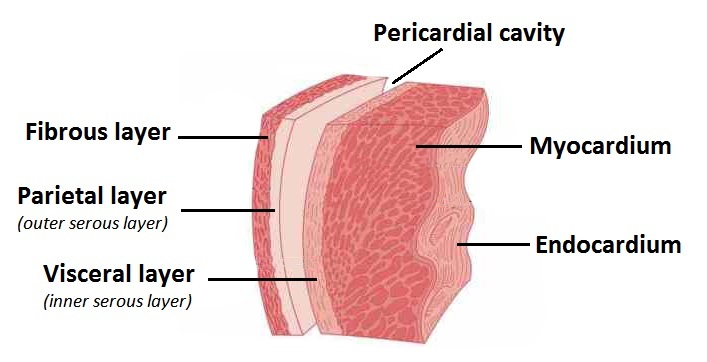
3 layers of the pericardium
- fibrous
- parietal
- visceral

fibrous pericardium
- very dense and non flexible connective tissue
- helps protect and anchor the heart

two divisions of serous pericardium
parietal layer
visceral layer
parietal layer
adheres to the fibrous pericardium
visceral layer (epicardium)
inner layer that adheres to the surface of the heart
epicardim contains _________ & _________ vessels
blood and lymphatic
myocardium
striated skeletal muscle
endocardium
lines the chambers
decreases friction
4 foundations of the fibrous skeleton of the heart
- wheres the heart valves attach
- point of intersection for cardiac muscle
- prevent overstetching of the valves
- acts as an electrical insulator
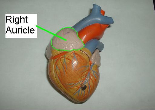
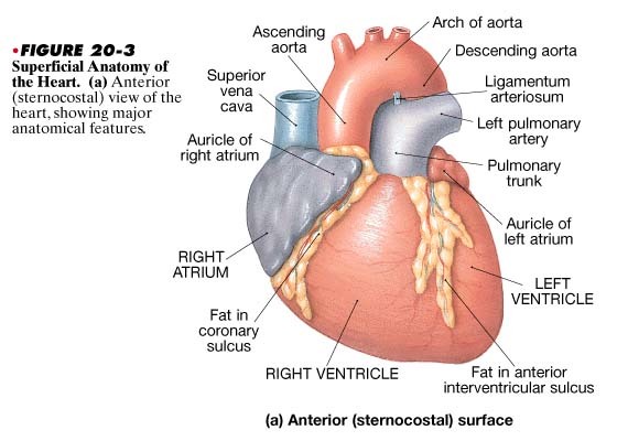
auricle
pouchlike structure on the anterior surface of the heart
Sulci
series of grooves on the surface of the heart
coronary sulcus
marks the external boundary between superior atria and inferior ventricle
(white branches)
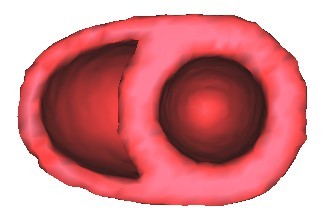
how is the right ventricle shaped
pouch shaped
right atrium receives blood from
superior vena cava
inferior vena cava
coronary sinus
fossa ovalis`
an oval depression of the septa
(an opening between the two atria at birth)
another name for the tricuspid valve
right atrioventricular valve
trabeculae corneae
bundle of cardiac muscles that convey part of the conduction system

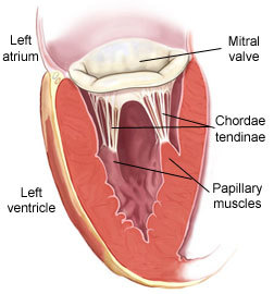
papillary muscles
the muscles that hold the cordae tendinae
ventricles have thicker walls because
they deliver blood under high pressure over longer distance


