Black & White Pathology Flashcards
What is an area of hematopoietic bone marrow that produces a radiolucency, typically in the posterior mandible?
Focal Osteoporotic Marrow Defect
What are the characteristics of benign neoplasms of bone?
- asymptomatic
- slow growth
- expands the cortex instead of going through it
- symmetrical
- does not metastasize
What are characteristic of malignant neoplasms of bone?
- symptomatic
- grows faster
- invades/destroys structures like the cortex
- asymmetrical
- poorly defined margins
- deposits bone outside of cortex
- can metastasize
This is a benign (non-neoplastic) radiographic finding in a female. These are usually seen in the posterior mandible. Dx?

focal osteoporotic marrow defect
if this were a focal osteoporotic marrow defect, what would be your next step?

Incisional biopsy is necessary to get definitive dx
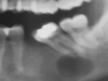
Don’t really know why this is here but what do you think it is?

Idiopathic osteosclerosis
What is an area of radiodensity with unknown cause and cannot be identified as anything else?
idiopathic osteosclerosis
New patient that hasn’t had an infection there. Dx?

idiopathic osteosclerosis
What is your possible differential dx?

- Condensing osteitis: associated with an infection
- Idiopathic osteosclerosis: unknown cause
- Focal cemento-osseous dysplasia: will have a radiolucent rim
- Cementoblastoma: fused with the tooth
*bolded pathology is definitive dx
Tooth has Hx of infection. Dx?

condensing osteitis
What is an asymptomatic radiolucent lesion that is usually seen crossing the midline of the mandible?
Central giant cell granuloma
What radiolucent lesion is usually seen in the posterior mandible of women?
Focal osteoporotic marrow defect
What lesion is usually seen across the anterior/midline of the mandible in women?
central giant cell granuloma
Female patient. Non-neoplastic lesion. Dx?

Central Giant Cell Granuloma
What is your differential? Female patient. Asymptomatic.

- Central Giant Cell Granuloma
- Brown Tumor (of hyperparathyroidism)
- Aneurysmal Bone Cyst
- Odontogenic Keratocyst
Upturned eyes and big, plump cheeks. Dx?

Cherubism
What syndrome features odontogenic keratocysts?

Gorlin Syndrome
What is your differential dx?

- odontogenic keratocyst
- aneurysmal bone cyst
- traumatic bone cyst
- Brown’s tumor
Pt recently experienced trauma. What’s your dx?

traumatic bone cyst (AKA simple bone cyst)
What radiographic feature is suggestive of traumatic bone cysts?

Scalloping of bone between the roots
Dx?

Traumatic bone cyst
What is your differential?

- Odontogenic keratocyst
- Traumatic bone cyst
- Aneurysmal bone cyst
- Ameloblastoma
*bolded pathology is definitive
What is an intraosseous blood-filled cavity surrounded by connective tissue lining?
Aneurysmal bone cyst
Why is an aneurysmal bone cyst not a true cyst?
It doesn’t have an epithelial lining. (lined by connective tissue)
What are three examples of benign fibro-osseous lesions?
- fibrous dysplasia
- cemento-osseous dysplasia
- ossifying fibroma
Fine, ground glass appearance. Dx?

Fibrous dysplasia
What two syndrome are associated with polyostotic fibrous dysplasia?
- McCune-Albright syndrome
- Jaffe-Lichtenstein syndrome
What is the most common fibro-osseous lesion encountered in clinical practice?
Cemento-osseous dysplasia
Dx?

Focal Cemento-Osseous Dysplasia
Lesion later calcifies. Dx?
Focal cemento-osseous dysplasia
Dx?

Focal cemento-osseous dysplasia
Asymptomatic teeth that test vital. Dx?

Periapical Cemento-Osseous Dysplasia
Dx?

Periapical cemento-osseous dysplasia
Dx? How do these lesions appear in early stages and late stages?

Dx: periapical cemento-osseous dysplasia
Early: radiolucent lesion
Late: radiodense lesion with radiolucent rim
Usually, occurs in the posterior mandible of women. Dx?

Florid cemento-osseous dysplasia
Differential dx?

- Florid cemento-osseous dysplasia
- Odontogenic keratocyst
- Traumatic bone cyst
*bolded pathology is definitive
What are the demographics of florid cemento-osseous dysplasia?
90% women, 90% black
Are men or women more likely to have cemento-osseous dysplasia?
90% women
These fibro-osseous lesions will expand the inferior cortex of the mandible. Dx?

ossifying fibroma
Is ossifying fibroma more likley in males/females and mandible/maxilla?
F>M
Md > Mx
Usually, these lesions are mixed RO/RL. The inferior border in expanding. Dx?

ossifying fibroma
These mixed RO/RL lesions are usually seen in women and in the mandible, but this time. Dx?

ossifying fibroma
Outline of root is obscured. Dx?

Cementoblastoma
What is your differential?

- Cementoblastoma: cannot see normal shape of tooth
- Condensing osteitis: follows infection
- Idiopathic osteosclerosis: unknown cause
- Focal cemento-osseous dysplasia: starts as purely RO lesion
*bolded is definitive dx
25% of the time these present with a sunburst appearance.

osteosarcoma
What is the most common malignancy to originate in the bone?
osteosarcoma
Pt has pain, unilateral swelling of the face, and moth eaten bone upon radiograph. Provisional dx?

osteosarcoma
What is the most common cancer involving bone?
Metastatic tumor of the bone
