BIOL 360 (Cell Biol) Flashcards

1
Q

Who came up with the cell doctrine?

A

Schwann, Schleiden, and Virchow.

2
Q

What is the cell doctrine?

A
  1. All organisms are made up of 1 or more cells; 2. Cells are distinct units with specific tasks; 3. 1 cell can only come from another cell by division
3
Q

Who was the first to observe living cells?

A

Antony van Leeuenhoek (17th-18th century).

4
Q

Who was the first to describe cells?

A

Robert Hooke (17th century).

5
Q

What are the 3 most highly conserved gene families among all 3 domains of life?

A
  1. Translation; 2. Amino acid transport and metabolism; 3. Coenzyme transport and metabolism
6
Q

What features are unique to archaea among all 3 domains of life?

A
  1. May have branched hydrocarbons in membrane lipids; 2. Some can live above 100 degrees C
7
Q

Which domain(s) include cells with a nuclear envelope?

A

Eukarya.

8
Q

Which domain(s) includes cells with membrane-enclosed organelles?

A

Eukarya.

9
Q

Which domain(s) includes cells with peptidoglycan in their cell walls?

A

Bacteria.

10
Q

Which domain(s) includes cells with branched membrane lipids?

A

Archaea.

11
Q

Which domain(s) includes cells with several kinds of RNA polymerases?

A

Archaea and Eukarya.

12
Q

Which domain(s) includes cells with only one kind of RNA polymerase?

A

Bacteria.

13
Q

Which domain(s) includes cells that use F-Met as an initiator for protein synthesis?

A

Bacteria.

14
Q

Which domain(s) includes cells that use unmodified Met as an initiator for protein synthesis?

A

Archaea and Eukarya.

15
Q

Which domain(s) includes cells with genes containing introns?

A

Archaea (a few) and Eukarya.

16
Q

Which domain(s) includes cells whose growth is inhibited by streptomycin and chloramphenicol (antibiotics)?

A

Bacteria.

17
Q

Which domain(s) includes cells that have histones associated with their DNA?

A

Archaea (some species) and Eukarya (all species).

18
Q

Which domain(s) includes cells with a circular chromosome?

A

Bacteria and Archaea.

19
Q

Which domain(s) includes cells that are able to grow at temperatures above 100 degrees C?

A

Archaea.

20
Q

What 4 basic features must all cells have?

A
  1. DNA; 2. Plasma membrane; 3. Ribosomes; 4. Cytosol
21
Q

What organelles are found in animal cells, but not in plant cells?

A

Centrosomes, centrioles, and lysosomes.

22
Q

What organelles are found in plant cells, but not in animal cells?

A

Cell walls, the central vacuole, chloroplasts, and plasmodesmata.

23
Q

What pH levels are found in different parts of the mitochondria?

A
  1. Matrix: pH 7.5-7.8; 2. Intermembrane space: 6.8-7.0 (relative to cytosol, pH 7-7.5)
24
Q

What pH levels are found in different parts of the chloroplasts?

A
  1. Stroma: pH 8; 2. Thylakoid space: pH 5 (relative to cytosol, pH 7-7.5)
25
Q

What is the main function of the cytosol?

A

To provide a liquid matrix for intracellular transport of materials.

26
Q

What is the main advantage of organizing protein complexes within organelle membranes?

A

The proteins needed for complementary intermediate reactions are all in one place, so the overall reaction is faster.

27
Q

How do pH differences among cell compartments help overall cell function?

A

By establishing pH gradients for more efficient transport of molecules across membranes.

28
Q

How do pH differences among cell compartments prevent cell damage?

A

If a protein optimized for a particular pH ends up in the wrong compartment, it may be deactivated before it can perform its normal function in the wrong place.

29
Q

How do we know mitochondria arose before chloroplasts?

A

Animal cells have mitochondria, but no history of chloroplasts, while plant cells have both mitochondria and chloroplasts.

30
Q

What do mitochondria and bacterial cells have in common?

A
  1. An inner and outer membrane, with intermembrane space; 2. Their own DNA; 3. Their own ribosomes (so they can synthesize their own proteins from their own genomes)
31
Q

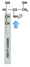
What is a hybrid genome?

A

The total gene content of a cell, including the nuclear genome and the genome(s) of any endosymbionts (mitochondria or chloroplasts).

32
Q

What is the simplest eukaryotic model organism?

A

Saccharomyces cerevisiae (yeast).

33
Q

What feature of Drosophila chromosomes makes Drosophila a good model for genetic studies?

A

Distinct banding patterns on the chromosomes for easy identification and tracking.

34
Q

What is the average number of genes in prokaryotes?

A

1000-6000.

35
Q

How does selective pressure favour prokaryotes having smaller genomes than eukaryotes?

A

Prokaryotes grow and divide as soon as they have enough nutrients, so it makes sense to pare down the genome as much as possible to replicate competitively; eukaryotic cells are not in the same constant state of competitive division, so they can keep more genes.

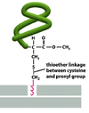
36
Q

What is the difference between prokaryotes and eukaryotes in terms of number of genes and genome size?

A

Eukaryotes have 3-30 times more genes, and 1000 times more DNA, than prokaryotes (including much more noncoding DNA).

37
Q

What is the significance of post-translational modification with respect to genome size?

A

1 gene can encode many gene products, so a cell may be much more complex than the size of its genome might suggest.

38
Q

Define polyploidy.

A

A state in which a cell replicates its DNA, but does not divide, resulting in a single cell with multiple copies of each of its chromosomes.

39
Q

How many chromosomes does the average eukaryotic cell have?

A

6 to 100.

40
Q

Define reciprocal chromosome translocation.

A

An event in which part of one chromosome is moved to another chromosome.

41
Q

How many protein-encoding genes are in the human genome?

A

~21,000.

42
Q

How many noncoding RNA genes are in the human genome?

A

~9,000.

43
Q

How many pseudogenes are in the human genome?

A

Over 20,000.

44
Q

What percentage of human DNA is found in exons?

A

1.5%.

45
Q

What percentage of human DNA is found in high-copy-number repetitive elements?

A

~50%.

46
Q

Define transposon (transposable element).

A

A DNA sequence that can replicate itself and change its position within the genome.

47
Q

Define microsatellite.

A

A DNA sequence made up of a variable number of repeats of a very short sequence (2-5 base pairs) of DNA.

48
Q

What is the main function of noncoding RNA genes in humans discovered so far?

A

Regulation of other genes.

49
Q

What is included in a gene for a protein product?

A
  1. Protein-encoding region; 2. Start/stop sites for transcription; 3. Introns; 4. Promoter region(s); 5. Other regulatory regions (i.e. binding sites for transcription factors)
50
Q

How big is the largest protein-encoding gene in the human genome?

A

2.4 million nucleotide pairs (vs. the average size, 27,000 nucleotide pairs).

51
Q

Define pseudogene.

A

A region of DNA that looks like a gene but is never expressed to make a functional gene product.

52
Q

What makes individual humans genetically different from one another?

A

SNPs or duplication/deletion events that change the genome without affecting overall gene function.

53
Q

How can transposons lead to the creation of new genes?

A

They may take neighbouring DNA with them when they move to a new position in the genome, resulting in a shuffling of DNA segments.

54
Q

How are gene families created?

A

A gene or whole-genome duplication event results in extra functional copies of the original gene(s), which are free to mutate to do other tasks while the original gene does its job.

55
Q

What are orthologs?

A

Similar genes in different species that perform the same function.

56
Q

What are paralogs?

A

Related genes within a single organism that have diverged to perform different functions.

57
Q

What are the main 4 events that result in the generation of new genes?

A
  1. Intragenic mutation; 2. Gene duplication; 3. DNA segment shuffling; 4. Horizontal transfer
58
Q

What do the members of the tomato terpene synthase gene family have in common?

A

They all use the same substrate (but make slightly different products).

59
Q

What are effector proteins?

A

Target proteins that lie at the end of signalling pathways and are altered in some way by an incoming signal to implement the appropriate change in cell behaviour.

60
Q

What is the conventional resolution limit of a light microscope?

A

200 nm.

61
Q

From the eye down, what are the basic parts of a light microscope?

A
  1. Eyepiece; 2. Tube lens; 3. Objective lens; 4. Stage (for specimen); 5. Condenser; 6. Iris diaphragm; 7. Light source
62
Q

What is the significance of Kaplan and Ewers’ work (2015) with nanobodies?

A

Nanobodies are small enough to cross cell walls without digestion, so they can be used instead of antibodies for visualization/tagging without damaging the cell.

63
Q

How does the wave-like behaviour of light determine which areas of an image look light or dark?

A

Light waves change phase when they interact with matter; the denser the matter, the more waves knocked out of phase relative to each other, and the difference between them results in lower amplitude of the combined wave, or dimmer light.

64
Q

What two factors determine resolution in microscopy?

A
  1. Wavelength of the light/electron source; 2. Numerical aperture of the microscope
65
Q

What two factors determine numerical aperture?

A
  1. Refractive index of the medium surrounding the specimen; 2. The angle between the condenser and the specimen field
66
Q

What is the relationship between resolution and limit of resolution?

A

As limit of resolution decreases, resolution increases–separate objects can be closer together before becoming indistinguishable from each other.

67
Q

What is the function of the condenser lens in a light microscope?

A

It focuses a cone of light rays from the light source onto each point of the specimen to be passed up to the objective lens.

68
Q

What is the function of the objective lens in a light microscope?

A

It collects a cone of light rays passing through the specimen from the condenser lens to form an image.

69
Q

What are the four basic types of light microscope?

A
  1. Bright-field; 2. Dark-field; 3. Phase contrast; 4. Differential-interference phase contrast
70
Q

What is the main problem with bright-field light microscopy?

A

Not enough contrast in the image.

71
Q

How is contrast improved for bright-field light microscopy?

A

Fixation and/or staining of specimens to enhance contrast in specific areas.

72
Q

How does dark-field microscopy work?

A

An opaque disc between the light source and the condenser ensures that light hits the specimen obliquely, not directly, so that the only light waves that reach the eyepiece are those that have been scattered by interacting with the specimen.

73
Q

What does a dark-field microscopy image look like?

A

A light object against a black background.

74
Q

How does phase contrast microscopy work?

A

The phase of each light wave changes according to the density of the part of the specimen it travels through; a computer detects and translates differences in phases to differences in brightness for better contrast.

75
Q

What does a phase contrast microscope image look like?

A

A high-contrast image against a dimmer background, with a “halo” of brighter light around the specimen.

76
Q

How does differential interference phase contrast microscopy work?

A

Like phase contrast microscopy (difference in phase translated to difference in brightness), but with a polarized light source to add an extra dimension for a 3D image.

77
Q

What does a differential interference phase contrast microscope image look like?

A

A greyscale 3D image showing very fine texture detail.

78
Q

What are the two main techniques used to detect objects too small to resolve with light microscopy?

A
  1. Fluorescence labelling; 2. Electron microscopy
79
Q

What are the three main categories of fluorescent probes used in microscopy?

A
  1. Fluorescent dyes; 2. Fluorescently labelled antibodies; 3. Fluorescent proteins (transgenic)
80
Q

How does fluorescence microscopy work?

A

A high-energy wavelength from the light source passes through the specimen and excites fluorescent particles specific to that wavelength; when they fall back to ground state, they emit a lower-energy wavelength that is selectively detected and allowed past the condenser.

81
Q

What fluorescent probe is used to label DNA (all bases)?

A

DAPI (fluorescent dye).

82
Q

What fluorescent probe is used to label proteins (all kinds)?

A

FITC (fluorescent dye).

83
Q

How are specific proteins typically labelled for fluorescence microscopy?

A

Fluorescently labelled antibodies or fluorescence proteins (most commonly GFP).

84
Q

What 3 filters does the light pass through between the source and the eyepiece in a fluorescence microscope?

A
  1. First barrier filter (lets through excitation wavelength only); 2. Dichroic mirror (lets through emission wavelength only); 3. Second barrier filter (cuts out unwanted fluorescence signals)
85
Q

How does confocal fluorescence microscopy work?

A

A laser shone through a pinhole excites 1 point at a time in the sample, and another pinhole past a dichroic mirror collects emission-wavelength light from 1 point at a time, assembling a full 3D image from many optical scans through thin sections.

86
Q

What are the two main advantages of confocal fluorescence microscopy for cell biology?

A
  1. Very fast; 2. Can analyze living organisms
87
Q

When is marker gene fusion used in fluorescence microscopy?

A

To visualize specific proteins.

88
Q

What is the difference between dyes and other classes of fluorescence probes?

A

Dyes detect a whole class of molecules (e.g. DNA or proteins), not a specific molecule (e.g. A vs G).

89
Q

How do primary and secondary antibodies work together in fluorescence microscopy with fluorescent-tagged antibodies?

A

The primary antibody comes from one animal (usually rabbits) and is specific to some antigen that has been introduced for tagging; the secondary antibody is from a different animal and recognizes any (rabbit) antibody as foreign.

90
Q

What is the purpose of the secondary antibody in fluorescent-tagged antibody microscopy?

A

To amplify the fluorescence signal by binding to any primary antibody bound on the specimen.

91
Q

What is the main advantage of gene fusion techniques over dyes and antibodies in fluorescence microscopy?

A

Dyes and antibodies have to cross cell membranes to bind to their targets; genetically engineered cells express gene fusion tags on their own.

92
Q

How do reporter genes work in gene-fusion microscopy?

A

The coding sequence for some protein of interest is replaced with the coding sequence for the reporter gene (e.g. GFP), so wherever that protein would be expressed, GFP is expressed instead, allowing visualization of when and where a specific protein is expressed in the cell.

93
Q

What can be detected using cis regulatory sequence-marker gene fusions?

A

Tissue- or cell-specific expression patterns (different regulatory sequences may be used to regulate the same gene in different locations).

94
Q

What gene fusion technique is used to track protein turnover inside a cell?

A

Protein-marker fusions that leave the protein intact but attach a reporter protein.

95
Q

What are signal peptide marker gene fusions used to detect?

A

Subcellular localization of specific proteins: the signal peptide tells the protein where to go in the cell, so the gene fusion can be used to follow the protein inside the cell.

96
Q

What is detected by photoactivation and photobleaching microscopy techniques?

A

Protein diffusion dynamics (based on how quickly proteins move out of activated areas or into bleached areas).

97
Q

How does photoactivation work?

A

Fluorescence tags are activated in specific spots by a point of light, and the dissipation of the fluorescent signal in the activated region indicates the movement of proteins away from the area.

98
Q

How does photobleaching (FRAP) work?

A

A point of light destroys the fluorescence signal in a specific area, and the rate of signal recovery (return of fluorescence) indicates the movement of new proteins into the bleached area.

99
Q

What does FRAP stand for?

A

Fluorescence recovery after photobleaching (a visualization technique for fluorescence-tagged proteins).

100
Q

What does FRET stand for?

A

Fluorescence resonance energy transfer (a visualization technique for protein-protein interactions).

101
Q

How does FRET work?

A

Two proteins are tagged with different fluorescent markers so that the emission wavelength of one is the excitation wavelength of the other. If the proteins interact with each other, lighting the sample with the excitation wavelength of the first protein will return the emission wavelength of the second.

102
Q

What does TIRF stand for?

A

Total internal reflection fluorescence (a visualization technique for tagged molecules within a sample).

103
Q

How does TIRF work?

A

An angled laser light source is aimed at the bottom of the sample so that only molecules at the sample surface are excited while the rest of the light reflects away, resulting in a finely focused image.

104
Q

What is the resolution limit of electron microscopy?

A

1 nm (vs 200 nm for light microscopy!).

105
Q

What is the difference between scanning (SEM) and transmission (TEM) electron microscopy?

A

SEM scans the surface of the specimen and creates detailed 3D images; TEM scans internal cross-sections of the specimen and creates images of its insides.

106
Q

Why does electron microscopy have a higher resolution than light microscopy?

A

Electrons have a much shorter wavelength than visible light (0.004 nm vs hundreds of nm).

107
Q

What are the “lenses” in an electron microscope?

A

Magnetic coils that focus the electrons as they run through the vacuum tube from the electron gun.

108
Q

Why do electron microscopes require a vacuum?

A

Air would scatter the electrons, reducing the focus of the image.

109
Q

How is the contrast created in an electron microscopy image?

A

The image is darker where electrons are dense, and lighter where the electrons can pass through the sample more easily.

110
Q

How are specimens prepared for SEM?

A

Dried, rapidly frozen, and coated with a thin layer of gold.

111
Q

What is the purpose of heavy metal salts in electron microscopy?

A

Atoms with a higher atomic number create more contrast, but biological molecules have low atomic numbers, so binding them to heavy metal salts increases their weight and therefore their contrast.

112
Q

Why is it important to freeze samples rapidly for electron microscopy?

A

Rapid freezing (e.g. with liquid nitrogen) ensures there is no time for water to form crystals, which would damage structures and create artefacts in the image.

113
Q

How does immunogold electron microscopy work?

A

Specific proteins are tagged with antibodies that are labelled with gold (instead of fluorescent labels), which increases the contrast where the gold-labelled proteins are localized within cells.

114
Q

Which two chemicals are used to fix specimens for electron microscopy?

A
  1. Glutaraldehyde; 2. Osmium tetroxide
115
Q

What is the typical (practical) resolution of SEM?

A

About 10 nm.

116
Q

What does STEHM stand for?

A

Scanning transmission electron holographic microscope.

117
Q

What is the resolution of STEHM?

A

40 *pm* (the diameter of a He atom is ~31 pm!).

118
Q

What can be determined from STEHM’s measure of phase in a specimen?

A

A more precise measure of where individual electrons are, which can be used to determine the specimen’s absolute composition, internal strain, electrostatic/magnetic fields, temperature, etc.

119
Q

What can be visualized with STEHM?

A
  1. Specimen surface; 2. Internal cross-sections; 3. Electron phase (mean inner potential for electron localization)
120
Q

What two materials are needed to disrupt cells before isolating them from tissues?

A
  1. Proteolytic enzymes (to digest extracellular matrix proteins); 2. EDTA (to bind Ca2+, needed for cell-cell adhesion)
121
Q

What does FACS stand for?

A

Fluorescence-activated cell sorter (equipment for isolating specific cell types from a cell suspension).

122
Q

How does FACS work?

A

A cell suspension is passed in single file through a laser at the excitation wavelength for a particular fluorescence-tagged cell in the suspension; a detector then flags any excited (tagged) cells with a negative charge and untagged cells with a positive charge; cells are sorted by charge into separate collectors.

123
Q

What happens to aggregates of cells in single droplets that pass through a FACS system?

A

They get no charge and are dumped into a waste beaker automatically.

124
Q

How does laser-capture microdissection work?

A

A high-energy laser beam cuts out a precise area of interest from a specimen slide, and a second laser beam from underneath the slide shoots the cut-out area up into a collection container.

125
Q

What is laser-capture microdissection used for?

A

To analyze very specific regions within larger samples (gene expression patterns, amino acid compositions, etc.).

126
Q

From beginning to end of centrifugation, starting with a cell homogenate, what does each pellet contain?

A
  1. Whole cells, nuclei, cytoskeletons; 2. Mitochondria, lysosomes, peroxisomes; 3. Microsomes, small vesicles; 4. Ribosomes, viruses, large macromolecules
127
Q

How can purity be improved when isolating organelles by centrifugation?

A

Repeating centrifugation steps for each pellet.

128
Q

How does ion-exchange chromatography work?

A

Beads in the column are charged to attract desired ions of the opposite charge while other ions pass through unaffected.

129
Q

How does gel-filtration chromatography work?

A

Column beads are porous: smaller particles move through the pores on their way down while larger particles move straight past the beads, so proteins are isolated based on size.

130
Q

How does affinity chromatography work?

A

The beads in the column are covalently attached to a substrate or antibody specific to the protein of interest; once the rest of the solution is washed out, only the protein of interest remains stuck to the beads.

131
Q

How are epitope tags used to isolate proteins?

A

A cell whose coding sequence is known for the protein of interest is genetically engineered to express a tag attached to the protein that is easily recognized and bound by affinity chromatography beads.

132
Q

Define primary culture.

A

Tissue isolated directly from an organism for the first time.

133
Q

Define secondary culture.

A

Cells taken from a primary culture and regrown in a new culture.

134
Q

What are the two main reasons for cells to stop replicating?

A
  1. Enough successive replications for telomeres to degrade;
  2. Cell cycle checkpoints trigger an end to replication
135
Q

What makes transformed (cancer) cells replicate indefinitely?

A

No more cell cycle checkpoints.

136
Q

Define hybridoma cell.

A

A hybrid cell formed from a heterokaryon by the fusion of the two parent nuclei, one of which is a transformed (cancer) cell.

137
Q

What is the application of hybridoma cells?

A

Because they are fused with transformed (cancer) cells, they replicate indefinitely, so they can be used to maintain a continuous culture (and a reliable source of protein products).

138
Q

What are ES cells?

A

Embryonic stem cells (which can differentiate into a full organism).

139
Q

How does the CRISPR system work in bacteria?

A
  1. Virus inserts DNA/RNA into host;
  2. Bacteria cleaves off a portion and inserts it into the CRISPR locus within the bacterial genome;
  3. CRISPR locus is transcribed, and resulting RNA is bound to Cas proteins (endonucleases);
  4. On new infection, RNA on Cas recognizes and binds incoming complementary sequences, tagging them for cleavage by Cas
140
Q

What is the difference between bacterial Cas and engineered Cas9?

A

Cas9 doesn’t cleave DNA–instead, it has a fused activation or repressor domain that targets genomic DNA to activate or deactivate specific genes.

141
Q

How does CRISPR-Cas9 work?

A

Cas9 is bound to guide RNA complementary to the genomic sequence of interest, which targets a specific location on the genome; the fused activation or repressor domain then targets the nearby genes to turn specific genes on or off.

142
Q

How many different lipids can be found in a membrane?

A

~5,000.

143
Q

What are the two most common ways that lipids move within membranes?

A
  1. Lateral diffusion;
  2. Rotation
144
Q

Where in the plasma membrane is phosphatidlyserine found?

A

Only on the cytosolic side; flipping to the extracellular side triggers phagocytosis during apoptosis.

145
Q

What is the general structure of a phospholipid?

A
  1. Polar head: glycerol group linked to phosphate group linked to other head group(s);
  2. Nonpolar tails: 2 fatty acid chains (18-24 C), one saturated (no double bonds), one unsaturated (1 or more cis C=C double bonds)
146
Q

What distinguishes phosphatidylserine from other phospholipids?

A

It is only ever found on the cytosolic side of the plasma membrane, and it has a net negative charge (not neutral).

147
Q

What molecule is this?

A

Phosphatidylcholine (a membrane phospholipid).

148
Q

What molecule is this?

A

Phosphatidylethanolamine (a membrane phospholipid).

149
Q

What molecule is this?

A

Phosphatidylserine (a membrane phospholipid).

150
Q

What is the general structure of a sphingolipid?

A

A molecule of sphingosine (which has 1 long fatty chain) attached to a fatty acid tail and a polar head group.

151
Q

What molecule is this?

A

Cholesterol.

152
Q

What molecule is this?

A

Sphingomyelin (a membrane sphingolipid).

153
Q

What molecule is this?

A

Sphingosine (the scaffold molecule for sphingolipids).

154
Q

What is the purpose of cholesterol in lipid membranes?

A

Decreases membrane permeability: rigid structure packs lipids more tightly, making it harder for other molecules to pass through.

155
Q

What molecule is this?

A

Galactocerebroside (a membrane glycolipid).

156
Q

What molecule is this?

A

GM1 ganglioside (a membrane glycolipid).

157
Q

What is the general structure of a glycolipid?

A

A sphingolipid with a sugar attached instead of a phosphate group.

158
Q

Where are glycolipids found in cell membranes?

A

Sugar groups are only found on the extracellular side of the plasma membrane.

159
Q

What are isoprenoid side chains?

A

15- or 20-carbon chains that branch off sideways from the fatty acid tail of some archaeal membrane lipids.

160
Q

What type of linkage is unique to archaeal membrane lipids?

A

Ether linkages where there is no fatty acid head group; bacteria and eukaryotes have ester linkages.

161
Q

How are prokaryotic cell membranes less complex than eukaryotic membranes?

A
  • Less variability (usually 1 main lipid, combined with a few others)
  • No cholesterol for permeability control
162
Q

In what type of membrane are glycolipids most common?

A

Myelin (surrounding nerve cells).

163
Q

What is the main lipid in E. coli plasma membranes?

A

Phosphatidylethanolamine.

164
Q

In what 2 ways can inositol phospholipids be activated in signalling cascades?

A
  1. Phosphorylated, with the 3 phosphate groups forming a docking site for intracellular proteins;
  2. Cleaved into fragments, with the fragments acting as signalling molecules within the cascade
165
Q

Why do glycerols and cholesterol esters arrange in a monolayer instead of a bilayer?

A

They are purely hydrophobic (no hydrophilic portions to arrange on the outside).

166
Q

Where do we see monolayer membranes in cells?

A

In lipid droplets (for storage and transport of purely hydrophobic molecules).

167
Q

What type of structure usually crosses the membrane in transmembrane proteins?

A

Alpha helix.

168
Q

What are two mechanisms for attaching a membrane protein to only one monolayer?

A
  1. Domains of the protein integrated into the membrane;
  2. Lipid anchors (e.g. GPI)
169
Q

In a hydrophobicity plot of a transmembrane protein, what do the peaks correspond to?

A

Hydrophobic portions, i.e. the domains of the protein that are embedded within the membrane; the number of peaks is equal to the number of transmembrane domains.

170
Q

What are the 3 main types of lipid anchors for transmembrane proteins?

A
  1. Myristoyl anchors (amide linkage);
  2. Palmitoyl anchors (thioester linkage);
  3. Farnesyl anchors (thioether linkage)
171
Q

Of myristoyl, palmitoyl, and farnesyl lipid anchors, which forms a reversible bond with its anchored protein?

A

Palmitoyl.

172
Q

Are linkages between proteins and membrane lipid anchors co- or post-translational?

A

Post-translational.

173
Q

True or false: Most transmembrane proteins are glycosylated.

A

True.

174
Q

What are the 2 main functions of the sugar groups on glycoproteins?

A
  1. Protective layer outside the plasma membrane: prevents chemical/mechanical damage by keeping molecules, other cells, etc. away from the cell;
  2. Recognition (cell-cell and self-vs.-nonself)
175
Q

What is the difference between glycoproteins and proteoglycans?

A

Proteoglycans have longer sugars that are attached differently.

176
Q

What structure is this?

A

A farnesyl anchor (an irreversible transmembrane protein anchor).

177
Q

What structure is this?

A

A myristoyl lipid anchor (for irreversibly anchoring transmembrane proteins).

178
Q

What structure is this?

A

A palmitoyl lipid anchor (for reversibly anchoring transmembrane proteins).

179
Q

Where can intrachain disulfide bonds be found in transmembrane proteins?

A

Only on the extracellular side of the membrane.

180
Q

What are two major functions of tight junctions between neighbouring cells?

A
  1. Keep materials from leaking into/out of cells;
  2. Keep localized membrane proteins from moving between different regions of the membrane
181
Q

What three types of molecules are found in greater concentrations within lipid rafts than in other regions of membranes?

A
  1. Cholesterol;
  2. Sphingolipids (with longer hydrophobic areas);
  3. GPI-anchored proteins
182
Q

What are lipid rafts?

A

Thick, dynamic “islands” within the membrane with specific protein compositions that can move around and quickly assemble or disassemble as needed.

183
Q

What are 2 ways in which membrane-bending proteins can deform bilayers?

A
  1. Inserting large proteins into 1 monolayer;
  2. Aggregating lipids with large head groups within 1 monolayer
184
Q

What are three types of interactions that limit protein mobility within membranes?

A
  1. Proteins interacting in large aggregates;
  2. Proteins attached to the membrane at either face;
  3. Protein-protein interactions between neighbouring cells
185
Q

How do small, hydrophobic molecules cross membranes?

A

Simple diffusion.

186
Q

How do ions and uncharged polar molecules cross membranes?

A

Transport proteins (channels or transporters).

187
Q

How do macromolecules cross membranes?

A

In vesicles (endo-/exocytosis).

188
Q

Of channels and transporters, which is involved in active transport and which is involved in passive transport?

A
  • Transporters: active or passive;
  • Channels: passive only
189
Q

Why are channels faster than transporters?

A

Transporters rely on conformational changes caused by binding and releasing different substrates; channels are either open or closed, with no conformational change or binding events required.

190
Q

What happens in cotransport (symport)?

A

Two molecules are transported at the same time in the same direction.

191
Q

What happens in exchange (antiport)?

A

Two molecules are transported at the same time in opposite directions.

192
Q

When does a transporter reach its maximum rate of transport?

A

When all of its binding sites are saturated with substrate.

193
Q

When does a channel reach its maximum rate of transport?

A

It doesn’t–channels are left open for molecules to move through until some external signal prompts it to close again.

194
Q

What three types of transporters are involved in glucose transport from the gut lumen to the extracellular fluid?

A
  1. Na+-driven glucose symport (to actively transport glucose into intestinal epithelial cells);
  2. Glucose transporter (to mediate passive transport of glucose into extracellular fluid);
  3. Na+-K+ pump (to pump the Na+ back out of the cell and maintain the Na+ gradient for more glucose symport)
195
Q

How does Na+ help to transport glucose from gut lumen into intestinal epithelial cells?

A

[Na+] is higher outside than inside the epithelial cell, so coupled glucose takes advantage of the passive transport of Na+ to move against its concentration gradient into the cell.

196
Q

Why do intestinal epithelial cells have Na+-K+ pumps on the cytosolic side (away from the gut lumen)?

A

Glucose transport is mediated by passive Na+ transport, so when Na+ accumulates in the cell from bringing in glucose, it needs to be pumped back out of the cell to maintain the [Na+] gradient for continued glucose transport.

197
Q

Why does it require active transport to move glucose into gut epithelial cells from the lumen, but only passive transport to move it from the cells into the extracellular fluid?

A

[Glucose] is lower in the cell than in either the lumen or the extracellular fluid, so it is moving up its concentration going into the cell but down it going out.

198
Q

What are the three classes of ATP pumps?

A
  1. P-type pump (for ions);
  2. ABC transporter (for small molecules);
  3. V-type/F-type pumps (for protons)
199
Q

What type of pump is the Ca2+ pump?

A

P-type ATPase.

200
Q

What is the difference between F-type and V-type pumps?

A

F-type is an ATP synthase that makes ATP by passive proton transport. V-type is an active proton pump that requires ATP to move protons against their concentration gradient.

201
Q

What is a major application of V-type proton pumps in the cell?

A

To increase pH of a compartment (e.g. lysosomes) by actively pumping in protons.

202
Q

When do Ca2+ pumps in the SR start to work?

A

When signalling events have resulted in release of Ca2+ into the cytosol (need to get Ca2+ back into the SR lumen to maintain the concentration gradient).

203
Q

What is the function of the nucleotide binding domain in a P-type ATPase (e.g. Ca2+ pump)?

A

To bind ATP, which activates the pump.

204
Q

What happens when ATP binds to a Ca2+ pump?

A
  1. Cavity opens: 2H+ out, 2Ca2+ in
  2. Ca2+ binds: pump closes up and brings pump domains together
  3. Phosphorylation domain (with Asp) takes phosphate group to turn ATP into ADP: ADP leaves
  4. ATP binds in vacant spot: shape change lets 2Ca2+, 2H+ in on opposite side
  5. H+ binds: pump closes up again
205
Q

How do Na+-K+ pumps create a membrane potential?

A

They pump in 2 K+ for every 3 Na+, maintaining greater positive charge outside than inside the cell (with low [Na+], high [K+] inside the cell).

206
Q

What does the ABC in ABC transporter stand for?

A

ATP-binding cassette.

207
Q

What is the difference between prokaryotic and eukaryotic ABC transporters?

A

Prokaryotic ABC transporters work to export and import molecules out of/into the cell; eukaryotic ABC transporters are mainly used in export only.

208
Q

In what organism were ABC transporters first identified?

A

E. coli (of course).

209
Q

Where were ABC transporters first identified in humans?

A

Cancer cells (especially drug-resistant cells, which can have many types of ABC transporters to export chemotherapy drugs).

210
Q

What are the three major functional domains of P-type ATPases?

A
  1. Nucleotide binding domain (binds ATP);
  2. Activation domain (phosphorylates ATP to ADP);
  3. Phosphorylation domain (contains the Asp residue to be phosphorylated during dephosphorylation of ATP)
211
Q

What are the three main functional domains of ABC transporters?

A
  1. Solute-binding site (binds the substrate to be transported);
  2. ATPase domains (bind ATP to drive conformational change for transport);
  3. Hydrophobic domains (contain the solute-binding site and provide a channel through the membrane on conformational change)
212
Q

What types of ion channels are involved in active transport?

A

None of them! All ion channels are passive.

213
Q

What are the four key characteristics of ion channels?

A
  1. High selectivity for one ion over all others;
  2. Gated transport (fully open or fully closed);
  3. High efficiency (millions of ions through per second);
  4. Always passive (never involved in active transport)
214
Q

What are the three main classes of ion channels?

A
  1. Voltage-gated (based on charge on either side of the membrane);
  2. Ligand-gated (activated by some other molecule);
  3. Mechanically gated (open/closed by conformational changes in the surrounding membrane)
215
Q

When does K+ stop diffusing through K+ leak channels in animal cell membranes?

A

When the positive charge outside the cell becomes high enough that the electrical gradient opposes K+ diffusion (even when the concentration gradient favours it).

216
Q

How selective are K+ channels?

A

K+ channels transport 10,000 times more K+ than any other ion.

217
Q

Why are ion channels so much faster than transporters?

A

Channels have no binding sites, so they can let through many more ions at once (millions per second!).

218
Q

How does the selectivity filter work in K+ channels?

A

It is a narrow channel leading from the pore opening, and it is lined with carbonyl oxygens that transiently bond with passing K+ to strip them of their hydrate shell, making them small enough to pass through.

219
Q

Why can’t Na+ fit through K+ channels, even though K+ is bigger than Na+?

A

K+ binds with 4 carbonyl Os lining the selectivity filter to overcome the energy cost of stripping its hydrate shell; Na+ is too small to interact with all 4 Os at once, so it can’t lose its shell (and a hydrated Na+ is bigger than a naked K+).

220
Q

How are K+ channels gated?

A

Mechanosensitive (osmotic pressure): when ion concentration is high inside the cell, there is more water too, which makes the cell swell; swelling stretches the membrane, which pulls the channel pore open to let ions out.

221
Q

Why are aquaporins important in cell membranes?

A

Water is needed to balance ion concentrations as ions move in and out of the cell constantly, so water-specific channels speed up the overall rate of diffusion for quick adjustment.

222
Q

How are hydrated ions kept out of aquaporins?

A

The hydrophilic parts of the aquaporin are within the narrowest part of the channel, which is too narrow for hydrated ions (water molecules pass through single-file).

223
Q

How are dehydrated ions kept out of aquaporin channels?

A

Even if they were small enough to fit through the channel, they are repelled by the hydrophobic side of the inner channel, and the hydrophilic side is occupied by water molecules.

224
Q

How are protons usually transported within cells?

A

By relay through a series of water molecules (converting H2O to H3O+ when it binds).

225
Q

How do aquaporins prevent protons from passing through?

A

H+ normally moves by relay, binding to successive O on H2O molecules; the hydrophilic side of the aquaporin channel has 2 strategically placed Asn residues that tie up the O on H2O passing through, so any H+ that makes it that far can only go back the way it came.

226
Q

What are the 3 stages of a voltage-gated Na+ channel?

A

Open, closed, and inactive (not closed, but not working).

227
Q

What is the key characteristic of the amino acid sequence of the voltage receptors in voltage-gated Na+ channels?

A

Many positively charged amino acid residues.

228
Q

What is the general structure of a voltage-gated Na+ channel?

A
  • 4 domains (each with a voltage sensor and a selectivity filter) forming a central channel;
  • 1 inactivation gate between domains 3 and 4;
  • Lateral pores opening from the central cavity into the cell membrane (hydrophobic portion)
229
Q

What effect do small, hydrophobic drugs have on voltage-gated channels?

A

They can block the lateral pores to prevent movement of ions between the membrane and the central cavity.

230
Q

What triggers the opening of a voltage-gated ion channel?

A

An action potential triggers the opening of some other channel in the membrane that changes the membrane potential, which triggers the voltage sensors on the voltage-gated channel to open that channel.

231
Q

Why is the inactivated state of voltage-gated Na+ channels important?

A

An open channel lets in Na+, causing a change in membrane potential that then activates nearby voltage-gated channels, propagating the signal; inactivating a channel immediately ensures that the signal is only propagated in one direction and limits the positive feedback activation of nearby channels.

232
Q

In what direction is the signal propagated within a membrane when a voltage-gated Na+ channel is opened?

A

Opening the channel allows Na+ in and depolarizes the membrane, with nearby channels activated by the incoming positive charge, so the signal is propagated from regions where the membrane has been depolarized toward regions where the membrane is still at resting potential.

233
Q

What are transmitter-gated ion channels?

A

Channels with very selective binding sites for specific neurotransmitters, with binding events either causing or inhibiting depolarization of the surrounding cell membrane by opening channels for specific ions.

234
Q

What happens when excitatory neurotransmitters bind to transmitter-gated ion channels?

A

Na+ channels open, causing membrane depolarization.

235
Q

What happens when inhibitory neurotransmitters bind to transmitter-gated ion channels?

A

Cl- or K+ channels open, preventing depolarization of the surrounding membrane.

236
Q

How does the opening of K+ channels by inhibitory neurotransmitters prevent membrane depolarization?

A

[K+] is usually high inside the cell, so K+ channels are open at resting potential; opening even more makes it harder to drive the cell away from resting potential, as K+ efflux (with [K+] gradient) balances Na+/Ca2+ influx (electrical gradient).

237
Q

Is acetylcholine an excitatory or inhibitory neurotransmitter?

A

Both, but usually excitatory (causes membrane depolarization).

238
Q

Is GABA an excitatory or inhibitory neurotransmitter?

A

Inhibitory (prevents membrane depolarization).

239
Q

What makes transmitter-gated ion channels insensitive to membrane potential relative to voltage-gated channels?

A

Transmitter-gated channels are open or closed based on highly specific binding events, not any change in charge, so they don’t respond to changes in voltage.

240
Q

Why aren’t transmitter-gated ion channels capable of producing the self-amplifying positive feedback excitation seen in voltage-gated channels?

A

Transmitter-gated channels are opened by highly specific binding events between substrate (neurotransmitter) and binding site, not by any change in membrane potential or other properties, so they can only be activated when the right neurotransmitter is made available to it by a different structure.

241
Q

How do transmitter-gated ion channels cause a change in membrane potential to generate an action potential?

A

Opening of transmitter-gated channels causes a brief increase in membrane permeability that allows ions through to depolarize the membrane, activating nearby voltage-gated ion channels that can then propagate an action potential.

242
Q

How does the opening of Cl- channels by inhibitory neurotransmitters prevent membrane depolarization?

A

[Cl-] is usually much higher outside than inside the cell, but membrane potential keeps it out; if the membrane starts to depolarize, Cl- can follow its concentration gradient without being opposed by the electrical gradient, buffering the membrane potential.

243
Q

How does strychnine work?

A

Strychnine binds to glycine receptors on inhibitory transmitter-gated ion channels, blocking the inhibitory action of the neurotransmitter (overexciting the cell) and causing muscle spasms, convulsions, and death.

244
Q

What are the 4 distinct intracellular compartment families?

A
  • Nucleus and cytosol
  • Secretory/endocytic pathway (ER, Golgi, lysosome, vesicles, etc.)
  • Mitochondria
  • Chloroplasts (plastids)
245
Q

What are the 3 main protein transport pathways?

A
  • Secretory transport
  • Transmembrane transport
  • Vesicle transport
246
Q

What is the function of a protein signal sequence?

A

To act as an “address” recognized by sorting receptors to determine where it will go in the cell.

247
Q

Do protein sorting receptors always recognize a specific amino acid sequence within protein signal sequences?

A

No–they often just recognize certain patterns (e.g. patterns of positive or negative charge).

248
Q

What is different about signal sequences in proteins destined for the nucleus (versus proteins that go to other organelles)?

A
  • The signal is mid-protein (not at either terminus);
  • The protein is transported fully folded, so the sequence must be exposed somewhere on the outside of the protein in its final conformation;
  • The signal sequence is usually not cleaved off after delivery
249
Q

Which proteins have a return signal sequence on their C-terminus?

A

Only proteins that are meant to stay in the ER (in case they accidentally get packed up with other proteins to go to the Golgi); otherwise, proteins always stay where they are once delivered.

250
Q

What feature of the amino acid sequence is unique to signal sequences in proteins addressed for import into the ER?

A

A long string (10 amino acids) of hydrophobic residues in the middle of the sequence.

251
Q

Why is it important that the return signal sequence for ER proteins is on the C-terminus?

A

Any signal on the N-terminus (directing it to the ER in the first place) was likely cleaved off once it got there; the C-terminal signal will still be there, so it can still direct the protein back to the ER.

252
Q

What two experimental techniques are commonly used to study protein signal sequences?

A
  • GFP tagging (where does it go in the cell?);
  • Truncation experiments (what does the signal absolutely require to function as a cellular address?)
253
Q

In 1970s experiments in vitro with mRNA, ribosomes, and microsomes, what was the significance of the finding that synthesized proteins were longer in tubes without microsomes than in tubes with microsomes?

A

Microsomes act like ER and cleave off signal sequences when proteins are directed to them; without microsomes, there is nothing to cleave sequences, so the proteins remain fully intact after synthesis.

254
Q

What is the difference between two proteins that are otherwise practically identical, except one has a signal sequence and the other does not?

A

The (longer) one with the signal sequence will be sent to some organelle; the (shorter) one without a signal sequence will stay in the cytosol and function there.

255
Q

Which portion of the nucleus is continuous with the ER?

A

The outer membrane of the nuclear envelope (which is often also studded with ribosomes).

256
Q

What is the rate of traffic through the nuclear pores in the nuclear envelope?

A

About 500 molecules per second, in both directions (in and out of the nucleus).

257
Q

What types of molecules are transported in and out of the nucleus?

A
  • RNAs involved in transcription, translation, splicing, etc.;
  • Proteins (fully folded)
258
Q

What structure is this? What are the different parts?

A

Nuclear pore complex (NPC):

  • A: membrane ring proteins;
  • B: nuclear basket;
  • C: disordered region of channel nucleoporin fibrils;
  • D: channel nucleoporins;
  • E: scaffold nucleoporins;
  • F: cytosolic fibrils
259
Q

How many different types of proteins can be found in a mammalian nuclear pore complex?

A

About 30.

260
Q

How many nuclear pore complexes can be found in a single mammalian cell nucleus?

A

3,000 to 4,000.

261
Q

What is the function of the channel nucleoporins in a nuclear pore complex?

A

The fibrils of the channel nucleoporins create a tangled mesh that inhibits free diffusion through the pore.

262
Q

What is the function of the membrane ring proteins in a nuclear pore complex?

A

To anchor the pore in the nuclear envelope.

263
Q

What two key functions are performed mainly in the cytosol?

A
  • Protein synthesis & degradation;
  • Intermediary metabolism (reactions providing building blocks for macromolecules)
264
Q

Why is the ER unique among organelles in having ribosomes attached to it?

A

It is the only organelle that receives proteins as they are being translated (all other organelles only receive complete proteins).

265
Q

How do the nucleus and the cytosol communicate?

A

Through nuclear pore complexes in the nuclear envelope.

266
Q

What makes protein translocation different from gated transport and vesicular transport in terms of cell compartment topology?

A

Gated and vesicular transport ferry proteins between compartments that are topologically equivalent; protein translocation by transmembrane transporters moves proteins from the cytosol into topologically different compartments.

267
Q

What signal sequence is characteristic of proteins destined for the mitochondria?

A

Positively charged amino acids alternating with hydrophobic ones.

268
Q

What two types of proteins have signal sequences at their C-terminus?

A
  • Peroxisome proteins;
  • ER resident proteins (return signal)
269
Q

True or false: The signal sequences of different proteins with the same destination are functionally interchangeable.

A

True–physical properties (e.g. hydrophobicity) seem to be more important than the exact amino acid sequence for directing proteins to the right place.

270
Q

How are organelles duplicated during cell division?

A

The parent cell incorporates new molecules into the organelles to enlarge them before they divide, so that each daughter cell inherits a complete set of specialized cell membranes from the parent cell.

271
Q

What is the nuclear lamina?

A

A protein meshwork bound to the inner nuclear membrane that provides structural support for the nuclear envelope and acts as an anchoring site for chromosomes and the cytoplasmic cytoskeleton (via protein complexes spanning the nuclear envelope).

272
Q

What proteins are found within the inner nuclear membrane that are not found in the outer nuclear membrane?

A

Proteins that act as binding sites for chromosomes and for the nuclear lamina.

273
Q

What type of localization signal is common in many nuclear proteins?

A

Signals consisting of 1 or 2 short sequences rich in positively charged Lys and Arg residues.

274
Q

What are nuclear import receptors?

A

A family of soluble cytosolic proteins that recognize, bind, and transport the subset of cargo proteins containing nuclear localization signals, binding to FG repeats in NPC nucleoporins to deliver the cargo.

275
Q

What are FG repeats?

A

Regions with repeated Phe/Gly residues in the fibrils of NPC nucleoporins that create a permeability barrier between the cytosol and the nucleus and provide binding sites for nuclear import receptors bringing in cargo proteins.

276
Q

How do nuclear import receptor-cargo complexes move through the NPC?

A

They repeatedly bind, dissociate, and re-bind to adjacent FG-repeat sequences in a random walk through the pore, disrupting weak interactions between repeats and locally dissolving the gel stage of the matrix by binding; the complex only dissociates if it successfully reaches the nuclear side of the NPC.

277
Q

What are the most abundant membrane lipids?

A

Phospholipids (and the most abundant phospholipids are phosphoglycerides).

278
Q

What is the general structure of a phosphoglyceride?

A
  • 3-carbon glycerol backbone;
  • 2 long-chain fatty acids ester-linked to adjacent C atoms on the glycerol;
  • Phosphate group attached to 3rd C of the glycerol;
  • 1 of several types of head group attached to phosphate group
279
Q

What are the 4 most abundant phospholipids in mammalian cell membranes?

A
  • Phosphatidylethanolamine;
  • Phosphatidylserine;
  • Phosphatidylcholine;
  • Sphingomyelin
280
Q

What is the main structural difference between sphingolipids and phospholipids?

A

Sphingolipids are built from sphingosine; phospholipids are built from glycerol.

281
Q

What is the general structure of a sphingolipid?

A
  • Sphingosine core: 1 long acyl (fatty) chain with an amino group and 2 OH groups at 1 end;
  • 1 fatty acid tail attached to amino group;
  • Phosphate group attached to terminal OH group;
  • Head group attached to phosphate group
282
Q

What is the most common sphingolipid?

A

Sphingomyelin.

283
Q

What head group is attached to the terminal hydroxyl group of sphingosine to make sphingomyelin?

A

Phosphocholine (the same head group as in phosphatidylcholine–just on sphingosine instead of glycerol).

284
Q

What is the difference between sphingolipids and glycolipids?

A

Both are based on sphingosine, but sphingolipids have a phosphate group attached to the terminal OH group, while glycolipids have 1 or more sugars attached.

285
Q

What is the difference between phosphoglycerides and glycolipids?

A

Phosphoglycerides are based on glycerol and have a phosphate group attached between glycerol and head groups; glycolipids are based on sphingosine and have sugars attached instead of phosphate.

286
Q

What is the general structure of cholesterol?

A

A rigid steroid ring structure, with 1 polar hydroxyl head group and 1 short nonpolar hydrocarbon chain attached (on opposite ends of the ring).

287
Q

How do cholesterol molecules orient themselves in the lipid bilayer?

A

With the polar hydroxyl head group close to the polar head groups of adjacent phospholipids.

288
Q

Why do hydrophilic molecules dissolve readily in water?

A

They contain charged groups or uncharged polar groups that form either favourable electrostatic interactions or hydrogen bonds with water molecules.

289
Q

How does cholesterol make membranes less permeable without reducing their fluidity?

A

Cholesterol’s rigid structure immobilizes portions of the fatty acid tails near the phospholipid head groups, so small molecules can’t slip through near the surface, but the rest of the phospholipid (and therefore the whole membrane) remains fluid below the surface.

290
Q

What class of proteins regulate most processes in eukaryotic cells?

A

GTP-binding proteins: monomeric & trimeric GTPases, regulated by GEFs (guanine exchange factors) and GAPs (GTPase-activating proteins).

291
Q

How do GAPs inactivate GTPases?

A

By triggering the hydrolysis of the GTPase’s bound GTP to GDP, which converts the GTPase back to its inactive form.

292
Q

How do GEFs activate GTPases?

A

By triggering the inactive GTPase to release its bound GDP, which is quickly replaced by a free GTP, converting the GTPase to its active GTP-bound form.

293
Q

Which monomeric GTPase is involved in transport of proteins between the nucleus and cytosol?

A

Ran-GDP/Ran-GTP.

294
Q

Where is the highest concentration of Ran-GDP in the cell?

A

In the cytosol.

295
Q

Where is the highest concentration of Ran-GTP in the cell?

A

Inside the nucleus.

296
Q

Where is the highest concentration of Ran-GEF?

A

In the nucleus, where it is anchored to chromatin and promotes conversion of Ran-GDP to Ran-GTP.

297
Q

Where is the highest concentration of Ran-GAP in the cell?

A

In the cytosol, where it promotes the conversion of Ran-GTP to Ran-GDP.

298
Q

What is lumen?

A

The interior space of any membrane-enclosed compartment.

299
Q

What is the major consequence for proteins that are transported between compartments whose lumen are topologically equivalent?

A

They don’t have to cross a membrane when transported between compartments.

300
Q

In the context of vesicular transport, what does “cargo” refer to?

A

Membrane components or soluble lumenal molecules carried within transport vesicles from one compartment to another.

301
Q

In vesicular transport, what is the direction taken by the secretory pathway?

A

Outward from the ER toward the Golgi and the cell surface, with a side route leading toward lysosomes.

302
Q

In vesicular transport, what is the direction taken by the endocytic pathway?

A

Inward from the plasma membrane.

303
Q

What is the purpose of retrieval pathways in vesicular transport?

A

To balance the flow of membrane portions between compartments by working in the opposite direction of the endocytic or secretory pathway, restoring membrane components removed by vesicle formation.

304
Q

What two levels of selectivity are required for proper functioning of transport vesicles?

A
  • Must take up only the appropriate molecules for transport;
  • Must fuse only with the appropriate target membrane
305
Q

What is the main feature that defines the character of an intracellular compartment?

A

The composition of its enclosing membrane, with a specific combination of marker molecules unique to each compartment.

306
Q

What are the 2 main functions performed by the 2 layers of a transport vesicle’s protein coat?

A
  • Inner coat layer concentrates specific membrane proteins to be transported into a patch forming the vesicle membrane;
  • Outer coat layer assembles into a curved lattice that deforms the membrane patch to shape the vesicle
307
Q

What are the 3 main types of coated transport vesicles?

A
  • Clathrin-coated;
  • COPI-coated;
  • COPII-coated
308
Q

In what transport steps are clathrin-coated vesicles involved?

A
  • Transport from the plasma membrane;
  • Transport between endosomal and Golgi compartments
309
Q

In what transport steps are COPI-coated vesicles involved?

A

Transport from Golgi compartments.

310
Q

In what transport steps are COPII-coated vesicles involved?

A

Transport from the ER.

311
Q

How does clathrin determine the geometry of a clathrin-coated vesicle?

A

Each clathrin subunit consists of 3 large and 3 small polypeptide chains that form a 3-legged triskelion; multiple triskelions spontaneously assemble into polyhedral basket-like structures, forming pits/buds on the cytosolic surface of membranes.

312
Q

What is the role of adaptor proteins in clathrin-coated vesicles?

A
  • Bind the vesicle membrane to the clathrin coat;
  • Trap transmembrane proteins (including cargo receptors) to package them into the vesicle;
  • Induce membrane curvature for formation and budding of the vesicle
313
Q

What type of membrane lipid do adaptor proteins bind to during transport vesicle formation?

A

Phosphoinositides (phosphorylated phosphatidylinositol lipids).

314
Q

How do adaptor proteins in transport vesicles act as coincidence detectors?

A

They must bind simultaneously to cargo receptors and phosphoinositide head groups, and simultaneous binding requires several other conditions to be met first, so adaptor proteins can only initiate vesicle formation at the right time and in the right place.

315
Q

What chemical reactions drive the regulatory functions of inositol phospholipids?

A

Rapid interconversion of phosphatidylinositol (PI) and phosphoinositides (PIPs) by phosphorylation and dephosphorylation with specific sets of PI/PIP kinases and PIP phosphatases.

316
Q

True or false: PIP distribution and composition can vary not just from organelle to organelle, but also from one region to another within a continuous membrane.

A

True: PIP variation within membranes creates specialized membrane domains.

317
Q

How is the steady-state distribution of specific PIP species determined within organelles and membrane domains?

A

By the distribution, regulation, and local balance of their corresponding PI/PIP kinases and PIP phosphatases.

318
Q

What are the two main functions of PIP-binding proteins?

A
  • Regulating vesicle formation and traffic;
  • Recruiting intracellular signalling proteins in response to extracellular signals
319
Q

What are BAR-domain proteins?

A

Membrane-bending proteins containing crescent-shaped BAR domains that help shape budding vesicles via electrostatic interactions.

320
Q

In what 3 ways can BAR-domain proteins shape vesicles?

A
  • The crescent-shaped BAR domain imposes its shape on the membrane via electrostatic interactions with the lipid head groups;
  • Proteins with amphiphilic helices can insert helices as wedges into the membrane to induce curvature;
  • Some proteins stabilize sharp membrane bends in the neck of a budding vesicle
321
Q

How does local assembly of actin filaments help vesicle formation?

A

The actin filaments introduce tension to help pinch off and propel a forming vesicle away from the membrane.

322
Q

What is dynamin?

A

A soluble cytoplasmic protein that helps in transport vesicle formation:

  • Recruits other proteins to destabilize the membrane at point of budding;
  • Regulates rate at which vesicles pinch off from membrane
323
Q

What are the 2 key functional domains of dynamin?

A
  • PI(4,5)P2-binding domain (tethers dynamin to budding vesicle membrane);
  • GTPase domain (regulates rate of vesicles pinching off)
324
Q

What 2 mechanisms can dynamin and its recruited proteins use to help bend membrane patches during vesicle formation?

A
  • Directly distorting the bilayer structure, and/or
  • Changing the membrane’s lipid composition by recruiting lipid-modifying enzymes
325
Q

How does the hsp70 chaperone protein help during clathrin-coated vesicle formation?

A

It functions as an uncoating ATPase, using energy of ATP hydrolysis to peel off the clathrin coat once the vesicle has pinched off from its parent membrane.

326
Q

What are ARF proteins?

A

Monomeric GTPases involved in assembly of clathrin and COPI vesicle coats at Golgi membranes.

327
Q

What is the Sar1 protein?

A

A monomeric GTPase responsible for assembly of COPII coats for transport vesicles at the ER membrane.

328
Q

Which proteins hydrolyze GTP to GDP in vesicle coat proteins during coat disassembly?

A

Sec23-24 and Sec 13-31.

329
Q

How does GTP hydrolysis of vesicle coat proteins lead to coat disassembly?

A

Converting GTP back to GDP causes a conformational change: the hydrophobic tail of the amphiphilic helix pulls out of the membrane, so the coat protein is no longer attached to the vesicle.

330
Q

What are Sec23/24 and Sec13/31?

A

Adaptor proteins that help during assembly of COPII protein coats for transport vesicles: Sec23/24 form the inner coat, with binding sites for cargo receptors, and Sec13/31 form the outer shell of the coat.

331
Q

What is the difference between COPII-coated vesicles and COPI- and clathrin-coated vesicles with respect to coat disassembly?

A

COPII coats are stable enough to stay with the vesicle even after GTP hydrolysis, so the coat is not disassembled until it reaches the target membrane (which has kinases for coat disassembly); COPI and clathrin coats disassemble as soon as the vesicle starts to pinch off.

332
Q

What are the 2 key functions of vesicle protein coats?

A
  • Selectively concentrating specific cargo proteins (by binding only certain ones);
  • Helping the vesicle membrane bend/curve into the right shape
333
Q

Why is it important that Arf1-GAP is activated by curved membranes?

A

GAPs hydrolyze GTP to GDP, deactivating coat proteins, so Arf1-GAP being activated by membrane curvature means that vesicles with COPI or clathrin coats (using Arf adaptors) begin to shed their coats as soon as the vesicle starts to pinch off from the membrane.

334
Q

What would happen to a vesicle coat protein if a polar substitution were made in its amphiphilic helix?

A

The binding of a coat protein to a membrane depends on hydrophobic interactions between the nonpolar part of the helix and the nonpolar part of the lipid bilayer, so a polar substitution would reduce the coat protein’s affinity for the membrane.

335
Q

What is a Rab cascade?

A

A sequence of assembly, disassembly, and replacement of different Rab protein domains that results in a change in identity of an organelle over time (based on changes to incoming & outgoing vesicular traffic and to orientation of the organelle within the cell).

336
Q

How do Rab proteins direct endosome maturation?

A

Early endosomes, marked by Rab5, eventually have their Rab5 domains replaced by Rab7 domains, marking them as late endosomes.

337
Q

Why is the process of endosome maturation unidirectional and irreversible?

A

The process is driven by a Rab cascade from Rab5 to Rab7, and Rab cascades are self-amplifying: Rab7 domains recruit effectors, kinases, and GEFs that are unique to Rab7 and that selectively enhance the recruitment and activation of more Rab7 domains in a positive feedback loop, so no Rab7 is converted back to Rab5.

338
Q

Where are v-SNARE and t-SNARE proteins found?

A
  • v-SNAREs: vesicle membranes;
  • t-SNAREs: target organelle membranes (secretory or endocytic)
339
Q

What is the main structural difference between t-SNARE and v-SNARE proteins?

A

A v-SNARE is a single polypeptide chain, while a t-SNARE is usually made of 3 proteins.

340
Q

What do t-SNARE and v-SNARE proteins have in common structurally?

A

Both have a characteristic helical domain (1 on the single v-SNARE polypeptide chain, and 1 on each of the 3 protein subunits of the t-SNARE).

341
Q

What is a trans-SNARE complex?

A

A very stable 4-helix bundle created when the helical domain of a v-SNARE on a vesicle membrane wraps around the helical domains of a t-SNARE on a target membrane to lock the two membranes together.

342
Q

How do SNARE proteins provide an extra level of specificity in vesicular transport?

A

Pairing between v-SNAREs and t-SNAREs is highly specific, and every organelle has a different type of SNARE, so only certain combinations of vesicles and organelle membranes will fuse.

343
Q

How do Rab proteins control vesicle-membrane fusion via SNARE proteins?

A

t-SNAREs in target membranes are often associated with inhibitor proteins that must be removed before they can function; Rab proteins and effectors can trigger the release of SNARE inhibitors to activate t-SNAREs for recognition and binding of the appropriate v-SNAREs.

344
Q

How do SNARE proteins catalyze membrane fusion during vesicular transport?

A

Binding of t-SNARE and v-SNARE to form the trans-SNARE complex releases free energy as the interacting helices wrap around each other, which the trans-SNARE complex uses to pull the two membranes together and squeeze out water molecules from between them, allowing the membranes to get close enough to each other for their lipid bilayers to merge.

345
Q

How close do 2 fusing membranes need to be for their lipid bilayers to merge?

A

1.5 nm.

346
Q

What are SNARE proteins?

A

Specialized fusion proteins on vesicle and organelle membranes that help to overcome the energy barrier involved in driving out water molecules between the two membranes, allowing them to get close enough to fuse.

347
Q

What happens if a membrane’s SNARE proteins are inactive?

A

They can’t interact with the SNARE protein on their complementary membrane surface, so no vesicle-membrane fusion can happen at that location on the membrane.

348
Q

What happens when a v-SNARE on a vesicle membrane and a t-SNARE on a target membrane bind to each other?

A

Water molecules are driven out from between the two membranes, allowing the compartments to fuse, and the v-SNARE and t-SNARE stay entwined in a stable, inactive complex until disassembled by NSF (a regulatory ATPase).

349
Q

What is the main advantage of vesicle and organelle membranes’ requirement for NSF-mediated reactivation of SNAREs by SNARE complex disassembly?

A

If SNAREs didn’t need to be actively turned back on after fusion, membranes could fuse indiscriminately whenever membranes with complementary t- and v-SNAREs happened to make contact with each other.

350
Q

What simple cell-signalling event is seen in S. cerevisiae when haploid cells switch over to sexual reproduction (resulting in diploid zygotes)?

A

One haploid individual secretes a peptide mating factor that signals cells of the opposite mating type to undergo a conformational change to prepare for mating.

351
Q

What is the main type of molecule used to mediate communication between cells in multicellular organisms?

A

Extracellular signal molecules.

352
Q

What two cellular processes rely most heavily on contact-dependent signalling?

A

Development and immune responses.

353
Q

How does contact-dependent signalling work?

A

Extracellular signal molecules stay bound to the signalling cell, and signal receptor proteins stay bound to the target cell, so a signal is transmitted only when the two cells come into direct contact with each other.

354
Q

What is paracrine signalling?

A

A cell signalling mechanism in which signalling cells secrete local mediators, which are released into the extracellular space and act only on neighbouring cells.

355
Q

What is autocrine signalling?

A

A form of cell signalling in which a cell responds to signal molecules that it secretes itself.

356
Q

What type of cell signalling is often seen in cancer cells?

A

Autocrine signalling: cancer cells produce extracellular signals that stimulate their own survival and proliferation.

357
Q

How does endocrine signalling work?

A

Signalling cells secrete their signal molecules (hormones) into the bloodstream, which carries the signal molecules to target cells that can be anywhere else in the body.

358
Q

Why do cells involved in endocrine signalling need to have especially sensitive receptors?

A

Hormones become diluted as they move from the signalling cell through the bloodstream to the target cell, so the target cell needs to be able to respond to a very low concentration of signal molecule.

359
Q

Why is endocrine signalling so much slower compared to other modes of intercellular signalling?

A

Signal molecules (hormones) are transmitted from signalling cells to target cells through the bloodstream and may travel from one part of the body to another; it can take minutes or hours for a hormone to diffuse such a long distance through the bloodstream.

360
Q

What happens when a cell no longer has any intercellular signals associated with it?

A

It activates its own suicide program (usually apoptosis).

361
Q

What is terminal differentiation?

A

Differentiation of a cell into a state in which it no longer divides, usually in response to a specific combination of intercellular signals that override its signals to divide.

362
Q

What is the effect of acetycholine on heart pacemaker cells, salivary gland cells, and skeletal muscle cells?

A
  • Heart: decreases the rate of action potential firing;
  • Salivary glands: stimulates production of saliva;
  • Muscle: causes cells to contract
363
Q

How can a single type of signal molecule result in several different possible responses?

A

Binding a signal molecule is just the first step in a series of many along a signal pathway, so even if the first step is the same, the other signalling proteins, effector proteins, and activated genes may be completely different.

364
Q

How do cell-surface receptors act as signal transducers during intercellular signalling events?

A

The act of binding an extracellular signal molecule on the outside of the cell membrane prompts conformational changes in the receptor that affect intracellular components, translating the ligand-binding event into an intracellular signal without letting the signal molecule into the cell.

365
Q

What are the 3 main classes of cell-surface signal receptor proteins?

A
  • Ion-channel-coupled receptors (a.k.a. transmitter-gated ion channels);
  • G-protein-coupled receptors;
  • Enzyme-coupled receptors
366
Q

How do G-protein-coupled signal receptors work?

A

They indirectly regulate the activity of a separate membrane-bound target protein: the receptor is activated when it binds a signal molecule, and in doing so it activates its associated G-protein, which then activates the target protein, which is generally either an enzyme or an ion channel.

367
Q

What is a G-protein?

A

A trimeric GTP-binding protein (GTPase).

368
Q

What generally happens when a G-protein-coupled receptor activates a target protein if the target protein is an enzyme?

A

The target enzyme changes the concentration of one or more small intracellular signalling molecules to propagate the signal within the cell.

369
Q

What generally happens when a G-protein-coupled receptor activates a target protein if the target protein is an ion channel?

A

The activated ion channel can change the permeability of the plasma membrane to ions, which can prompt changes within the cell that activate further signalling proteins.

370
Q

What is the general structure of most enzyme-coupled signal receptors?

A

They are either protein kinases themselves or are associated with protein kinases, which phosphorylate specific sets of proteins in the target cell when activated.

371
Q

What are second messengers?

A

Small chemicals generated in large amounts in response to cell-surface receptor activation that diffuse away from their source to spread the signal to other parts of the cell.

372
Q

What are the 2 most common water-soluble second messengers?

A

Cyclic AMP and Ca2+.

373
Q

What is the difference in the path of diffusion between water-soluble and lipid-soluble second messengers?

A

Water-soluble second messengers diffuse through the cytosol; lipid-soluble second messengers diffuse within the plane of the plasma membrane.

374
Q

How do second messengers propagate extracellular signals?

A

By diffusing away from the signal source and binding with specific signalling or effector proteins, altering their behaviour.

375
Q

What are the two most common methods that signalling proteins use to relay a signal into the cell?

A
  • Generating second messengers;
  • Activating the next signalling or effector protein in the pathway
376
Q

What is the largest class of molecular switches?

A

Proteins that are activated or inactivated by phosphorylation.

377
Q

What determines the activity of any protein regulated by phosphorylation?

A

The balance between the activities of the kinases that phosphorylate it and of the phosphatases that dephosphorylate it.

378
Q

What proportion of human cells contain covalently attached phosphate?

A

~30-50%.

379
Q

What are the 2 main types of protein kinase?

A
  • Serine/threonine kinases (which phosphorylate OH groups of serines or threonines in their targets);
  • Tyrosine kinases (which phosphorylate OH groups of tyrosines in their targets)
380
Q

What are the two main classes of molecular switches?

A
  • Molecular switches controlled by phosphorylation & dephosphorylation;
  • GTP-binding proteins (G-proteins and monomeric GTPases)
381
Q

How does double-negative activation work in signalling systems?

A

A sequence of two inhibitory steps in a signalling pathway can have the same effect as one activating step (e.g. inhibiting an inhibitor to activate a protein).

382
Q

What kind of extracellular signalling molecule is involved in contact-dependent cell signalling?

A

Membrane-bound signal molecules.

383
Q

What kind of extracellular signalling molecule is involved in paracrine signalling?

A

Local mediators.

384
Q

What kind of extracellular signalling molecule is involved in synaptic signalling?

A

Neurotransmitters.

385
Q

What kind of extracellular signalling molecule is involved in endocrine signalling?

A

Hormones.

386
Q

What type of cell signalling involves membrane-bound signal molecules?

A

Contact-dependent signalling.

387
Q

What type of cell signalling involves local mediators?

A

Paracrine signalling.

388
Q

What type of cell signalling involves neurotransmitters?

A

Synaptic signalling.

389
Q

What type of cell signalling involves hormones?

A

Endocrine signalling.

390
Q

What is the difference between paracrine and autocrine signalling?

A

In paracrine signalling, the signalling and target cells are of different types; in autocrine signalling, cells respond to signals that they have produced themselves.

391
Q

How are most enzyme-coupled receptor proteins activated?

A

A signal molecule in the form of a dimer promotes the dimerization of the corresponding receptor proteins, resulting in the interaction and activation of their cytoplasmic domains.

392
Q

What is the difference between extracellular signalling molecules that interact with extracellular receptors and those that interact with nuclear (intracellular) receptors?)

A

Extracellular signals are hydrophilic and stay outside the cell, excluded by the membrane; nuclear signals are small and hydrophobic, so they can diffuse freely through the membrane to get to intracellular receptors.