Bioenergetics Flashcards
Free energy
the energy available that can be converted to do work
Gibbs free energy (G)
maximum amount of non-PV work that can be performed within a closed system in a completely reversible process at a constant temperature and pressure. ◦ Indicates the total amount of chemical potential energy available in a system ◦ Reflects the overall favorability of the rxn that it describes
∆G equation
∆G = ∆H - T∆S
-∆G
spontaneous reaction = exergonic = will release energy that can be used to perform work in the surroundings
+∆G
non-spontaneous reaction = endergonic = require work to be put in to make them go forward
∆G vs ∆G˚
while ∆G = max free energy that can be produced, ∆G˚ = standard free energy change = the free energy that occurs if the concentration of reactants and products is 1 M; temp = 298K ∆G = this is the change of Gibbs free energy for a system. It is the max free energy that can be produced; applicable under nonstandard conditions ∆G˚ = this is Gibbs energy change for s system under standard conditions; it’s the free energy that occurs if the concentration of reactants and products is 1 M
Equilibrium; ∆G˚ =
Equilibrium- ratio of products:reactants = Keq ◦ ∆G˚= -RTlnKeq ◦ R = ideal gas constant (8.314 J/mol・K); T = temperature in K; Keq = equilibrium constant ◦ ↑ Keq value = more + ln value = more (-) ∆G˚
Not at equilibrium; ∆G =
∆G = ∆G˚ + RTlnQ R = ideal gas constant; T = temperature in K; Q = reaction quotient = the ratio of products to reactants @ a given time
Law of mass action:
the rate of the chemical rxn is directly proportional to the product of the activities or [reactants]; explains & predicts behaviors of solutions in dynamic equilibrium. Only includes gases & aqueous species.
Keq:
The ratio of products to reactants @ equilibrium; each species is raised to its stoichiometric coefficient Keq = products / reactants
∆H in a spontaneous/endothermic rxn
decreasing
∆H in a nonspontaneous/exothermic rxn
increasing
∆S in a spontaneous rxn
increasing
∆S in a nonspontaneous rxn
decreasing
When Q < Keq, ∆G
∆G < 0 (proceeds in the forward direction)
When Q > Keq, ∆G
∆G > 0 (proceeds in the reverse direction)
When ∆G˚ is — , what direction and what’s favored?
Forward direction, favors products w/ a Keq > 1
When ∆G˚ is +, what direction and what’s favored?
Reverse direction, factors reactants w/ a Keq < 1
Le Chatlier’s Principle
if an equilibrium mixture is disrupted, it will shift to favor the direction of the reaction that best facilitates a return to equilibrium
increase concentration of reactants shifts equilibrium….
right
decrease concentration of products shifts equilibrium…
right
increase concentration of products shifts equilibrium….
left
decrease concentration of reactants shifts equilibrium…
left
increase the temp of an endothermic reaction shifts equilibrium …
Right
How is ATP formed?
1 adenosine + 3 phosphate groups Adenosine is a nucleoside (adenine, a nitrogenous base, and a 5-carbon sugar ribose) Bonds b/t the phosphates = phosphoanhydride bonds (high energy) Formed from substrate-level phosphorylation & oxidative phosphorylation; ∼30 kJ/mol of energy
FAD is the (oxidized/reduced) form and (accepts/donates) electrons in glycolysis
FAD is the (oxidized/form and (accepts) electrons in glycolysis
Flavoproteins
Flavoproteins are electron carriers in oxidation-reduction reactions = FAD, FMN
FAD
electron carrier (FADH₂ = reduced form) that is oxidized @ the 2ⁿᵈ complex in the ETC
FAD⁺
an oxidizing agent in the TCA
FMN
a cofactor for the ETC’s Complex I enzymatic activity.
Simple monomeric sugars generic formula
Cn(H₂O)n
Complex sugars (water loss occurs) generic formula
Cn(H₂O)m
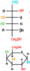
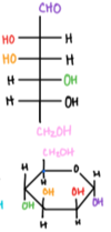
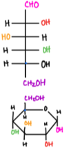
Nomenclature of all sugars = D- & L- forms of glyceraldehyde
◦ Aka- the absolute configuration for a carbohydrate is assigned based on the last chiral carbon in the chain as compared to the configurations of glyceraldehyde; L and D are based on the chiral carbon furthest from the carbonyl group. ◦ Those with the highest-#d chiral carbon with the OH group on the right in a Fischer projection = D sugars (naturally occurring) ◦ OH group on left = L sugars
Same D- and L- forms of the same sugar =
enantiomers
D/L =
◦ D/L = absolute configuration, assigned based on the chirality of the carbon atom furthest from the carbonyl group ◦ every chiral center in D-glucose has the opposite configuration of L-glucose
α/β = anomeric configuration
‣ The α form = anomeric carbon is in the axial position (OH below the plane) ‣ The β form = anomeric carbon is in the equatorial position (OH above the plane)
Diastereomers
nonsuperimposable configurations of molecules w/ similar connectivity; differs at least one but not all chiral carbons
epimers
different configuration @ exactly 1 chiral carbon
anomers
subtype of epimers that differ at the chiral, anomeric carbon
Aldoses
carbohydrates that contain an aldehyde group as their most oxidized functional group
Ketoses
has a ketone as their most oxidized functional group
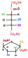
Pyranose
6-membered hexagonal shaped
Furanose
5-membered pentagonal shaped
For classification as a carbohydrate, 3 criteria:
◦ At least a 3-carbon backbone ◦ An aldehyde or a ketone group ◦ At least 2 hydroxyl groups
Simplest smallest carbohydrates
glyceraldehyde & dihyrdroxyacetone
Fructose forms ____ when carbon 5 attacks the carbonyl carbon
Fructose forms furanose when carbon 5 attacks the carbonyl carbon
Glucose forms _____ when carbon 5 attacks the carbonyl carbon
Glucose forms pyranose when carbon 5 attacks the carbonyl carbon
Haworth Diagram
• Down right • Up left • As you fill in the substituents, those on the right side of the Fischer diagram will point down, and those on the left side will point up. -OH group on the anomeric carbon (Fischer carbonyl) can be either UP (β) or down (α) • The CH₂OH group on absolute configuration carbon (C-5) points UP for D and down for L. • In planar conformation, everything = eclipsed • In chair conformation, everything = staggered • Everything in between = partially eclipsed
Glycosidic linkage
an acetal linkage that chains together monomers to form disaccharides, oligosaccharides, and polysaccharides; also terms the linkage b/t sugar and base in nucleotides
Monosaccharides:
simple sugars; formula (CH₂O)n; typically have 3-7 C.
- Position of the carbonyl group (C=O) can be used to categorize the sugars (aldose vs ketone)
- They are named according to their # of carbons (trioses = 3 C; pentoses = 5 C; hexoses = 6 C)

glucose
(aldohexose); main fuel source for the organism

Fructose (ketohexose); commonly used as sweetener; produced by many plants/fruits

Galactose (aldohexose); found in dairy products & sugar beets; can be rapidly converted to glucose

Mannose (aldohexose)
Sugars that can be oxidized
Sugars that can be oxidized = reducing agents; can be detected by reacting w/ Tollens’ or Benedict’s reagents
Sugars + COOH and COOH derivatives→
Sugars + COOH and COOH derivatives → esters
Phosphorylation- phosphate phosphate group from ATP + Sugar =
Phosphorylation- phosphate phosphate group from ATP + Sugar = phosphate ester
Glycoside formation
Glycoside formation: basis for building complex carbs; requires the anomeric carbon to link to another sugar
Disaccharides
formed when two monosaccharides join together via a dehydration rxn (aka condensation rxn or dehydration synthesis). Here, the hydroxyl group of one monosaccharide combines with the H of another, releasing a molecule of water and forming a covalent bond = glycosidic linkage

Sucrose: made from α-glucose and β-fructose joined @ the hydroxyl groups on the anomeric carbons (creating acetals); glycosidic bond is formed b/t C1 of the α-anomer of glucose & C2 of the β-anomer of fructose; Glu(α1→β2)Fru

Lactose: requires lactase to be hydrolyzed; made from β-galactose and α/β-glucose joined by a 1,4-linkage; Gal(β1→4)Glu

Maltose: two glucose molecules; produced when amylase breaks down starch; Glu(α1→4)Glu
Polysaccharides
long chains of monosaccharides linked by glycosidic bonds
Starch
main energy storage form for plants; made of glucose molecules joined by α-1,4-linkages
Amylose
linear polymer of glucose; connected by α-1,4-glycosidic bonds; 20%-30% of starch
Amylopectin
made of α-1,4-glycosidic bonds and α-1,6-glycosidic branches every 24-30 units; 70%-80% of starch
Glycogen:
main energy storage form for animals; has α-1,4-glycosidic bonds and α-1,6-glycosidic bonds
◦ Humans store glycogen in:
‣ Liver- hepatocytes; regulates BGL and provides cells w/ energy ‣ Muscle- can be broken down to power glycolysis
◦ More heavily branched than amylopectin; branching occurs every 8-12 units
Cellulose
main structural component for plant cell walls; main source of fiber for humans; made of repeating β-1,4glycosidic bonds
Peptidoglycan
major component of bacterial cell walls; polymer of carbohydrates that have been modified w/ amino acids
Chitin
made of N-acetylglucosamine; connected by β-1,4-glycosidic bonds; major component of cell walls in the exoskeletons of crustaceans, insects, and fungi
Glycolysis converts ______ to _____.
It takes place in the ______
Key enzymes include (3)
_____ are made for every _____
Glycolysis converts glucose (6-C) to 2 molecules of pyruvate (3-C each).
Takes place in the cytosol.
Key enzymes: hexokinase, phosphofructokinase, pyruvate kinase.
2 NADH made for every glucose
Steps in the investment phase of glycolysis (5) with their enzymes

Steps in the payoff phase of glycolysis (5) with their enzymes

Aerobic decarboxylation
- Aerobic decarboxylation occurs in the mitochondrial matrix. Converts pyruvate (3-C) to an acetyl group (2-C). (Pyruvate decarboxylation / pyruvate oxidation)
- Key enzyme: pyruvate dehydrogenase
- 1 NADH made for every pyruvate
- Only occurs in the presence of oxygen
- Acetyl group attaches to Coenzyme A to make acetyl CoA
Fermentation– mammalian and alcohol
oxygen absent
Mammalian cells: Lactate dehydrogenase oxidizes NADH → NAD⁺ which replenishes the oxidized coenzyme for glyceraldehyde-3-phosphate dehydrogenase.
Lactic acid fermentation: Pyruvate [reduction] → lactate
Alcohol fermentation: Pyruvate [reduction] → ethanol
Gluconeogenesis: _____ –> _____
Mitochondria
Gluconeogenesis: 2 molecules of pyruvate (noncarbohydrate source) → glucose
Mitochondria: pyruvate carboxylase converts pyruvate → oxaloacetate by adding a COO- group. Oxaloacetate is briefly converted to malate for transport out of the mitochondria where it is then converted immediately back to oxaloacetate. In the cytosol, PEP carboxykinase converts oxaloacetate to PEP.
The steps in glycolysis that are bypassed in gluconeogenesis (3)
Glycolysis step 10: PEP → enolpyruvate (tautomerize) → pyruvate ‣ Goal: bypass pyruvate kinase
‣ Pyruvate carboxylase converts pyruvate → oxaloacetate → phosphoenolpyruvate ‣ Acetyl-CoA from β-oxidation activates pyruvate carboxylase ‣ Glucagon & cortisol activate PEPCK
Glycolysis step 3: (F6P + ATP → F1,6BP)
‣ Frutose 1,6-bisphosphate → (catalysis of the hydrolysis of C1 phosphate group of F1,6BP) → fructose-6-phosphate = isomerized → glucose 6-phosphate (G6P) ‣ Rate limiting step of gluconeognesis ‣ Activated by ATP (directly) & glucagon (indirectly from ↓ levels of fructose-2,6-bisphosphate) ‣ Inhibited by AMP (directly) & insulin (indirectly from ↑ levels of F2,6BP)
Glycolysis step 1: (Glucose + ATP → G6P)
‣ Goal: bypass glukokinase ‣ Glucose 6-phosphate → → free glucose
Gluconeogenic substrates: (3)
◦ Glycerol 3-phosphate (from triacylglycerols)
◦ Lactate (from anaerobic glycolysis)
◦ Glucogenic amino acids (from muscle proteins)
Gluconeogenic intermediates → glycolytic intermediates (3)
◦ Lactate → → pyruvate
◦ Alanine → → pyruvate
◦ Glycerol 3-phosphate → → DHAP
Gluconeogenic enzymes: (4)
Pyruvate carboxylase: pyruvate → oxaloacetate
Phosphoenolpyruvate carboxykinase (PEPCK): oxaloacetate → phosphoenolpyruvate
Fructose 1,6-bisphosphatase: Fructose 1,6-bisphosphate → fructose 6-phosphate
Glucose 6-phosphatase: glucose 6-phosphate → free glucose
Glycolysis 3 enzymes and steps
Hexokinase; 1: glucose & glucose 6-phosphate
Phosphofructokinase; 3: fructose 6-phosphate & fructose 1,6-bisphosphate
Phosphofructokinase; 10: phosphoenolpyruvate & pyruvate
Glycolysis
- What happens
- Location
- Triggers
- Initial materials
- Final products
- Importance
Glycolysis
- What happens: the partial oxidation/breakdown of glucose → 2 pyruvate through 10 enzyme-mediated rxns
- Location: occcurs in the cytosol or cytoplasm and is common for both aerobic and anaerobic respiration
- Triggers: High BGL; insulin
- Initial materials: Glucose
- Final products: Pyruvate, ATP & NADH
- Importance: Energy production
Glycogenesis
- What happens
- Location
- Triggers
- Initial materials
- Final products
- Importance
Glycogenesis
- What happens: formation fo glycogen from excess glucose by liver cell sw/ the help of insulin; excess glucose = controlled by insulin
- Location: cytosol/cytoplasm
- Triggers: postprandial/well-fed state
- Initial materials: Glucose & ATP
- Final products: (glucose)n+1, ADP, Pi
- Importance: creates glycogen from glucose, storing the excess energy for future use
Glycogenolysis
- What happens
- Location
- Triggers
- Initial materials
- Final products
- Importance
Glycogenolysis
- What happens: conversion/breakdown of glycogen to release glucose by liver cells w/ the help of glucagon; deficient glucose = glucagon
- Location: cytosol/cytoplasm
- Triggers: fasting or b/t meals
- Initial materials: glycogen
- Final products: glycogenin-1 residues & glucose-1-phosphate
- Importance: energy production & BGL maintenance
Gluconeogenesis
- What happens
- Location
- Triggers
- Initial materials
- Final products
- Importance
Gluconeogenesis
- What happens: formation of glucose (or glycogen) fro noncarbohydrate sources (amino acids, fatty acids, glycerol)
- Location: takes place in the liver, kidneys, and striated muscles
- Triggers: low BGL, glucagon
- Initial materials: pyruvate
- Final products: glucose
- Importance: allows other sources (noncarbhydrates) to be converted into glucose
Purpose of the PPP
the pentose phosphate pathway (PPP), AKA hexose monophosphate shunt, HMP; it’s 1 of the 3 main ways the body creates molecules w/ reducing power; accounting for ∼60% of NADPH production; occurs in the cytoplasm/cytosol; ratelimiting enzyme = glucose-6-phosphate dehydrogenase (activated by NADP⁺ & insulin; inhibited by NADPH)
Primarily anabolic role, using the energy stored in NADPH to synthesize large complex molecules from small precursor ones
Oxidative phase of the PPP (reactant, enzyme, product)

NonOxidative phase of the PPP (reactant, enzyme, product)

Allosteric regulation of glycogenesis & glycogenolysis is done through
Allosteric regulation of glycogenesis & glycogenolysis is done through the regulation of glycogen synthase & glycogen phosphorylase.
Hormonal regulation of glycogenesis & glycogenolysis is done through
Hormonal regulation of glycogenesis & glycogenolysis is done by insulin and glucagon
Rate-limiting enzyme of glycogenesis
Rate-limiting enzyme = glycogen synthase. This forms the α-1,4-glycosidic bonds found on the linear glucose chains
Glycogenesis is stimulated by and inhibited by
It is stimualted by glucose-6-phosphate & insulin and inhibited by epinephrine & glucagon through a protein kinase cascade
Glycogenesis & branching enzyme
Branching enzyme = introduces the α-1,6-linked branches; hydrolyzes one of the α-1,4 bonds, releasing a block of oligoglucose which is then moved and added to a slightly different location
glycogen synthase =
Glycogen synthase makes a linear α-1,4-linked polyglucose chain
6 steps, overall, and enzymes of glycogenesis

3 steps and enzymes of glycogenolysis

What is Glycogenolysis
Glycogenolysis = the catabolism of glycogen (glycogen break down); is induced by adrenaline
Enzyme: Glucokinase
Action:
Activated by:
Enzyme: Glucokinase
Action: converts glucose –> glucose 6-phosphate
Activated by: present in the pancreatic β-islet cells; is responsive to inuslin in liver
Enzyme: Hexokinase
Action:
Activated by:
Inhibited by:
Enzyme: Hexokinase
Action: converts glucose –> glucose 6-phosphate in peripheral tissues
Activated by: insulin
Inhibited by: G6P (feedback inhibition) Glucagon
Enzyme: Phosphofructokinase-1 (PFK-1)
Action:
Activated by: (3)
Inhibited by: (4)
Enzyme: Phosphofructokinase-1 (PFK-1)
Action: phosphorylates fructose 6-phosphate –> fructose 1,6-bisphosphate (rate-limiting step of glycolysis)
Activated by: (3)
- AMP (indicates that energy is required -> glycolysis is activated)
- Fructose 2,6-bisphosphate
- Insulin (postprandial state); indirect stimulation by increasing these levels
Inhibited by: (4)
- ATP (energy is plentiful -> slows glycolysis)
- citrate (indicates plentiful supply of intermediates for producing energy)
- low pH
- In the liver only = glucagon = indirectly inhibits glycolysis by decreasing levels of fructose 2,6-bisphosphate during a fasting state
Enzyme: Phosphofructokinase-2 (PFK-2)
Action:
Activated by:
Enzyme: Phosphofructokinase-2 (PFK-2)
Action: makes F2,6-BP
Activated by: insulin
Enzyme: Glyceraldehyde-3-phosphate dehydrogenase
Action:
Enzyme: Glyceraldehyde-3-phosphate dehydrogenase
Action: makes NADH which can feed into the ETC
Enzyme: 3-phosphoglycerate kinase
Action:
Enzyme: 3-phosphoglycerate kinase
Action: substrate-level phosphorylation, putting Pi onto ADP -> ATP
Enzyme: Pyruvate kinase
Action:
Activated by:
Inhibited by:
Enzyme: Pyruvate kinase
Action: substrate-level phosphorylation, putting Pi onto ADP -> ATP
converts PEP -> pyruvate
Activated by: Fructose 1,6-bisphosphate (feed-forward stimulation)
Inhibited by: ATP; alanine
Enzyme: Pyruvate dehydrogenase
Action:
Activated by:
Inhibited by:
Enzyme: Pyruvate dehydrogenase
Action: complex of enzymes that converts pyruvate -> acetyl CoA
Activated by: Insulin
Inhibited by: acetyl-CoA
Enzyme: Glycogen synthase
Action:
Activated by:
Enzyme: Glycogen synthase
Action: creates α-1,4 glycosidic links b/t glucose molecules
Activated by: insulin
Enzyme: Branching enzyme
Action:
Enzyme: Branching enzyme
Action: moves a block of oligoglucose from one chain and adds it to the growing glycogen as a new branch using an α-1,6 glycosidic link
Enzyme: Glycogen phosphorylase
Action:
Activated by: (2)
Enzyme: Glycogen phosphorylase
Action: removse a single glucose 1-phosphate molecule by breaking α-1,4 glycosidic links
Activated by:
- liver- glucagon to prevent low blood sugar
- skeletal muscle– epinephrine & AMP to glucose for the muscle
Enzyme: Debranching enzyme
Action:
Enzyme: Debranching enzyme
Action: moves a block of oligoglucose from one branch and connects it to the chain using an α-1,4-glycosidic link and removes the branchpoint
Aerobic decarboxylation occurs in where? converts what to what? key enzyme is what?
Aerobic decarboxylation occurs in mitochondrial matrix
Converts pyruvate to an acetyl group
key enzyme is pyruvate dehydrogenase
Mammalian cells: lactate dehydrogenase oxidizes ______ to _____ which replenishes the oxidized coenzyme for _______.
Mammalian cells: lactate dehydrogenase oxidizes NADH to NAD+ which replenishes the oxidized coenzyme for glyceraldehyde-3 phosphate dehydrogenase.
Lactic acid fermentation: ______ [reduction] –> ______
Lactic acid fermentation: pyruvate [reduction] –> lactate
Alcohol fermentation: ______ [reduction] –> ______
Alcohol fermentation: Pyruvate [reduction] –> ethanol
How to go from pyruvate to acetyl Co-A?
- A carboxyl group is removed from pyruvate; CO₂ molecule is released; leaves behind a 2-C molecule
- The 2C molecule is oxidized, and the e⊖ are accepted by NAD+ reducing it → NADH
- The oxidized 2C molecule is now an acetyl group attached to Coenzyme A (organic molecule derived from vitamin B5) and this forms acetyl-CoA.
Acetyl-CoA can be produced in the 4 ways:
Acetyl-CoA can be produced in the following ways
• Glycolysis
◦ Pyruvate is transformed into acetyl-CoA in the mitochondria. Pyruvate dehydrogenase makes pyruvate lose a carbon, producing a new 2C molecule called acetyl-CoA. The carbon that’s removed takes two O from private with it, exits the body as CO₂
• Aerobic decarboxylation
◦ Done in the mitochondrial matrix; converts pyruvate (3C) → an acetyl group (2C)
◦ 1 NADH for every pyruvate; requires oxygen; acetyl group attaches to Coenzyme-A → acetyl-CoA
• Fat metabolism
◦ In β-oxidation, fatty-CoA is broken down, 2C at a time, to make acetyl-CoA which feeds into the TCA
◦ β-oxidation also produces FADH₂ & NADH. These feed into the ETC
• Protein metabolism
◦ The catabolism of some amino acids (phenylalanine, tyrosine, leucine, lysine, and tryptophan) can make acetyl-CoA
Large influx of glucose, stimulates ____, promotes ____.
Large influx of glucose, stimulates insulin, promotes glycolysis.
Large influx of oxaloacetate, stimulates ____, promotes ____.
Large influx of oxaloacetate, stimulates glucagon, promotes gluconeogenesis.
the regulation step of gluconeogenesis
F1,6BP is regulated by the activation of citrate (↑ citrate = ↑ enzyme activity) & inhibited from ↑ AMP or ↓ ATP
Citric acid cycle products: fate of 1 turn of cycle (1 pyruvate molecule) but keep in mind there’s 2 pyruvate molecules
In total for one round of the cycle (aka 1 pyruvate molecule)
◦ 1 GTP
◦ 3 NADH
◦ 1 FADH₂
◦ 2 CO₂
◦ 1 regenerated oxaloacetate molecule
8 steps of the TCA
• Step 1— Citrate Formation
◦ Acetyl-CoA & oxaloacetate [condensation rxn] → citric-CoA
◦ The hydrolysis of citric-CoA → citrate & CoA-SH ; catalyzed by citrate synthase ‣ Synthases = enzymes that form new covalent bonds w/o needing significant energy
• Step 2 — Citrate Isomerized → Isocitrate
◦ Achiral citrate → isomerized: 1 of 4 possible isomers of isocitrate ‣ 1: citrate binds @ 3 points to the enzyme aconitase ‣ 2: water is lost from citrate → cis-aconitate ‣ 3: water is added back to form isocitrate
◦ Enzyme is a metalloprotein, requires Fe²⁺
◦ Results in a switching of a H and a hydroxyl group
• Step 3—α-Ketoglutarate and CO₂ Formation:
◦ Isocitrate: oxidized → oxalosuccinate by isocitrate dehydrogenase (the rate-limiting enzyme of the CAC); the first of the 2 carbons from the cycle is lost here; this is also the first NADH produced from intermediates in the cycle
◦ Oxalosuccinate: decarboxylated → α-ketoglutarate & CO₂
• Step 4—Succinyl-CoA and CO₂ Formation
◦ These rxns are carried out by the α-ketoglutarate dehydrogenase complex; formation of succinyl-CoA, α-ketoglutarate & CoA
= 1 CO₂
◦ The CO₂ represents the 2ⁿᵈ and last carbon lost form the cycle; NAD⁺: reduced → NADPH ‣ Dehydrogenases are a subtype of oxidoreductases (enzymes that catalyze an oxidation–reduction reaction).
‣ Dehydrogenases transfer a hydride ion (H − ) to an electron acceptor, usually NAD + or FAD. Therefore, whenever you see dehydrogenase in aerobic metabolism, be on the lookout for a high-energy electron carrier being formed!
• Step 5—Succinate Formation
◦ Hydrolysis—thioester bond on succiny-CoA → succinate & CoA-SH & is coupled to the phosphorylation of GDP → GTP
◦ Rxn is catalyzed by succinyl-CoA synthetase ‣ Synthetases: creates new covalent bonds w/ energy input
◦ Phosphorylation of GDP → GTP = driven by the energy released by thioester hydrolysis
◦ GTP is formed: nucleosidediphosphate kinase = catalyzes phosphate transfer GTP → ADP thus producing ATP (this is the only tie in the entire CAC where ATP is produced directly); ATP production is predominantly w/in the ETC ‣ Citrate synthase doesn’t require energy input in order to form covalent bonds, but succinyl-CoA synthetase does.
• Step 6—Fumarate Formation:
◦ Occurs in the inner membrane
◦ Succinate: oxidation → fumarate (catalyzed by succinate dehydrogenase = a flavoprotein b/c it is covalently bonded to FAD, the electron acceptor in this rxn)
◦ As succinate is oxidized to fumarate → FAD is reduced to FADH₂ = 1.5 ATP ‣ NADH = 2.5 ATP
◦ FAD = the electron acceptor in this rxn b/c the reducing power of succinate is not great enough to reduce NAD⁺
• Step 7—Malate Formation
◦ Fumarase: catalyzes hydrolysis of the alkene bond in fumarate → malate (only L-malate forms in this rxn)
• Step 8—Oxaloacetate Formed Anew
◦ Malate dehydrogenase: catalyzes oxidation: malate → oxaloacetate
◦ 3ʳᵈ and final molecule of NAD⁺: reduced → NADH
◦ Newly formed oxaloacetate is ready to take part in another turn of the CAC; we’ve gained all of the high-energy electron carriers possible from 1 turn of the cycle
describe the main function and typical structure of a fatty acid
- Fatty acids are amphipathic molecules that have a polar carboxylate-head group and nonpolar hydrocarbon tail. They are longchain carboxylic acids. They are generally considered insoluble in water while longer hydrocarbon chains have a lower solubility.
- Main function = energy provision & precursors for the synthesis of other molecules
The 4 common forms of fatty acids include
triglycerides, phospholipids, cholesterol, and eicosanoids
Simply explain fatty acid oxidation
Fatty acid oxidation (β-oxidation) = catabolic breakdown of fatty acids → energy
◦ Can completely degrade saturated fatty acids
◦ Requires the input of the enzymes enoyl-CoA isomerase & 2,4-dienoyl CoA for the complete degradation of unsaturated fatty acids
◦ Occurs in the mitochondrial matrix; converts their fatty acid chains → 2 C units of acetyl groups, producing NADH & FADH₂ which are then used by the ETC.
Steps of β-oxidation
- The fatty acid is activated. Coenzyme A is added to the end of a long-chain fatty acid, after which the activated fatty acyl-CoA enters the β-oxidation pathway
- fatty acyl-CoA (oxidation) → enoyl-CoA
A. A trans double bond is introduced into the acyl chain
- Enoyl-CoA (hydration) → an alcohol
- Alcohol (oxidation) → carbonyl
- Carbonyl → cleaves off acetyl-CoA from the oxidized molecule; also yields an acyl-CoA that is 2 C shorter than the original molecule that entered the β-oxidation pathway
- Cycle repeats until the fatty acid has been completely reduced to acetyl-CoA, which is fed through the citric acid cycle to yield cellular energy in the form of ATP.
Carbohydrates break down into _______ which go through _____.
Fats break down into _____ which go through ______.
Proteins break down into _____ which go through ______.
Carbohydrates break down into glucose (sugar) which go through glycolysis.
Fats break down into fatty acids and glycerol which go through β-oxidation.
Proteins break down into amino acids which go through transamination.
Acetyl-CoA (2C; inner mitochondrial matrix) = precursor for__________. Acetyl-CoA is converted to ________ so that the _____________ can transport it into the ____________, where its broken back up to _____________ & ____________. For __________ to return to pyruvate, a C is lost as CO₂ and _________ is reduced to ___________
Acetyl-CoA (2C; inner mitochondrial matrix) = precursor for fatty acid synthesis. Acetyl-CoA is converted to citrate so that the citrate shuttle can transport it into the cytoplasm, where its broken back up to oxaloacetate & acetyl-CoA. For OAA to return to pyruvate, a C is lost as CO₂ and NADP⁺ is reduced to NADPH
Acetyl-CoAs are linked together → a large fatty acid chain = ____________ = primary product of fatty acid synthesis in the body. Requires ____________ (since they’re 2 C each). ____ ATP molecules are required for this rxn.___ NADPH are required to reduce the carbonyl group in the acetyl-CoA to just C-C bonds; end up with __ ADP & Pi & ____ NADP⁺
Acetyl-CoAs are linked together → a large fatty acid chain = Palmitic acid (16C) = primary product of fatty acid synthesis in the body. Requires 8 acetyl-CoA molecules (since they’re 2 C each). 7 ATP molecules are required for this rxn. 14 NADPH are required to reduce the carbonyl group in the acetyl-CoA to just C-C bonds; end up with 7 ADP & Pi & 14 NADP⁺
First enzyme that carries out the activation step in fatty acid synthesis = ___________ which adds a carboxy group to _________ It is the rate-limiting step of the entire fatty acid synthesis pathway, so it’s regulated through allosteric and hormonal regulation. Citrate is an allosteric activator; ________ activates this pathway. _______________ can inhibit this enzyme through product inhibition. __________ is a hormonal inhibitor–it signals adipose cells to release fatty acids and for our cells to break down.
First enzyme that carries out the activation step in fatty acid synthesis = acetyl-CoA carboxylase which adds a carboxy group to acetyl-CoA. It is the rate-limiting step of the entire fatty acid synthesis pathway, so it’s regulated through allosteric and hormonal regulation. Citrate is an allosteric activator; insulin activates this pathway. Long-chain fatty acids can inhibit this enzyme through product inhibition. Glucagon is a hormonal inhibitor–it signals adipose cells to release fatty acids and for our cells to break down.
Fatty acid synthase
the enzyme that polymerizes the manlonyl-CoA subunits together.
The actual digestion of proteins begins in the
The actual digestion of proteins begins in the stomach. HCl & pepsin begin the process of breaking down the proteins. As chyme enters the small intestine, it mixes w/ bicarbonate (neutralizes the HCl) & digestive enzymes (breaks down the proteins into smaller polypeptides & AA).
Enteropeptidase
Enteropeptidase = enzyme in the wall of the small intestine; activates trypsin which then activates chymotrypsin.
what are the 5 substrates and 4 products of oxidative phosphoryaltion?
Substrates:
- NADH (reduced form)
- FADH2 (reduced form)
- O2
- ADP
- Pi
Products:
- ATP
- NAD+ (oxidized form)
- FAD (oxidized form)
- H2O
The final stage in cellular respiration is _______which is made up of two interdependent processes: flow of e⊖ through the_______ down to oxygen & ________.
The final stage in cellular respiration is oxidative phosphorylation which is made up of two interdependent processes: flow of e⊖ through the ETC down to oxygen & chemiosmotic coupling.
Describe the Glycerol 3-phosphate shuttle
E⊖ are transferred from NADH → DHAP making glycerol-3-phosphate. These e⊖ can then be transferred to mitochondrial FAD making FADH₂
Describe the Malate-aspartate shuttle
E⊖ are transferred from NADH → oxaloacetate making malate which can then cross the inner mitochondrial membrane and transfer the e⊖ to mitochondrial NAD⁺ making NADH.
Complex I of the ETC
NADH-CoQ oxidoreductase & NADH dehydrogenase; 4 protons are transferred from the mitochondrial matrix -> intermembrane space
converts NADH -> NAD+
Overal rxn: NADH + H+ + Q -> NAD+ + QH2
NADH transfers its e⊖ to CI. The part of complex I that receives the e⊖ = a flavoprotein (a protein w/ an FMN molecule attached). FMN is a prosthetic group (a non-protein molecule tightly bound to a protein and required for its activity). It’s the FMN that actually accepts e⊖ from NADH. FMN passes the e⊖ to the Fe-S protein (another protein inside complex I ) which in turn transfers the e⊖ to ubiquinone (Q).
Complex II of the ETC
Succinate-CoQ oxidoreductase
Succinate dehydrogenase
No proton pumping
specifically deals w/ FAD/FADH2
Succinate dehydrogenase = a membrane-bound enzyme that catalyzes the conversion of succinate -> fumarate in the TCA, generating FADH2 which delivers more e- to Q -> QH2
FADH₂ deposits its electrons in the ETC via CII. The enzyme that reduces FADH₂ in the TCA & FADH₂ are embedded in the inner mitochondrial membrane. FADH₂ transfers its e⊖ to the iron-sulfur proteins w/in CII, which then passes the e⊖ to ubiquinone (Q); same as in CI.
Complex III of the ETC
CoQH2
cytochrome c oxidoreductase
e- are passed from QH2 -> cytochrome c, regenerating Q, resulting in teh reduced form of cytochrome c, Q carries 2 e-, cytochrome c carries 1
4 protons are translocated into teh intermembrane space per molecule of QH2
As e⊖ move through complex III, more H ions are pumped across the membrane, and the e⊖ are ultimately delivered to cytochrome C (cyt C) which carries the e⊖ to complex IV (CIV) where a final batch of H ions is pumped across the membrane. CIV passes the e⊖ to O₂ which splits into 2 oxygen atoms & accepts protons from the matrix to form water. 4 e⊖ are required to reduce each molecule of O₂, and 2 H₂O molecules are formed.
Complex III (CIII): has both the Fe-S protein as well as cytochromes (family of related proteins that have heme prosthetic groups w/ iron ions). In CIII, e⊖ are passed from one cytochrome to the Fe-S protein to a 2ⁿᵈ cytochrome, and then finally transferred out of the complex to cytochrome C (e⊖ carrier). CIII also pumps protons from the matrix → intermembrane space
Complex IV
Cytochrome C oxidase
2 protons are translocated
Cytochrome C delivers the e⊖ to the last complex in the ETC, CIV. Here, the e⊖ are passed through 2 more cytochromes, and the 2ⁿᵈ cytochrome, w/ the help of a copper ion, transfers the e⊖ to O₂, splitting oxygen to form 2 H₂O molecules. The protons used to form H₂O come from the matrix, contributing to the H ion gradient, and CIV also pumps protons from the matrix to the intermembrane space.
In eukaryotes, ETC is in the __________ while in prokaryotes it’s in the ________.
In eukaryotes, ETC is in the inner mitochondrial membrane while in prokaryotes it’s in the plasma membrane.
prosthetic group
a non-protein molecule tightly bound to a protein and required for its activity
Oxidized form of Q is ______ while the reduced form is _____
Oxidized form of Q is ubiquinone while the reduced form is ubiquinol (QH2)
How much ATP do you get from NADH vs how much you get from FADH2?
You get 2.5 ATP from NADH and 1.5 ATP from FADH2.
NADH vs NADPH
◦ NADH is a very good e⊖ donor (its e⊖ are at a high energy level)
◦ NADPH is primarily produced in the oxidative part of the pentose phosphate pathway and is used in anabolic synthesis to make cholesterol, fatty acids, transmitter substances & nucleotides
◦ NADH & NADPH are both reductive agents
Flavoproteins
proteins that have both a nucleic acid derivative of riboflavin (flavin adenine dinucleotide / flavin mono nucleotide)
Located on the matrix face of the inner mitochondrial membrane; functions as a specific e⊖ acceptor for 1˚ dehydrogenase, transferring the e⊖ to the ubiquinone pool in the inner mitochondrial matrix
Cytochromes
◦ Compounds made of heme bound to a protein
◦ Functions as e⊖ transfer agents
◦ Cytochrome c transfers e⊖ from CIII → CIV in the ETC; can only carry 1 e⊖ at a time
Chemiosmotic coupling
the process that links the ETC (which creates an electrochemical gradient across the inner mitochondrial membrane) to the production of ATP through ATP synthase.
Chemiosmotic coupling allows the chemical energy from the gradient to be harnessed to phosphorylate ADP → ATP (endergonic)
Conformational coupling
the relationship b/t the proton gradient and ATP synthesis is indirect; ATP is released by the synthase as a result of a conformational change caused by the gradient. The F₁ portion of ATP synthase harnesses the gradient energy for chemical bonding from the spinning that occurs when protons go through it.
- The F₀ portion is the portion of ATP synthase that spans the membrane. It functions as an ion channel, so protons travel through it along their gradient back into the matrix.
- The F₁ portion utilizes the energy released from the electrochemical gradient to phosphorylate ADP → ATP.
- When the proton-motive force is dissipated through the F₀ portion, (free energy change of the rxn) ∆G˚’ = -220 kj/mol (highly exergonic)
Direct products and ultimate ATP yield of the following:
Glycolysis
Pyruvate oxidation
TCA
Total

Pyruvate dehydrogenase complex
Pyruvate dehydrogenase complex converts pyruvate → acetyl-CoA and this is upregulated by high levels of AMP, CoA, and NAD⁺ (signs that the cell needs to make more energy).
TCA is regulated @ 3 major exergonic steps:
◦ 1) formation of citrate
‣ Upregulated: high levels of ADP
‣ Downregulated: ATP, NADH, [citrate, and succinyl-CoA = negative feedback]
◦ 2) Conversion of isocitrate → α-ketoglutarate
‣ Upregulated: ADP
‣ Downregulated: ATP
◦ 3) α-ketoglutarate → succinyl-CoA
‣ Upregulated: Ca²⁺
‣ Downregulated: NADH & [succinyl-CoA = negative feedback]
significance of Mitochondria
Mitochondria play a significant role in initiating apoptosis:
◦ 1st steps = ↑ permeability of the outer mitochondrial membrane.
‣ Regulated by proteins in the BCL-2 family (which includes both pro-apoptotic proteins and anti-apoptotic proteins)
◦ When the mitochondrial membrane becomes more permeable, cytochrome C (the shuttle b/t CIII & CIV of the ETC) is released from the intermembrane space → cytoplasm
◦ Once in the cytoplasm, cyt c activates the caspases (enzymes that act as proteases to initiate large-scale protein degradation) and the nucleases to degrade DNA
◦ Together, the above processes result in regulated cell breakdown; from there the components can be phagocytosed and reused
Hormones and the 3 classes
hormones are signals produced in the endocrine system & released into bodily fluids to be transferred to the target cells
3 classes of hormones (which are based on their chemical structure):
◦ Steroids
◦ Amines (amino acid-derived)
◦ Peptide hormones (includes peptides & proteins)
Steroid hormones
Steroid hormones are lipid-derived so they are lipid soluble and water insoluble
- They remain in circulation longer
- They have high-affinity receptors, work @ low concentrations, and affects gene expression & metabolism.
- These hormones and some of the amines can diffuse through the cell membrane with ease, but once they do so, they need transport proteins to get to their target cells
- Target cell receptors for lipid-soluble hormones = cytoplasm or nucleus
- When the lipid-soluble hormone forms a complex w/ its receptor, they bind together the DNA and induce the expression of certain genes. So the action of steroid hormones is slower b/c they need to promote the translation & transcription of these mRNA @ the target gene
Peptide Hormones & Amines
- Includes insulin, growth hormone, and MOST of the amines (like epinephrine)
- They are water-soluble molecules (so they can’t pass through the cell membrane) so their receptors are on the surface of the target cells.
- These hormones are secreted by exocytosis and travel to the destination via the blood
- When the water-soluble hormone binds to its target receptor, this induces a cell-signaling pathway that either (1) causes a prompt cellular response, or (2) induces changes in the expression of certain genes
- IE— When epinephrine (adrenaline) binds to its receptor, this activates a cytoplasmic enzyme that inhibits glycogen synthesis.
- Since peptide hormones must bind to the membrane receptors, they rely on cascades of kinases to transmit their message to the target cell, having a faster effect.
synthesis of glycogen from glucose
Glycogenesis
breakdown of glycogen → glucose
Glycogenolysis
synthesis of glucose from non-carbohydrate sources
Gluconeogenesis
production of ketone bodies from acetyl-CoA (insulin absent)
Ketogenesis
breakdown of fatty acids to generate acetyl-CoA
β-oxidation
lipid & ribose synthesis
Pentose phosphate pathway (PPP)
Postprandial state– Liver: Glucose
Glucose is transported to the liver; can be stored as glycogen or converted to triglycerides; glucose must be converted into glycerol & fatty acids; triglycerides are mostly stored in adipose tissues and can’t be transported in the blood, so they have to be converted into VLDL which can be exported from the liver into blood.
the liver breaks down AA -> keto acids. These α-keto acids give off ammonia when it’s excreted as waste (urea)
if the liver needs energy during the absorptive state, the keto acids can be broken down -> acetyl-CoA which then goes through TCA & ETC to produce ATP
After a meal, [glucose] in the portal blood is ↑. The liver extracts excess glucose and uses it to replenish the glycogen stores.
Remaining glucose is then converted to acetyl-CoA for fatty acid synthesis. This ↑ in insulin s/p a meal stimulates both glycogen synthesis & fatty acid synthesis in the liver. Fatty acids → triacylglycerols & released into the blood as VLDL
• Well fed state: liver derives most of its energy from oxidizing excess AA
Postprandial state– Adipose tissue: Glucose
glucose needs to be converted into triglycerides. The VLDL from the liver will be turned into fatty acids which are then combined w/ glycerol to form triglycerides.
- After a meal, the ↑ insulin levels stimulate glucose uptake by adipose tissue. Insulin triggers fatty acid release from VLDL and chylomicrons.
- Lipoprotein lipase = enzyme found in the capillary bed of adipose tissue; induced by insulin
- The fatty acids released from lipoproteins are taken up by adipose tissue and re-esterified to triacylglycerols (storage)
- Insulin can suppress the release of fatty acids from adipose tissue
Postprandial state– Muscle: Glucose
Glucose can be taken up & has 2 pathways:
- can convert to glycogen (storage form)
- can convert to pyruvate (if energy is needed)
- muscle can take up AA from proteins and store them as proteins in the muscle
- Major fuels = glucose & fatty acids
- S/p a meal, insulin promotes glucose uptake, replenishing glycogen stores & AA used for protein synthesis. Excess glucose & AA can be oxidized for energy.
Postprandial state– Brain: Glucose
glucose from blood -> pyruvate makes ATP so brain can work
Postabsorptive state– Liver
Glycogen is broken down to glucose which can then be exported out of the liver into the blood. AA are taken up by the liver & converted -> keto acids and can be used to make glucose, broken down to acetyl-CoA
Triglycerides -> glucose
Fatty acids = 2nd rxn so they can be broken down -> ketones; can be used in the brain
- In the fasting state: the liver releases glucose into the blood and this ↑ promotes glycogen degradation & gluconeogenesis.
- Lactate (from anaerobic metabolism), glycerol (from triacylglycerols), and AA = provide the carbon skeletons for glucose synthesis
Postabsorptive state– Adipose tissue
triglycerides broken down -> glycerol & fatty acids; can be exported into the blood and brought to the liver to make glucose
• Fasting state: ↓ insulin & ↑ epinephrine = activates hormone-sensitive lipase in fat cells, allowing fatty acids to be released into circulation
Postabsorptive state– Muscle
Protein -> AA -> liver to be converted to α-keto acids -> glucose
Glycogen -> glucose -> pyruvate -> acetyl coA & produce energy for the muscles. If muscles low on O2, glucose can produce lactate
- Fasting state: resting muscle uses fatty acids derived from free fatty acids; ketone bodies may be used in prolonged fasting state (Active Muscle)
- Fuel depends on magnitude & duration of exercise;
◦ Short-lived source of energy (2-7 s) comes from creatine phosphate (transfers a phosphate group to ADP → ATP)
- Skeletal muscle has glycogen stores (and some triacylglycerol stores) so both blood glucose & free fatty acids may be used
- Short bursts, high intensity exercise = anaerobic glycolysis
- Moderately high-intensity, continuous = oxidation of glucose & fatty acids
- S/P 1-3 hours of continuous exercise = oxidation of fatty acids
Postabsorptive state– Brain
brain takes glucose from the blood -> pyruvate; goes thorugh cellular respiration to produce ATP
ketones from liver can be used by the brain
↑ BGL = _______ is released → promotes _______, _______ synthesis (liver & skeletal muscles); ________ (liver), and ________ (adipose tissue).
↑ BGL = insulin is released → promotes glycolysis, glycogen synthesis (liver & skeletal muscles); fatty acid synthesis (liver), and fatty acid storage (adipose tissue).
_____________ triggers glycogen breakdown; __________ promotes gluconeogenesis & ↑ BGL
Epinephrine triggers glycogen breakdown; cortisol promotes gluconeogenesis & ↑ BGL
Insulin = released by the ______; stimulates glucose uptake by target cells; is released upon the entry of glucose into _________ through the ________ glucose transporters.
Insulin = released by the β cells of the pancreas; stimulates glucose uptake by target cells; is released upon the entry of glucose into pancreatic β cells through the GLUT2 glucose transporters.
When glucose enters the Krebs cycle in the β cells, the ΑΤP:ADP ratio ↑ → _________→ __________ → ____________
Entry of Ca²⁺ ions triggers a signaling cascade → ____________ This ↑ in the intracellular [Ca²⁺] →_______________.
When glucose enters the Krebs cycle in the β cells, the ΑΤP:ADP ratio ↑ → closing the ATP-sensitive potassium channel → buildup of intracellular potassium ions → depolarization & opening of voltage-gated calcium channels w/in the membrane of β cells. Entry of Ca²⁺ ions triggers a signaling cascade → release of additional calcium ions from the ER. This ↑ in the intracellular [Ca²⁺] → a release of previously stored insulin from secretory vesicles
Other substances stimulating insulin release
Other substances stimulating insulin release = AA arginine, leucine; parasympathetic release of acetylcholine; cholecystokinin (CKK); glumoagon-like peptide-1 (GLP-1) & glucose-dependent insulinotropic peptide (GIP)
In insulin responsive tissues (like skeletal muscle & fat cells) insulin binds w/ insulin receptors → triggers a__________ → reduces ________
In insulin responsive tissues (like skeletal muscle & fat cells) insulin binds w/ insulin receptors → triggers an intracellular signaling cascade, promoting more GLUT4 receptors coming in; presence of more GLUT4 in the plasma membrane promotes the uptake of glucose into the target cells → reduces BGL
↑ the concentrations of circulating insulin ______ proteolysis (protein breakdown) & _____ AA uptake
↑ the concentrations of circulating insulin ↓ proteolysis (protein breakdown) & ↑ AA uptake
• Glucagon = synthesized by ________; compensatory ↑ in BGL by causing the liver to convert ________ → ________ which is then released into the bloodstream.
___________ antagonizes insulin’s action on BGL (aka _____ and ______ forms a feedback system that ensures blood glucose homeostasis if properly regulated)
• Glucagon promotes _______ (breakdown of triglycerides) by activating ________ → activates ________ → releases free fatty acids & glycerol into the bloodstream. ______ can enter the TCA (s/p conversion to ______ by _______) in the liver & kidneys
- Glucagon = synthesized by α-cells of the pancreas; compensatory ↑ in BGL by causing the liver to convert stored glycogen → glucose which is then released into the bloodstream. Glucagon antagonizes insulin’s action on BGL (aka glucagon and insulin forms a feedback system that ensures blood glucose homeostasis if properly regulated)
- Glucagon promotes lipolysis (breakdown of triglycerides) by activating protein kinase A → activates hormone-sensitive lipase → releases free fatty acids & glycerol into the bloodstream. Glycerol can enter the TCA (s/p conversion to glycerol 3-phosphate by glycerol kinase) in the liver & kidneys
Pancreatic β-cells vs Pancreatic α-cells
Pancreatic β-cells secrete insulin (high glucose) while Pancreatic α-cells secrete glucagon (low glucose, high AA levels)
Catecholamines
promotes glycogenolysis & ↑ basal metabolic rate through sympathetic nervous system activity
The primary hormones involved in body mass regulation:
Ones that ↓ food intake & ↑ energy expenditure = insulin & leptin.
‣ Insulin stimulates the production of leptin by adipose tissue, ↓ appetite & gives satiety feeling ‣ Leptin ↓ insulin secretion & ↑ tissue sensitivity to insulin → glucose uptake for energy utilization/storage.
• It lives in the adipose tissue and goes to the hypothalamus
‣ Insulin resistance = when he body doesn’t use the stored fat as an energy source ‣ Leptin resistance = dysfunction in the feeling of satiety
◦ Other hormones that act on the satiety center in the brain:
‣ Ghrelin: secreted by the stomach wall; ↑ feelings of hunger ‣ Peptide YY (PYY): secreted by the small intestine; acts on brain receptors to ↓ appetite
Orexin
further ↑ appetite; also involved in alertness & the sleep-wake cycle; triggered by hypoglycemia