Benign Non-Odontogenic Lesions Flashcards

1
Q

Cemento-Ossifying Fibroma clinical features

A

3-4th decade 5:1 female predilection Common in mandible

2
Q

Cemento-Ossifying Fibroma radio appearance

A

Well-demarcated lesion Radiolucent to mixed appearance

3
Q

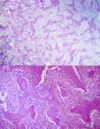
Cemento-Ossifying Fibroma histo appearance

A

Potato-like mass that usually comes out in one chunk Lots of connective tissue and bone/cementum interspersed. NO inflammation seen

4
Q

Cemento-Ossifying Fibroma treatment

A

Enucleation This is a TRUE neoplasm.

5
Q

Fibrous Dysplasia Types of Lesion

A

Polyostotic Monostotic

6
Q

Fibrous Dysplasia clinical features

A

MAXILLA more common Bones have fracture risk because they are weaker. Can have a hockey stick discrepancy. One leg longer than the other Will see SWELLING over time. Can see expansion.

7
Q

Fibrous Dysplasia radio appearance

A

Ground glass appearance with ill-defined borders Narrowing PDL space and the lamina dura hard to make out

8
Q

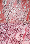
Fibrous Dysplasia histo appearance

A

Irregular shaped woven bone (Chinese characters) Bone has NO OSTEOBLAST around it. Distinctive feature. Bone arises in slide via metaplasia from fibroblasts Woven bone becomes more lamellar over time

9
Q

Fibrous Dysplasia treatment

A

Resection, but not in children. Lesion will regress over time maybe to 70% of size. Remove at skeletal maturity. Radiation is CONTRAINDICATED. It may actually cause malignant transformation to osteosarcoma.

10
Q

Fibrous Dysplasia age

A

Stage of fetal development determines if polyostotic or monostotic (MOST cases are monostotic) 1-2nd decade (YOUNGER) population

11
Q

Fibrous Dysplasia types of polyostotic involvement

A

Jaffe Type

  • Cafe au lait spots on the trunk and thigh
  • Multiple bone involvements

McCune-Albright Type

  • Cafe au lait spots
  • Endocrine hyperfunction
  • Precocious puberty
12
Q

Types of cemento-osseous dysplasias

A

Periapical

Focal

Florid

13
Q

Clinical features of periapical osseous dysplasias

A

14:1 Black female predilection

30-50 years old

14
Q

Clinical features of focal osseous dysplasias

A

80% in Caucasian females

4-5th decade

15
Q

Clinical features of florid osseous dysplasias

A

Black females

16
Q

Location of periapical osseous dysplasias

A

Mandibular anterior

Multiple lesions

17
Q

Location of focal osseous dysplasias

A

Posterior mandible ONE lesion

18
Q

Location of florid osseous dysplasias

A

More than one quadrant

19
Q

Radiograph of periapical osseous dysplasias

A

Start as RL then add more osteoid and become RO

20
Q

Radiograph of focal osseous dysplasias

A

RL to RO. RL rim remains over time

21
Q

Radiograph of florid osseous dysplasias

A

Multiple “cotton-wool” radiopacities

22
Q

Treatment of periapical osseous dysplasias

A

NONE needed. No biopsy

23
Q

Treatment of focal osseous dysplasias

A

Biopsy to rule out cemento-ossifying fibroma

24
Q

Treatment of florid osseous dysplasias

A

MAINTAIN DENTITION. Normal alveolar bone will resorb, but dysplastic bone will not and osteomylitis could occur.

25
Clinical features of all osseous dysplasias
Occur in tooth-bearing areas. Not outside the jaws Reactive lesions with cause unknown All types have the same histology, but are a spectrum along the process. No expansion seen Asymptomatic NO teeth displacement and teeth remain vital
26
Histo of all osseous dysplasias
Come out as coffee grounds
27
Clinical features of osteoblastoma
Pain NOT relieved by aspirin
28
Clinical features of osteoid osteoma
NOCTURNAL PAIN relieved by aspirin
29
Radiograph of osteoid osteoma
**Well-circumscribed** with radiolucent rim and central radiopacity. Less than 2cm in diameter
30
Radiograph of osteoblastoma
Similar location to OO (outside the jaws) Greater than 2cm Can have a RL rim with a mixed center
31
Histo of osteoid osteoma and OO
Lots of woven bone, NOT lamellar bone in a fibrovascular stroma Both can resemble low-grade osteosarcoma (malignancy) This similarity contributes to their misdiagnosis
32
TX of osteoid osteoma and OO
Local excision Curettage
33
Types of osteoma
Periosteal Endosteal
34
Clinical features of osteoma
Simply a benign bone lesion Paranasal sinuses are the most common, but can happen in bone
35
Radiograph of osteoma
Radiopaque
36
Histo of osteoma
Dense bone combined with cancellous bone
37
TX of osteoma
Surgical excision or just observation over time
38
Clinical presentation of Gardner syndrome
Epidermoid cysts GI polyps with 50% becoming malignant by age 30 Desmoid tumors: locally aggressive benign fibrous proliferations at incision site of colectomy Ocular -- hypertrophy retinal pigment Oral -- impacted supernumerary teeth and odontomas
39
Key indicator of Gardner syndrome
If you see multiple osteomas think about this lesion
40
Radiograph of Gardner syndrome
Osteomas NO radiolucent rim Supernumerary teeth
41
Heredity of Gardner syndrome
Autosomal dominant condition
42
TX of Gardner syndrome
Prophylactic colectomy as GI polyps will become malignant
43
Age of central giant cell granuloma
2-4th decade with a 2:1 female predilection
44
Location of central giant cell granuloma
Presents in posterior mandible 70% of time CROSSES MIDLINE
45
Radiograph of central giant cell granuloma
Radiolucency Start off as unilocular and over time become more multilocular and expansile Displacement of teeth Resorption of teeth possible
46
Histology of central giant cell granuloma
Granulation tissue Giant cells (frequently around the periphery) Reactive bone around periphery → body is trying to wall off the lesion; bone not made by lesion Extravasated RBC’s
47
DDX of central giant cell granuloma
ABC * Typically in children and doesn’t cross the midline Has blood-filled spaces in lesions with GC around the periphery of lesion Brown tumor * Need to rule out hyperparathyroidism Have calcium levels checked Giant cell tumor of bone * Most common in distal tibia and femur area not jaws
48
TX of central giant cell granuloma
Curettage Resection if the lesion recurs
49
Clinical features of Hemangioma of Bone (Vascular Malformation)
Pain and swelling when larger Sulcular bleeding Bruit or pulse if has a artery component. If only venous, no bruit present.
50
Location of Hemangioma of Bone (Vascular Malformation)
Seen in posterior mandible
51
Age of Hemangioma of Bone (Vascular Malformation)
2nd and 3rd decades most common
52
Radiograph of Hemangioma of Bone (Vascular Malformation)
Multilocular Presents as a mixed to radiolucent lesion Occasional sunburst pattern Cortical expansion
53
Histology of Hemangioma of Bone (Vascular Malformation)
Cavernous and capillary vessels
54
TX of Hemangioma of Bone (Vascular Malformation)
Make sure to aspirate the lesion before biopsy Pre-surgery embolization with resection, curettage
55
Types of Langerhans Cell Disease
Acute disseminated (Letterer-Siwe Disease) Chronic disseminated (Hand-Schuller-Christian Disease) Eosinophilic granuloma of bone
56
Features of Letterer-Siwe Disease
Most severe manifestation of disease Seen in infants and very young children. Usually FATAL. Lesions seen on (SOB) skin, organs, and bone
57
Features of Hand-schuller-Christian Cell Disease
(SOB) skin, organs, bone also involved, but organ involvement NOT as severe TRIAD: Bone lesions Exophthalmos → histiocytic cells accumulate around the eyeball Diabetes insipidus → histiocytic proliferation involves the posterior pituitary gland
58
Features of Eosinophilic granuloma of bone
MOST COMMON presentation of this disease BONE ONLY (solitary or multiple lesions) No skin or visceral involvement
59
Location of Langerhans Cell Disease
Skull and mandible in the head/neck area
60
What is Langerhans Cell Disease
Benign neoplasm consisting of histiocytic cells
61
What does Langerhans Cell Disease look like on radiograph
Radiolucency Usually well-defined and non-corticated Significant bone destruction Mobile teeth from bone loss
62
Histology of Langerhans Cell Disease
Large number of mononuclear histiocytic cells Large numbers of eosinophils Diagnosis NOT made only on light microscopy. Need immunohistochemistry with S100 stain. Histiocytes can appear as a coffee bean Look for birbeck cells in histiocytes (Langerhans cells)
63
TX of Langerhans Cell Disease
Curettage Low dose radiation of intralesional chemo NO ORGAN INVOLVEMENT WINS Acute is fatal. Chronic not as much.
64
Types of Osteomyelitis
ACUTE and CHRONIC
65
Common site of Osteomyelitis
Mandible
66
Clinical features of acute osteomyelitis
Will see acute inflammatory response Fever, leukocytosis, lymphadenopathy, pain, and swelling Will see chunks of bone out of lesion Drainage of pus
67
Clinical features of chronic osteomyelitis
Granulation tissue seen (Fibroblasts and BV’s) Swelling, pain, and discharge seen
68
Etiology of Osteomyelitis
Odontogenic infections Fractures Immunocompromised patients (poorly controlled diabetics) Decrease vascularity of bone
69
Radiograph of acute osteomyelitis
Typically radiolucent POORLY defined
70
Radiograph of chronic osteomyelitis
Mixed radiolucent, radiopaque lesion
71
Histo of acute osteomyelitis
Lots of neutrophils (HALLMARK cell of acute inflammation) Necrotic bone
72
Histo of chronic osteomyelitis
Chronic inflamed fibrous tissue Sequestra
73
TX of acute osteomyelitis
Oral antibiotics -- amoxicillin
74
TX of chronic osteomyelitis
IV antibiotics. Oral antibiotics can’t reach the tissue. Harder to treat because granulation tissue can encase the necrotic bone
75
Clinical features of Condensing Osteitis
Usually asymptomatic A bony reaction to pulpal inflammation.
76
What is Condensing Osteitis
This is a reaction to a low grade infection (large caries or non-vital teeth)
77
Radiograph of Condensing Osteitis
Periapical radiopacity
78
TX of Condensing Osteitis
Treat source of infection and it will regress
79
Clinical features of Proliferative Periostitis (Garres Osteomyelitis)
Reactive bone formed along the cortex of bone Seen with caries or periodontal infection
80
Ages of Proliferative Periostitis (Garres Osteomyelitis)
Seen in younger children
81
Radiograph of Proliferative Periostitis (Garres Osteomyelitis)
Onion skin radiopacity This ALSO seen in Ewing’s sarcoma
82
Histo of Proliferative Periostitis (Garres Osteomyelitis)
Cellular reactive bone seen
83
TX of Proliferative Periostitis (Garres Osteomyelitis)
NO Biopsy performed Resolve the infection and this goes away
84
Age and predilection of Osteosarcoma of Jaws
2:1 MALE predilection 34 years old
85
Clinical features Osteosarcoma of Jaws
MOST common primary malignancy of bone Pain and swelling Pick up on widening of the PDL before other symptoms
86
Etiology of Osteosarcoma of Jaws
Previous radiation Usually occur 10 years after radiation Osteitis deformans (Paget’s Disease) If the patient is over 50 and has osteosarcoma, check if they have Paget’s Disease. Very rare to not have Paget’s.
87
Radiograph of Osteosarcoma of Jaws
Uniform widening of the PDL Mixed lesion on radiograph Resorption, but not displacement of teeth Sun burst pattern
88
Clinical features Osteosarcoma of Jaws
Plump spindle-shaped cells (osteoblasts) Haphazard osteoid in sample Malignant osteoid Three cellular types
89
TX of Osteosarcoma of Jaws
Radical surgery Maxillary tumors worse prognosis than mandible because harder to resect the full tumor Best predictor of success is clean resection Can do preoperative chemo to shrink the tumor and then resect Check to see if tumor is susceptible to chemo
90
Age of Chondrosarcoma
50-60 years old (OLDER than osteosarcoma) Rare in less than 45 years old
91
Clinical features of Chondrosarcoma
2nd most common malignancy of the jaws More commonly painless than osteosarcoma Maxilla more than mandible Can show nasal obstruction and nose epitaxis
92
Radiograph of Chondrosarcoma
Poorly defined radiolucency and can have calcified material develop over time Lower grade have more Radiopaque Widened PDL and resorption
93
Histo of Chondrosarcoma
Histo determines prognosis Grade 1: mimic chondroma, prominent calcification/ossification Grade 2: increased cellularity and nuclear size; myxoid matrix Grade 3: even more cellular; spindle shaped cells; cartilage rare Variants Clear cell (low grade) Dedifferentiated (high grade)
94
TX of Chondrosarcoma
RADIO AND CHEMO RESISTANT Surgical excision Difficult in the maxilla
95
Age of Ewing's Sarcoma
Usually in KIDS (60% male)
96
Location of Ewing's Sarcoma
Posterior mandible most common site
97
Genetic cause of Ewing's Sarcoma
85% have a t(11;22) translocation
98
What is Ewing's Sarcoma
Poorly-differentiated PNET → “small round cell blue tumor”
99
Clinical features of Ewing's Sarcoma
Fever and leukocytosis (high WBC’s) Increased ESR Tend to penetrate the cortex and cause loose teeth
100
Radiograph of Ewing's Sarcoma
Ill-defined radiolucency Onion skin Resorption of teeth not displacement
101
Histo of Ewing's Sarcoma
Sheets of small round cells Necrosis and hemorrhage Cytoplasmic glycogen granules CD99 staining also see in Ewing’s Must do FISH to diagnose it Shows the translocation of genes
102
TX``` of Ewing's Sarcoma
Surgery Radiation and chemo. Usually MULTI-agent chemo Lung, liver, lymph node, bone mets are common metastatic sites Distal lesions have better prognosis since radical excision ca`n be accomplished easier
103
Predilection of multiple myeloma
63 average age African American males most common
104
What is multiple myeloma
Plasma cell neoplasm in which a monoclonal Ig light chain is produced (Lambda or Kappa)
105
Clinical symptoms of multiple myeloma
Bone pain most common presenting symptom Swelling + expansion Pathologic fracture Paresthesia and loose teeth Weakness and anemia → thrombocytopenia and lymphopenia Amyloidosis deposition (WAXY nodules or plaques)
106
Test results of multiple myeloma
Bence-Jones proteinuria: monoclonal light chains in your piss Can see an “M spike” in monoclonal gammopathy
107
Radiograph of multiple myeloma
Multiple well-defined punched out radiolucencies
108
Histo of multiple myeloma
Sheet-like monoclonal proliferation of plasma cells
109
TX of multiple myeloma
Chemo, steroids, radiation Can do a bone marrow transplant in healthier patients
110
Predilection of solitary plasmacytoma
50 years average age Male predilection
111
Clinical features of solitary plasmacytoma
Rare in jaws, but angle of mandible is most common site HIGH risk to develop multiple myeloma Pain swelling and pathologic fracture seen ONE LESION
112
Test results of solitary plasmacytoma
NO M spike Most cases do not have IgG in piss like in MM Normal peripheral blood picture
113
Radiograph of solitary plasmacytoma
Well-defined radiolucency. Lytic lesion of the bone Destruction of cortical bone possible
114
Histo of solitary plasmacytoma
Identical to multiple myeloma, but there is only 1 lesion
115
TX of solitary plasmacytoma
Complete bone scan to make sure not MM Radiation to the area Curette the lesion
116
Criteria for diagnosis of metastatic disease
Histologically verified primary tumor Metastatic deposit same as primary No possibility of direct extension Important in salivary gland tumors
117
Age of metastatic disease
50 years or older
118
Location of metastatic disease
Mandible (60%) maxilla (25%) of lesions
119
Radiograph of metastatic disease
Usually poorly defined radiolucency Radiopacity if breast of prostate cancer
120
Histo of metastatic disease
Variable depending on primary tumor Usually a carcinoma, NOT sarcoma
121
TX of metastatic disease
Palliative as these lesions are STAGE 4
122
122
122
122
What is this?
Hemangioma of bone
122
What is this?
Cemento-ossifying fibroma
123
What is this?
Fibrous dysplasia
124
What is this
Hemangioma of bone
125
What is this
Central giant cell granuloma
126
What is this?
Central giant cell granuloma
127
What is this?
Cemento-osseous dysplasia
128
What is this
Langerhans Cell Disease
129
What is this
Electron microscope Langerhans Cell Disease
130
What is this
Osteosarcoma
131
What is this
Chronic osteomyelitis
132
What is this?
Acute osteomyelitis
133
What is this?
Ewing's sarcoma
134
What is this?
Chondrosarcoma
135
What is this?
Ewing's sarcoma
136
What is this?
Multiple myeloma
137
What is this?
Chondrosarcoma
138
What lesions have onion skin appearance on radiograph?
1. Garres osteomyelitis (proliferative periostitis) 2. Ewings sarcoma
139
what is this
cemento ossifying fibroma
140
what is this
fibrous dysplasia
141
what is this
fibrous dysplasia
142
what is this
focal osseous dysplasia
143
what is this
hemangioma of bone
144
what is this
langerhans cell disease
145
what is this
acute osteomyelitis
146
what is this
osteosarcoma
147
what is this
ewing's sarcoma
148
what is this
metastatic cancer
149
CK7 positive histo means what
Carcinoma from the lung
150
what immunostain is used for langerhans cell disease
CD1aaaaa
151
what does langerhans cell disease look like on radiograph
large bone destruction multiple **radiolucent** lesions in the skull and jaws
152
patient complaints with langerhans cell disease
Pain, swelling, and loose teeth