Basic organisation of the nervous system Flashcards
(29 cards)
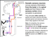
The nervous system is divided structurally into two parts:
Name these two parts and state what they consist of
Central nervous system (CNS)- Brain & spinal cord
Peripheral nervous system (PNS)- Nerves and ganglia (clusters of neuronal cell bodies) - outside the brain and spinal cord
What is the PNS functionally divided into?
Somatic PNS
Autonomic Nervous System

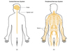
What is the role of the somatic PNS?
Controls motor and sensory function for the body wall, e.g. skin (sensory neuron), skeletal muscles (motor neuron)

What is the role of the autonomic nervous system?
Regulates function of the viscera: internal organs, smooth (involuntary) muscle, pupils, sweating, blood vessels, bladder, intestine, glands etc, and controls heart contraction rate.
Has sympathetic & parasympathetic arms.

What is the Autonomic nervous system also know as?
Visceral PNS
Vegatative NS
Involuntary NS
What are the two arms of the Autonomic nervous system?
Has Sympathetic and Parasympathetic arms
What does the term afferent axon mean?
propagate action potentials towards the brain & spinal cord from the PNS (e.g. sensory neurons, both somatic and ANS).

What does the term efferent axon mean?
Propagate action potentials from the brain and spinal cord to the periphery (e.g. motor neurons, both somatic and ANS)

What are internuerons?
Interneurons - CNS neurons that synapse with other CNS neurons within the brain and spinal cord

What is the function of the cerebellum?
Controls coordination of movement

What is the function of the brain stem?
Regulates vital functions (e.g. consciousness, breathing).
Damage here usually serious, can be fatal.

What is grey matter?
Grey matter, which has a pinkish-grey color in the living brain, contains the cell bodies, dendrites and axon terminals of neurons, so it is where all synapses are.

What is white matter?
White matter is found in the deeper tissues of the brain (subcortical).
It contains nerve fibers (axons), which are extensions of nerve cells (neurons).

Where does the CNS end?
The CNS ends at the margins of the spinal cord.

What are the Dorsal and Ventral roots that emerge from the spinal cord apart of?
PNS
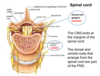
Lable the diagram below of a spinal nerve

The dorsal (posterior) or sensory root bears a dorsal root ganglion (DRG) containing the cell bodies of the sensory neurons.
In anatomy and neurology, the ventral root or anterior root is the efferent motorroot of a spinal nerve. At its distal end, the ventral root joins with the dorsal root to form a mixed spinal nerve.

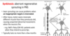
Lable the diagram below of a nueron


What is the function of a nueron?
To transmit and receive action potentials, or stimulate target tissue, e.g. to induce contraction of skeletal/smooth muscle, secretion from gland.

Distinguish between the regenerative capacities of injured axons in the central nervous system (CNS) and the peripheral nervous system (PNS).
Axons in peripheral nerves can regenerate after injury.
Axons in the CNS are unable to regenerate over long enough distances to be useful.

Why is recovery often compramised in the regenration of peripheral nerves?
What can this lead to?
Recovery is often compromised by non-specific target reinnervation & aberrant axon sprouting – e.g. can lead to neuropathic pain.
Axons in the CNS are unable to regenerate over long enough distances to be useful.
Why?
Inhibitory molecules in CNS but not PNS (e.g. myelin differences)
Absence of guidance cues that stimulate axon growth during development
Some loss of intrinsic axon growth capability by neurons

White matter comprises …………….. and …………….. axon tracts to and from the brain?
White matter comprises ascending and descending axon tracts to and from the brain.

Lable the diagram below


List the steps for how sensory imput percieved?
Use these key terms:
grey matter
Sensory cortex
ascending tracts
Perception of a sensory stimulus: sensory inputs activate sensory neurons in the spinal cord grey matter that transmit action potentials upward to the sensory cortex of the brain (ascending tracts).
Look at the image at the right