Anemias Flashcards
What is Plummer-Vinson Syndrome?
(1) Iron deficiency anemia with (2) esophageal web and (3) glossitis
Where is iron absorbed?
The duodenum via DMT1 receptors
How does iron get absorbed?
(1) Transported across enterocytes by ferroportin (2) Carried in blood by transferrin (3) Stored in liver and bone marrow macrophages by ferritin



What is an adaptation to slow loss of blood?
Increase in 2,3-bisphosphoglycerate, decreasing Hb affinity for oxygen to allow increased oxygen delivery to tissues
What are four causes of non-megaloblastic macrocytic anemia?
(1) Aplastic anemia
(2) Chronic liver disease
(3) Alcoholism
(4) Reticulocytosis
What are causes of megaloblastic macrocytic anemia?
(1) B12 deficiency
(2) Folate deficiency
(3) Myelodysplasia
(4) Erythroleukemia
(5) Drugs
(6) Orotic aciduria
What is performed in the initial lab assessment?
(1) CBCs
(2) Reticulocyte count
(3) Peripheral blood film exam (to identify abnormalities in number, morphology, or maturity)
What are four causes of iron deficiency anemia?
(1) Inadequate intake
(2) Increased need (pregnancy, infancy, pubertal growth)
(3) Decreased absorption
(4) Blood loss
What is the term for spoon shaped nails from IDA?
Koilonychia
How is IDA diagnosed?
(1) Low serum ferritin
(2) Low serum iron
(3) High TIBC
(4) Low transferrin saturation
What are the three stages of IDA?
(1) Iron deficient state (ID) - low ferritin
(2) Iron deficient erythropoiesis - serum iron, TIBC, %Tf sat, and RDW changing
(3) Iron deficiency anemia - decreased Hb, Hct, MCV, increased retic
How long after oral iron therapy does the reticulocyte count go up?
1 week
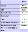
What happens in anemia of chronic disease?
Due to inflammatory state, (1) hepcidin goes up, which (2) blocks ferroportin and (3) inhibits erythropoiesis, (4) trapping iron in liver and bone marrow macrophages
What are the four major causes of sideroblastic anemia
(1) Congenital defect
(2) Alcoholism
(3) Lead poisoning
(4) Vitamin B6 deficiency
What step of protoporphyrin synthesis does congenital sideroblastic anemia involve?
Defective aminolevulinic acid synthetase (succinyl CoA to aminolevulinic acid, vit b6 as cofactor)
What does aminolevulinic acid dehydrogenase (ALAD) do?
Converts aminolevulinic acid to porphobilinogen
What attaches protoporphyrin to iron to create heme?
Ferrochelatase
What is the process of protoporphyrin synthesis?
(1) Succinyl CoA to ALA by ALA synthetase with vitamin B6 cofactor (rate limiting, affected in congenital sideroblastic anemia)
(2) ALA to porphobilinogen by ALA dehydrogenase
(3) Porphobilinogen to protoporphyrin in many steps
(4) Protoporphyrin to heme by ferrochelatase within mitochondria
Why does lead poisoning cause sideroblastic anemia?
Lead inhibits ferrochelatase and ALAD, leading to build up of ALA
What characteristic cells are seen in sideroblastic anemia?
Ringed sideroblasts with iron-laden mitochondria surrounding the nucleus
What are erythropoietic porphyrias and how are they clinically characterized?
Mutations in enzymes involved in heme synthesis, characterized by photosensitivity and anemia
What are the two types of hepatic porphyria?
(1) Porphyria cutanea tarda - photosensitivity and tea-colored urine
(2) Acute intermittent porphyria - polyneuropathy, abdominal pain, psych symptoms and red urine
What are hemochromatoses?
Iron overload due to ineffective excretion of iron
How does acquired hemochromatosis occur?
Repeated transfusions to treat severe, chronic anemias such as thalassemia major
How does hereditary hemochromatosis occur?
Dysregulation of iron metabolism, no feedback to reduce iron absorption when iron levels are high
Hereditary hemochromatosis (HFE) protein, which regulates hepcidin production in liver, is mutated
What are vitamin B6 and folate required for?
Synthesis of thymidine triphosphate (TTP) for DNA replication
(1) Folate circulates as methyl-THF
(2) Vitamin B12 transfers methyl group to homocysteine
(3) Methionine is formed by methyl + homocysteine, THF is free to enter nucleotide synthesis
What are five characteristics of megaloblastic anemia?
(1) Pancytopenia (decreased RBCs, WBCs, platelets)
(2) Increased MCV
(3) Decreased reticulocytes
(4) RBC inclusions (Howell-Jolly bodies)
(5) Hypersegmented (>5 segments) neutrophils
Where is folate absorbed?
The jejunum, uptake requires B12
What are five symptoms of folate deficiency?
(1) Weakness, pallor
(2) Jaundice
(3) Glossitis (high cell turnover requires folate for DNA synthesis)
(4) Gastritis (same as above)
(5) Neural tube defects
What drugs can cause folate deficiency?
(1) Methotrexate
(2) Trimethoprim
(3) Phenytoin
(4) 5-fluoruracil (5-FU)
Where is vitamin B12 absorbed?
The ileum, uptake requires binding by intrinsic factor secreted by parietal cells in the stomach
How is vitamin B12 absorbed?
(1) In the stomach, vitamin B12 is released from food after digestion by stomach acid
(2) Vitamin B12 is bound to R-binder protein secreted in the salivary gland
(3) Pancreatic proteasees detach vitamin B12 from R-binder
(4) Vitamin B12 binds intrinsic factor produced in the stomach and the complex is absorbed in the ileum
What are the causes of vitamin B12 deficiency?
(1) Inadequate intake (vegans)
(2) Increased requirement (pregnancy, infancy, pubertal growth, cancer)
(3) Impaired absorption (decreased gastric acidity, malabsorption due to disease, or competition with Diphyllobothrium latum tapeworm, H. pylori infection that destroys parietal cells and reduces IF secretion)
What can be used to differentiate folate and vitamin B12 deficiency?
Serum methylmalonic acid - normal in folate deficiency, high in vitamin B12 deficiency because B12 responsible for conversion to succinyl CoA
What is the cause of pernicious anemia?
(1) Autoimmune disease in which (2) autoantibodies to parietal cells or IF (3), preventing IF from binding vitamin B12
What is the Schilling Test?
Diagnostic test for pernicious anemia
Stage 1: Oral dose of vitamin B12 labeled with Co-57, measure Co in urine (<8% oral dose excreted in urine = PA)
Stage 2: Oral dose of vitamin B12 labeled with Co-57 + intrinsic factor, measure Co in urine (>8% oral dose excreted in urine)
What are some characteristic symptoms of vitamin B12 deficiency?
(1) Neuropsychiatric disorders (depression, dementia)
(2) Demyelinating neuropathies (loss of balance, peripheral neuropathy) because B12 required for fatty acid metabolism and myelination of spinal cord and peripheral nerves
Damage may be permanent
What is the normal retic count?
0.5-2.5%
What are three cellular characteristics of reticulocytes?
(1) Larger than RBCs
(2) No central pallor
(3) Bluish tinge from residual RNA


What is the body’s response to acute hemorrhage?
(1) First few hours, normal Hb, HCT, RBC, then movement of extravascular fluid into vascular space
(2) Untreated, increased retic count after 2-5 days, up to 15%
What serves as evidence for RBC regeneration in hemolytic anemia?
(1) Decreased erythroid:myeloid stem cell ratio in bone marrow
(2) Increased retics in peripheral blood ~3 days after hemolytic event
(3) Nucleated RBCs in severe anemia
(4) Elevated MCV because retics are larger than normal RBCs
What serves as evidence for hemolysis?
(1) Unconjugated bilirubin 2-3 days after hemolytic event
(2) Decreased serum haptoglobin because it binds hemoglobin
(3) Hemoglobinuria - if haptoglobin and hemopexin are depleted, Hb can’t be reabsorbed after filtration
(4) Increased lactate dehydrogenase, released from RBCs
What are some manifestations of acute hemolysis?
(1) Malaise
(2) Vomiting
(3) Platelet activation/thrombosis
(4) Renal failure
(5) Brown urine from methemoglobinuria in intravascular hemolysis
What are some manifestations of chronic hemolysis?
(1) Fatigue
(2) Jaundice
(3) Cholelithiasis (gallstones from bilirubin)
(4) Kernicterus in newborns (unconjugated bilirubin passes blood-brain barrier)
(5) Splenomegaly
(6) Bone deformities in children (excessive erythropoiesis)
What are three categories of intrinsic hemolysis?
(1) Membrane structural/functional defects (hereditary spherocytosis)
(2) Enzyme deficiencies (G6PD deficiency)
(3) Hemoglobinopathies
What is the difference between vertical and horizontal hereditary membrane defects?
Vertical defect - mutation in protein that disrupts linkage between transmembrane and cytoskeletal proteins
Horizontal defect - mutation in cytoskeletal protein that disrupts cytoskeleton
What causes hereditary spherocytosis?
(1) Defect in ankyrin, spectrin, or band 3 RBC cytoskeleton-membrane tethering proteins causing (2) membrane blebs that (3) get consumed by splenic macrophages
What are the clinical features of hereditary spherocytosis?
(1) Anemia
(2) Jaundice
(3) Splenomegaly
What are alternative causes of spherocytes?
(1) Burns that damage RBC membranes and (2) immune hemolytic anemia
What is the cause of paroxysmal nocturnal hemolysis (PNH)?
(1) Acquired membrane defect due to a (2) somatic mutation in the (3) PIGA gene, which (4) codes for glycosylphosphatidylinositol (GPI), (4) an anchor for CD55 (DAF, c3 convertase inhibitor) and CD59 (MIRL, blocks MAC formation)
Without GPI, CD55 and CD59 cannot bind to the cell membrane wall of RBCs, leading to destruction by complement
What are the clinical features of paroxysmal nocturnal hemolysis?
(1) Hemolytic anemia
(2) Pancytopenia
(3) Venous thrombosis
How does G6PD deficiency cause hemolysis?
(1) G6PD regenerates NADPH, which (2) reduces glutathione, (3) an antioxidant that neutralizes H2O2
Without G6PD, decreased NADPH, decreased reduced glutathione, leading to oxidative injury by H2O2 and intravascular hemolysis
Episodes of acute hemolytic anemia when challenged with oxidative stress
What are some causes of oxidative stress that lead to hemolysis in G6PD deficiency?
(1) Infections
(2) Fava beans
(3) G6PD IS PAIN:
Isoniazid, sulfonamides, primaquine, aspirin, ibuprofin, nitrofurantoin
What are the cellular features of G6PD deficiency?
Hemoglobin precipitates as (1) Heinz bodies that adhere to inner membrane of RBCs
(2) Bite cells form from HB consumption by splenic macrophages
How does pyruvate kinase deficiency lead to hemolysis?
(1) Pyruvate kinase converts pyruvate to lactate
(2) Without PK, RBCs are ATP-depleted
(3) ATP-depletion leads to rigidity and hemolysis
(4) Burr cells seen on blood smear

What are the two categories of extrinsic causes of hemolysis?
(1) Non-immune (microangiopathic hemolytic anemia, macroangiopathic, infectious agents, injury by drugs, chemicals, venoms, burns)
(2) Immune (autoimmune hemolytic anemia, transfusion reactions, hemolytic disease of fetus)
What are the three possible causes of microangiopathic hemolytic anemia (MAHA)?
(1) Thrombotic thrombocytopenic purpura (TTP)
(2) Hemolytic uremic syndrome (HUS)
(3) Disseminated intravascular coagulation (DIC)
What microorganism can be responsible for hemolytic anemia?
(1) Plasmodium
(2) Babesia
(3) C. perfringens
(4) Bartonella
What can lead to autoimmune hemolytic anemia?
(1) Chronic lymphocytic anemia (CLL), systemic lupus erythematosus (SLE), drugs cause warm-reactive antibodies to RBCs
(2) CLL, Mycoplasma pneumoniae, mononucleosis cause cold-reactive antibodies to RBCs
What do the direct and indirect antiglobulin tests look for?
(1) Direct looks for RBCs with bound anti-RBC IgG (add anti-human Ig to washed RBCs and look for agglutination)
(2) Indirect looks for anti-RBC IgG in serum (add RBCs to serum, wash, then add anti-human Ig and look for agglutination)
What are the causes of acquired aplastic anemia?
(1) Idiopathic
(2) Drugs (chloramphenicol, antiinflammatories)
(3) Viruses (Hep, EBV, HIV, Parvo B19)
What are the causes of inherited aplastic anemia?
(1) Fanconi anemia (FA) - mutations in DNA repair genes with risk of leukemia and tumors
(2) Dyskeratosis congenita (DK) - mutations in tolemerase complex genes with risk of epithelial cell cancers
What is pure red cell aplasia (PRCA)?
Decreased reticulocytes and erythroid precursors only, WBC and platelet counts normal
Acquired - idiopathic
Inherited - Diamond-Blackfan Anemia, mutations in ribosomal protein genes in stem cells
What are two causes of hypoproliferative states?
(1) Myelophthisic process - bone marrow pathologically replaced by abnormal cells, leading to anemia and teardrop cells
(2) Anemia of chronic kidney disease - decreased EPO production and RBC survival
What are three types of polycythemia and their causes?
(1) Polycythemia vera - myeloproliferative neoplasm, usually with JAK2 mutation
(2) Secondary polycythemia - pulmonary disease, high altitude, tumors with EPO production
(3) Relative polycythemia - decreased plasma volume from dehydration


How can the category of anemia (decreased production, increased destruction, blood loss) be quickly diagnosed?
Looking at retic count
How is anemia of chronic disease treated?
Treat cause of inflammation and give IV iron (oral iron not absorbed), which will increase RBC formation and decrease hepcidin